

Книги по МРТ КТ на английском языке / The Embryonic Human Brain An Atlas of Developmental Stages. Third Edition. 2006. By Ronan O'Rahilly
.pdf
ABBREVIATIONS
a. |
Artery |
Aff. |
Afferent |
A.-H. |
Adenohypophysis |
Amyg. |
Amygdaloid region, nuclei, complex |
Bas.a. |
Basilar artery |
BM |
Basement membrane |
BV |
Blood vessel |
C |
Cervical |
Cbl |
Cerebellar plate, cerebellum |
Ch. |
Chiasmatic plate |
Ch.tymp. |
Chorda tympani |
CN |
Cranial nerve |
CNS |
Central nervous system |
Comm. |
Commissural plate |
C-R |
Crown-rump length |
D1, D2 |
Diencephalic neuromeres |
Di |
Diencephalon |
Dors.f. |
Dorsal longitudinal fasciculus |
Eff. |
Efferent |
End.div. |
Endolymphatic diverticulum |
Ep. |
Epiphysis |
Ext.cbl |
External cerebellar swelling |
Ggl |
Ganglion |
GL |
Greatest length |
Hab.-interp.tr. |
Habenulo-interpeduncular tract |
Hem. |
Cerebral hemisphere |
Hipp. |
Hippocampus |
Hyp.-th.sulc. |
Hypothalamic sulcus |
Hyp.-th.-teg.-tr. |
Hypothalamotegmental tract |
Infund. |
Infundibulum |
Int.cbl |
Internal cerebellar swelling |
Interest.nu. |
Interstitial nucleus |
Isth. |
Isthmus rhombencephali |
Isth.rec. |
Isthmic recess |
I.-v.f. |
Interventricular foramen |
Lat.f. |
Lateral longitudinal fasciculus |
Lat.em. |
Lateral ventricular eminence |
L.l.f. |
Lateral longitudinal fasciculus |
LMP |
Last menstrual period |
Long.fiss. |
Longitudinal fissure |
L.t. |
Lamina terminalis |
M or Mes. |
Mesencephalon |
M1, M2 |
Mesencephalic neuromeres |
M.l.f. |
Medial longitudinal fasciculus |
Mam. |
Mamillary region, nuclei, body |
Mam. teg.tr. |
Mamillotegmental tract |
Med. fore.b |
Medial forebrain (or prosencephalic) |
|
bundle |
Med.tectobulb.tr. |
Medial tectobulbar tract |
Med.em. |
Medial ventricular eminence |
Mes. tr. 5 |
Mesencephalic tract of trigeminal |
|
nerve |
n. |
Nerve |
Nas. |
Nasal plate, pit, sac, septum |
N.-H. |
Neurohypophysis |
Not. |
Notochord |
NTD |
Neural tube defect |
Nu.mes.5 |
Mesencephalic trigeminal nucleus |
Nu.post.X |
Nucleus of posterior commissure |
Olf.bulb |
Olfactory bulb |
Olf.n. |
Olfactory nerve |
Olf.tub. |
Olfactory tubercle |
Opt. |
Optic sulcus, vesicle, cup |
Opt.stalk |
Optic stalk |
Ot. |
Otic plate, pit, vesicle |
Par. |
Parencephalon |
Post.-lat.fiss. |
Posterolateral fissure |
Post.X |
Posterior commissure |
Pros. |
Prosencephalon |
Red. nu. |
Red nucleus |
Rh. or Rhomb. |
Rhombencephalon; rhombomere |
Rh. A to D |
Specifically identified rhombomeres |
Rh. 1 to 4 |
Specifically numbered rhombomeres |
Rh.lip |
Rhombic lip |
Sp.ggl |
Spinal ganglion |
Subarach. |
Subarachnoid space |
Subth.nu. |
Subthalamic nucleus |
Sulc.lim. |
Sulcus limitans |
Supramam.X |
Supramamillary commissure |
Supra-opt.X |
Supra-optic commissure |
Syn. |
Synencephalon |
Tel. |
Telencephalon |
Tel.med. |
Telencephalon medium or impar |
Tent. |
Tentorium cerebelli |
Th.d. or Tha1.d. |
Dorsal thalamus |
Th.v. or Tha1.v. |
Ventral thalamus |
Tr. |
Tract(us); transversum |
V |
Ventricle |
V3, V4 |
Third ventricle, fourth ventricle |
Velum tr. |
Velum transversum |
V.-n. |
Vomeronasal nerve, ganglion |
X3 |
Commissure of the oculomotor nerves |
X4 |
Commissure of the trochlear nerves |
X sup.coll. |
Commissure of the superior colliculi |
1–12 |
Cranial nerves |
5i |
Intermediate trigeminal fibers |
5m |
Motor trigeminal fibers |
5s |
Sensory trigeminal fibers |
7i |
Nervus intermedius |
8c |
Cochlear component of |
|
vestibulocochlear nerve |
8v |
Vestibular component of |
|
vestibulocochlear nerve |

C H A P T E R 1
HISTORICAL ASPECTS
The Greek anatomist and physician, Erasistratus (ca. 300–250 BC), distinguished two convoluted parts of the brain, a greater and a lesser. Later, it became
clear that “the cerebrum and cerebellum are different in function as in form,” as Charles Bell wrote in 1811.
Although Malpighi in the 1670s observed the neural folds in the chick embryo, their significance escaped him, and he thought that the brain developed between them (Adelmann, 1966). Even von Baer was unaware that the neural folds are the primordium of the CNS, a role that was first appreciated by Rusconi and Reichert. Pander, however, understood that the neural folds form a tube. Nevertheless, the CNS was thought to be merely a fluid at first, and “the monotonous chronicle of vesicles and bubbles” (Adelmann) continued into the nineteenth century—if not, indeed, beyond it.
The vesiculae cerebrales, said to have been first noted by Coiter in 1573 (Tiedemann, 1816), were promoted by Meckel the younger, von Baer, and Bischoff, although the concept is largely a myth (Streeter, 1927). The current acceptance of three main divisions in the brain (prosencephalon, mesencephalon, and rhombencephalon) dates mainly from von Baer, who also identified the five subdivisions that appear a little later (telencephalon, diencephalon, mesencephalon, metencephalon, and myelencephalon). In 1893, Wilhelm His, senior, the founder of human embryology, added a sixth part, the isthmus rhombencephali, which becomes very evident during development.
SPECIFIC REFERENCES TO THE PRENATAL HUMAN BRAIN
Some key studies are mentioned here for convenience, but the list is not intended to be complete.
One of the earliest specific accounts of the prenatal human brain (Fig. 1–1) is that by Friedrich Tiedemann (1816), which was translated into French (1823) and English within a decade. The monograph is arranged according to nine prenatal months, but early examples of the human embryonic brain were inevitably lacking.
In 1896, the detailed atlas of the gross morphology of the brain by Gustaf Retzius appeared. In addition to the adult organ, more than 35 plates included the fetal brain from “the third to the tenth month,” the smallest being from two 35 mm fetuses. Retzius referred to the work of His on the embryonic brain; and he also cited Hochstetter.
In 1900 and 1901, Florence Sabin prepared solid (Born) reconstructions of the brain stem of a newborn, and the published illustrations were accompanied by a detailed description of some 90 pages.
In 1904, Wilhelm His, the real founder of human embryology (O’Rahilly, 1988), wrote an account of the prenatal human brain from 3.1 to 160 mm CR. His had been very interested in photography, and a striking feature of his book is the high quality of the photomicrographs. It is to be regretted that neither this work nor his Anatomie menschlicher Embryonen has been translated into English.
The Embryonic Human Brain: An Atlas of Developmental Stages, Third Edition. By O’Rahilly and Muller¨ Copyright C 2006 John Wiley & Sons, Inc.
1

2 |
C h a p t e r 1 : HISTORICAL ASPECTS |
Figure 1–1. Two early works on the prenatal development of the human brain: Tiedemann (1816) and His (1904).
In 1919, Ferdinand Hochstetter published a wellknown monograph on the human brain from 23 somitic pairs (stage 12) to 125 mm CR. It includes attractive illustrations of solid (Born) reconstructions and a series of photomicrographs of high quality. In 1923, the same author wrote an account of the development of the epiphysis cerebri. In 1929, he produced a further work, on the mesencephalon and rhombencephalon, based on a study of embryos from 6 mm GL to 250 mm CR. The illustrations include solid (Born) reconstructions and many photomicrographs of first-class sections. Hochstetter wrote also on the choroid plexuses (1913) and on the development of the meninges (1939).
A classical study of the neuromeres in the human was completed by Bartelmez in 1923, and many further details were provided in a neglected study by Bartelmez and Evans in 1936. The neuromeres were investigated again in the 1950s and 1960s by Bergquist and Kall¨ en´ .
In 1938, a detailed investigation of the development of the human CNS was written by Barbe´. This work, extending to 340 pages, covers the range from 14 mm CH to 470 mm CH (320 mm CR). Many photographs and photomicrographs are included. Unfortunately neither His nor Hochstetter is mentioned.
In 1944, a richly illustrated work on the external features of the later fetal brain (from “4 months”) was published by Fontes. A few years later (n.d.), some excellent external photographs (from “3 months”) were included in
a comparative study by Friant. The work of Fontes was unknown to Friant, as it still is to most authors.
In 1948, a very valuable series of reconstructions of the cranial arteries (in relation to the cranial nerves) was published by Padget, who subsequently (1957) produced a comparable work on the cranial venous system. The stages of the Carnegie embryos she studied are known. It needs to be appreciated that her “age groups” (of Streeter) are now the Carnegie stages and that her “stages” are phases of vascular development (Table 16–1).
In 1962, the very important study by Bartelmez and Dekaban appeared and was based on the Carnegie Collection. This was the first detailed investigation of the internal structure of the human brain in staged embryos, from stage 10 to stage 22, with the omission of stages 9, 18, 21, and 23. The account is more complete than that of Streeter (revised by O’Rahilly and Muller,¨ 1987a) and was not surpassed until the study of many embryos at each stage by Muller¨ and O’Rahilly began to be published in 1981–1990.
In 1965, an excellent and well-illustrated study of the globus pallidus and the corpus striatum was published by Richter. This important work was based on 13 embryos and 35 fetuses from 18.5 to 370 mm CR.
In 1969, a very valuable and well-illustrated investigation of the development of the human cerebral hemispheres was published by Kahle. The work is arranged from the “second month” to the “eighth month” and includes examples from 3 mm GL to more than 275 mm CR.

SPECIFIC REFERENCES TO THE PRENATAL HUMAN BRAIN |
3 |
In 1970, Windle published a noteworthy study of the “development of neural elements in human embryos of four to seven (postovulatory) weeks.” The examples range from about 3 to 16.5 mm GL. Although Streeter’s “horizons” were stated to be “helpful,” the embryos studied were unfortunately not staged. On the positive side, however, the use of Ranson’s pyridine–silver method enabled early neurons and neurofibrillary differentiation to be investigated thoroughly.
In 1975, much useful information concerning the normal and abnormal development of the human nervous sys-
tem was consolidated by Lemire, Loeser, Leech, and Alvord. Streeter’s “horizons” and postovulatory ages were used.
Important studies that have appeared since 1980 will be cited in appropriate places in the various chapters of this text.
The present work, published originally in 1994, is the first book devoted to the staged, embryonic human brain. It should be stressed, however, that very much further work on the fetal brain needs to be undertaken before a level of detail comparable with that now available for the embryonic brain can be attained.

C H A P T E R 2
TECHNIQUES
Anumber of matters related to human embryology in general, which also have particular relevance to human neuroembryology, will be summarized
in the first few chapters.
The present work is based largely on personal investigations of the embryonic brain, stage by stage, carried out over several decades and published in more than 50 research articles. The source material was mostly the Carnegie Embryological Collection and involved a careful study of 340 serially sectioned embryos (including 51 instances of silver impregnation), the preparation of graphic reconstructions from 89 brains, and the examination of 55 solid, three-dimensional reconstructions (modified method of Born). No similarly detailed and adequately illustrated documentation of the developing human brain had been previously attempted, and comparable detail for the fetal brain is not yet available. Moreover, because most studies of the developing brain of other mammalian species are based on timing, but not on staging, comparative tabulation in neuroembryology is still in its infancy, and a standard for the human is an advisable preliminary.
When the first morphological indication of the nervous system is distinguishable, the greatest length of the embryo is only 1 mm. Hence histological study is essential. Throughout the embryonic period, serial sections of excellent quality are indispensable. The examination of isolated or haphazard sections, or of those of poor histological quality, is almost useless and is frequently misleading.
At the end of the embryonic period proper, the diameter of the cerebral hemispheres is of the order of only
1 cm. This, coupled with the circumstance that many features are not at first sight identifiable (although most are present), as well as the rapidity with which changes occur, necessitates the preparation of enlarged reconstructions in order to investigate topographical relationships.
TRIDIMENSIONAL SERIAL RECONSTRUCTIONS
These are made by projecting serial sections at a previously determined magnification (Fig. 2–1). Many different techniques have been used (Gaunt and Gaunt, 1978) and several kinds of particular relevance here will be mentioned briefly.
(A)Solid reconstructions are those that can “sit on a table.” They are associated with the name of Gustav Born, who established the technique in 1876. It was adopted enthusiastically by Wilhelm His, the founder of human embryology (O’Rahilly, 1988). Such a reconstruction consists of stacked plates, each representing one or more enlarged serial sections. The laminae can be made of various substances. Originally wax was chosen, whereas those used here are made of plaster and were prepared by Mr. Osborn O. Heard. An example is shown in Figure 2–2. According to Carnegie usage, precise reconstructions are never referred to as “models.”
(B)Plane reconstructions, those designed to be reproducible on paper, are of several varieties.
The Embryonic Human Brain: An Atlas of Developmental Stages, Third Edition. By O’Rahilly and Muller¨ Copyright C 2006 John Wiley & Sons, Inc.
5
6 |
C h a p t e r 2 : TECHNIQUES |
A |
B |
C |
Figure 2–2. Example of a plaster reconstruction (made by Mr. |
|
Osborne Heard using a modified Born technique), showing a right |
|
lateral view of the brain at stage 17 (6 weeks). The rounded part in the |
|
lower right-hand corner is the right cerebral hemisphere. The large |
|
“V” in the upper middle portion is the fourth ventricle. The numerous |
|
horizontal lines show the plane of the histological sections and the |
|
way in which the plates were stacked. |
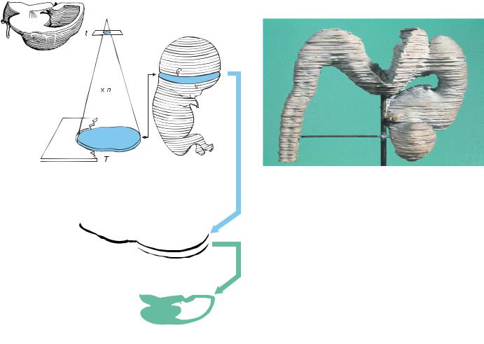
Figure 2–1. (A) The principle involved in solid three-dimensional reconstruction from serial sections. If each section, of thickness t, be projected at a magnification n, then each plate of the reconstruction should have a thickness T equal to t x n. (B) The plates were originally, and sometimes still are, made of wax and were stacked as shown in an embryo of stage 23 (8 weeks). Individual organs and structures can be reconstructed by this wax-plate technique (of Born). (C) The same principle used for a graphic reconstruction (made by F. Muller¨ with Perspektomat) showing a right lateral view of the brain at stage 23. The right cerebral hemisphere has been sectioned to reveal the lateral ventricle. The numerous horizontal lines indicate the plane of the histological sections. The bar represents 1 mm.
1.Point plotting graphic reconstructions, which were also used extensively by His and later by Florian (as well as by Beau, 1939, for the developing mammalian rhombencephalon), are made by superimposing the outlines of structures—the contours being obtained by projecting serial sections. Hidden lines are omitted. An example is shown in Figure 10–5. All the median views in the embryonic period and many of the lateral views in that period in this book are carefully prepared graphic reconstructions based on detailed examination of serial sections. These drawings and reconstructions are
the work of one of the authors (F.M.). A further technical advance, based on a modified pantograph, was introduced by Forster (1968) under the name Perspektomat and is patented. It was applied to human embryology by Muller¨ and O’Rahilly (1980, Figs. 5– 9) and is particularly useful for complicated structures, for correcting a markedly oblique plane of section, and for dissected views (Fig. 2–1).
2.Computerized graphic reconstructions are being used increasingly. Although most of those so far published are inadequate in precision and refinement, the standard may be expected to improve considerably.
It cannot be emphasized too strongly that firstclass histological quality is essential for precise reconstructions, and that reconstructions are absolutely necessary for a valid three-dimensional interpretation. Moreover, examination of sections taken in the three major planes is needed in order to follow certain structures, especially tracts, which disappear completely from view when sectioned through a right-angled bend.
3.Ultrasonic reconstructions can be prepared from images obtained in vivo by ultrasound (Fig. 23–35), as demonstrated by Blaas and associates (e.g., Blaas et al., 1995a, 1995b, 1998). Moreover, this procedure can be used in instances of prenatal anomalies, even during the first trimester (e.g., Blaas et al., 2000a, 2000b).
Techniques other than those concerned with reconstruction include (1) histochemistry and electron

TRIDIMENSIONAL SERIAL RECONSTRUCTIONS |
7 |
microscopy, which so far have been used to only a limited extent in human neuroembryology, (2) immunocytochemistry applied to staged embryos and correlated with in situ hybridization for expression of genes, (3) ultrasonic imaging and magnetic resonance imaging (MRI), which are used extensively in clinical practice. The results of these various techniques confirm the findings of embryological investigations and reconstructions.
Important
Imaging is valuable in prenatal diagnosis, but knowledge of the time of first appearance of various features, as well as the detailed morphology of the developing brain, depends on precise reconstruction from serial sections.