

Книги по МРТ КТ на английском языке / The Embryonic Human Brain An Atlas of Developmental Stages. Third Edition. 2006. By Ronan O'Rahilly
.pdf198 |
C h a p t e r 2 1 : THE FIRST APPEARANCE OF THE CORTICAL PLATE IN THE CEREBRAL HEMISPHERES |
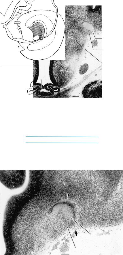
Figure 21–14. The ventral part of the lateral geniculate body at the level of the sulcus medius, and the entopeduncular nucleus of the subthalamus. The entopeduncular nucleus (globus pallidus internus) is at first at a more
caudal level than the globus pallidus externus.
21 – 14 
15
Lat. geniculate body
Marginal ridge
 Zona limitans intrathalamica
Sulcus
medius
Thal. v.
Entopeduncular nucleus
202 |
C h a p t e r 2 1 : THE FIRST APPEARANCE OF THE CORTICAL PLATE IN THE CEREBRAL HEMISPHERES |
|
|
Stage |
15 |
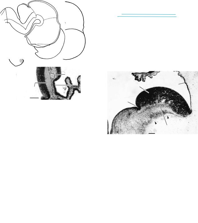
Figure 21–21. Right lateral views to show the |
|
13 |
|
1 |
1 |
|
ventricular cavities at approximately 4 /2–7 |
/2 |
|
|
|
weeks. The ventricles at stage 23 are illustrated |
|
|
|
in Figure 23–27. |
|
|
|
Growth of the medial and lateral ventricular |
|
|
|
eminences transforms the formerly |
|
|
|
hemispherical lateral ventricle into a C-shaped |
|
|
|
cavity, which is continuous rostrally with the |
|
|
|
olfactory ventricle. The first indication of |
|
|
|
anterior and inferior horns can be seen in stage |
|
|
|
21 in the lateral ventricle (O’Rahilly and Muller,¨ |
|
|
|
1990). |
|
17 |
|
21 |
|
External
contour of brain
Internal
THE FIRST APPEARANCE OF THE CORTICAL PLATE IN THE CEREBRAL HEMISPHERES |
203 |
A |
|
B |
C |
|
|
|
D |
|||
|
AICA
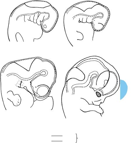
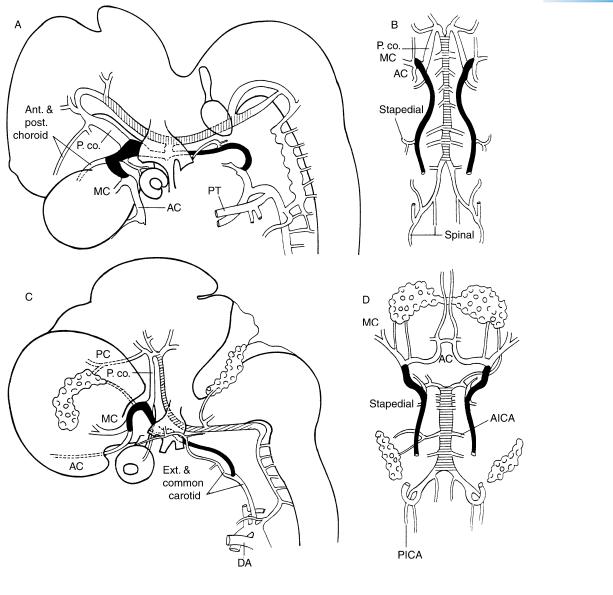
Figure 21–22. The arterial system at about 6 to 7 postfertilizational weeks. The internal carotid artery is shown in black, and the basilar by horizontal hatching. (A) and (B) The arteries at stage 17. New vessels that can now be identified include the anterior and middle cerebral (AC, MC), and the anterior and posterior choroid arteries. The vertebral artery is formed by anastomoses between the cervical segmental arteries.
(C) and (D) The arteries at stage 21 showing the anterior, middle, and posterior cerebral vessels. The anterior communicating artery is present, so that the circulus arteriosus is now complete. The choroid plexuses of the lateral and fourth ventricles are included. These drawings are based on graphic reconstructions made by Padget (1948), whose work should be studied for further details. The venous system at stages 18, 19, 20, and 21 has been illustrated by Padget (1957, Figs. 6, 9, 11, and 10). The arterial system at the end of the embryonic period is shown in Figures 23–33 and 23–34.
Abbreviations: AC, anterior cerebral; AICA, anterior inferior cerebellar artery; DA, ductus arteriosus; MC, middle cerebral; PC, posterior cerebral; P.co., posterior communicating; PICA, posterior inferior cerebellar artery; PT, pulmonary trunk.
204 |
|
C h a p t e r 2 1 : THE FIRST APPEARANCE OF THE CORTICAL PLATE IN THE CEREBRAL HEMISPHERES |
|
||||||||
|
|
|
|
|
|
|
|
|
|
|
|
|
|
4 weeks |
|
5 |
|
|
|
6 |
|
7 |
|
|
9 |
Stages 10 11 12 13 |
14 |
15 |
16 |
|
17 |
18 |
19 |
20 |
21 |
|
|
|
|
|
|
|
|
|
|
|
|
|
|
Inter- |
|
|
|
|
Post. spinal |
|
|
|
|
|
|
segmental |
|
Vertebral |
|
|
|
|
|
||
|
|
Longitudinal |
|
|
Ant. spinal |
|
PICA |
|
|
||
|
|
neural |
|
|
Basilar |
|
|
Labyrinthine |
|
|
|
|
|
|
|
|
|
Sup. |
|
|
|
|
|
|
|
|
|
|
|
AICA |
|
|
|
|
|
|
|
Trigeminal |
|
|
|
cerebellar |
|
|
|
|
|
|
|
|
|
|
|
|
|
|
|
||
|
|
|
CAROTICO- |
|
|
|
|
|
|
|
|
|
|
Otic |
|
BASILAR |
|
regresses |
|
|
|
|
|
|
|
|
ANASTOMOSIS |
|
|
|
|
||||
|
|
|
|
|
|
|
|
||||
|
|
|
|
|
|
|
|
|
|
||
|
Aortic |
|
|
|
|
|
|
|
|
|
|
|
arch |
Hypoglossal |
|
|
|
|
|
|
|
||
1 |
|
|
|
|
|
|
|
|
|
|
|
|
|
2 |
|
|
|
|
|
|
|
|
|
|
|
3 |
|
|
|
|
Ext. carotid |
|
|
|
|
|
|
|
|
|
|
|
|
|
|
||
|
|
Hyoid |
|
|
|
|
Stapedial |
|
|
|
|
|
|
c |
|
|
|
|
Post. choroid |
|
|
|
|
|
|
Post. communicating |
|
|
|
|
|
|
|||
|
|
Int. |
|
|
|
|
Post. cerebral |
|
CIRCULUS |
|
|
|
|
|
|
|
|
|
|
|
|||
carotid |
ARTERIOSUS |
|
|
|
Middle cerebral |
rOlfactory
Ant. cerebral |
Ant. communicating |
Ant. choroid |
|
Hyaloid |
New ophthalmic |
Ophthalmic |
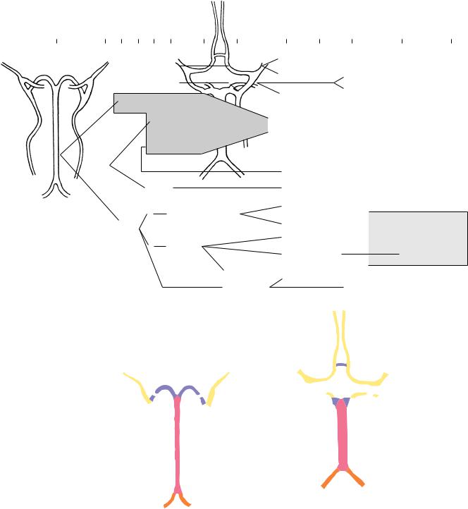
Development of circulus arteriosus
Stage 19 |
Stage 21 |
Figure 21–23. Above, summary of the arterial development. c, caudal division; r, rostral division. Below, the completion of the circuit by the formation of the anterior communicating artery.

THE INTERNAL CAPSULE AND THE OLFACTORY BULBS |
207 |
a
b
c |
(a) Sulcus limitans |
|
(b) Hypothalamic sulcus |
|
(c) Marginal ridge |
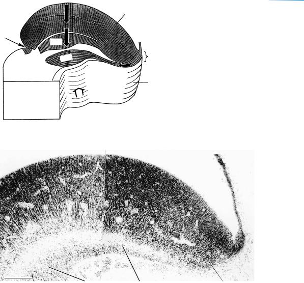
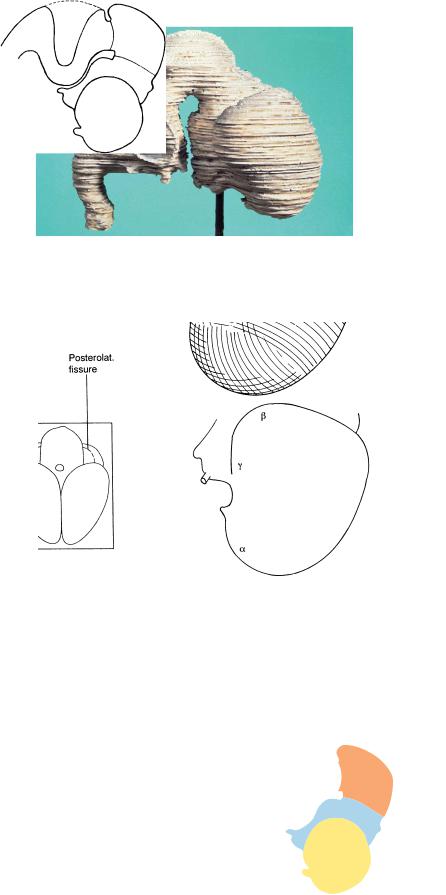
Figure 22–3. Graphic reconstruction prepared from transverse sections to show a median view of the brain. The asterisk indicates the junction with the spinal cord. The internal relief is shown, and the structures of the medial wall of the left hemisphere are projected as interrupted lines: the caudal extent of the cerebral hemispheres, the area dentata, and the hippocampus. The hippocampus extends between the occipital pole and the prosencephalic septum. The marginal ridge separates the diencephalon into halves: the dorsal thalamus on one side, and the ventral thalamus, subthalamus, and hypothalamus on the other. The pineal recess (arrow at right) is still undivided. The isthmic groove (upper arrow) and the isthmic recess (lower arrow) are also marked. The commissural plate has become thicker. No fusion takes place during the embryonic period proper between the cerebral hemispheres, as was clearly understood and stated by Hochstetter (1919).1 The approximate levels of Figures 22–7 to 22–12 are indicated, although the fit does not correspond completely, because these are from different embryos. (For example, in Figure 22–7 the interpeduncular fossa is slightly more dorsal than in Figure 22–3.) A three-dimensional reconstruction, prepared ultrasonically, of the ventricles in vivo in an embryo of 24 mm is illustrated by Blaas et al. (1995b).
1“Nicht der kleinste Umstand aber deutet darauf hin, dass die zu beobachtende Dickenzunahme etwa auf eine Verwachsung der unmittelbar an die Kommissurenplatte anschliessenden Abschnitte der medialen Hemispharenw¨ ande¨ zuruckzuf¨ uhren¨ ware”¨ (p. 109). See also definition of Massa commissuralis in the Glossary.