

Книги по МРТ КТ на английском языке / The Embryonic Human Brain An Atlas of Developmental Stages. Third Edition. 2006. By Ronan O'Rahilly
.pdf148 |
C h a p t e r 1 8 : THE FUTURE CORPUS STRIATUM, THE INFERIOR CEREBELLAR PEDUNCLES |
Sulcus limitans
Somatic & visceral afferent
Visceral efferent
Somatic efferent
H |
|
|
||
G |
|
|
||
E, F |
|
|
||
C |
|
|
||
D |
|
|
||
|
|
|
|
B |
|
|
|
|
A |
Figure 18–19. (Continued ) |
|
|
||
|
|
|
|
|
TABLE 18–1. Longitudinal Zones of the Diencephalon |
|
|
||
|
Zone |
Fig.a |
Sulcus |
|
|
|
|
|
Di-telencephalic sulcus |
|
|
|
|
|
|
1. Epithalamus |
|
|
|
|
|
|
|
|
|
2. Dorsal thalamus |
16–7E |
|
|
|
|
|
17–15 |
|
|
|
|
18–6 |
|
|
|
|
|
|
Thalamus |
17–16 |
Sulcus medius, marginal ridge, |
||
|
|
|
18–6 |
zona limitans intrathalamica |
|
|
|
18–7 |
|
|
|
|
|
|
|
3. Ventral thalamus |
16–7E |
|
|
|
|
|
18–6 |
|
|
|
|
|
|
|
|
|
|
Hypothalamic sulcus |
|
|
|
|
|
|
4. Subthalamus |
18–6 |
|
|
|
|
|
|
|
|
|
|
|
Faint, unnamed sulcus |
|
|
|
|
|
Hypothalamus sensu lato |
|
|
||
|
5. Hypothalamus sensu stricto |
18–6 |
|
|
|
|
|
|
|
a The figure references are to photomicrographs. Further details are given in Table 20–1.

C H A P T E R 19
STAGE 19: THE CHOROID
PLEXUS OF THE FOURTH
VENTRICLE AND THE
MEDIAL ACCESSORY
OLIVARY NUCLEUS
Approximately 16–18 mm in Greatest Length;
Approximately 44 Postfertilizational Days
Caution needs to be exercised in assigning an embryo to stages 19–23, and details of internal structure are particularly important here. The embryo
(which is human since the time of fertilization) now has a recognizably human face (Padget, 1957).
The olfactory nerve, bulb, and tubercle are welldeveloped. The nucleus accumbens appears. The cerebral hemispheres have grown rostrally, so that the prosencephalic septum has become larger. A paraphysis may be present and is telencephalic. Optic fibers arrive in the chiasmatic plate. The globus pallidus externus has developed in the diencephalon. The dorsal and ventral thalami are demarcated by the marginal ridge, and the dorsal thalamus shows an intermediate layer. The habenular region and its connections are well formed. Many bundles are identifiable, including the thalamostriatal and the stria medullaris thalami. The fourth ventricle possesses lateral recesses and a choroid fold. Cerebellar connections are increasing. The medial accessory olivary nucleus is
beginning to form. Electrical activity can be detected in the brain stem at about stages 18 and 19 (Borkowski and Bernstine, 1955). Three semicircular ducts are isolated. The basicranium comprises occipital and sphenoidal parts.
Precision
Basal nuclei is the correct term, not basal ganglia, because, by definition, ganglia are found only in the peripheral nervous system.
Important
The isthmus contains the trochlear decussation and its alar lamina contributes to the cerebellum and the rostral medullary velum. Its site in the adult is indicated by the trochlear nerves.
The Embryonic Human Brain: An Atlas of Developmental Stages, Third Edition. By O’Rahilly and Muller¨ Copyright C 2006 John Wiley & Sons, Inc.
149

150 C h a p t e r 1 9 : CHOROID PLEXUS OF THE FOURTH VENTRICLE AND THE MEDIAL ACCESSORY OLIVARY NUCLEUS
Figure 19–1. An embryo of stage 19 surrounded by the intact amnion (limiting the amniotic cavity) and the bisected leafy mass of the chorion (limiting the extra-embryonic coelom). The fourth ventricle is clearly visible “behind” the cerebellar plate. The umbilical vesicle appears as a small sphere at the lower right-hand corner. Villous stems are visible in the future chorion frondosum. Photography by Mr. Chester F. Reather.
CHOROID PLEXUS OF THE FOURTH VENTRICLE AND THE MEDIAL ACCESSORY OLIVARY NUCLEUS |
151 |
V4
Int. & ext. cerebellum
12 |
5 |
|
|
|
9 |
11
10
M
Di.
g b
T
a
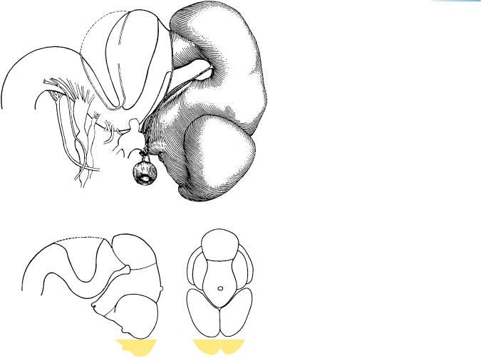
Figure 19–2. A lateral view of the brain. An occipital pole (β in the key drawing) can be clearly identified for the first time. The olfactory bulb and tubercle are visible. All cranial nerves are present, although the well-developed olfactory nerve (Figs. 19–12 and 19–14) is not shown here. The roof of the fourth ventricle is indicated by an interrupted line. The cerebellar plate is “vertical” and at almost a right angle to the longitudinal axis of the rest of the rhombencephalon. The inset (end-on view) demonstrates that the cerebellar plates are still wide and that their width equals that of the cerebral hemispheres. It is of interest to mention that the insular region was identified by Rager (1972, Fig. 7) in an intact embryo of 17 mm. Partly from a drawing by Mary M. Cope (Gilbert, 1957).
152 C h a p t e r 1 9 : CHOROID PLEXUS OF THE FOURTH VENTRICLE AND THE MEDIAL ACCESSORY OLIVARY NUCLEUS
a
(a) Sulcus
limitans
(b) Hypothalamic
sulcus
b
(c) Marginal ridge
c
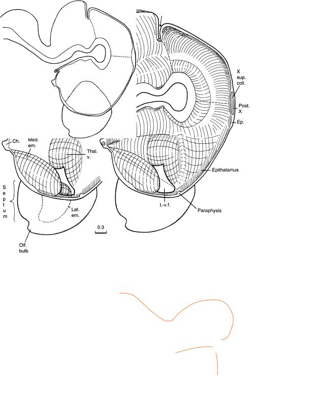
Figure 19–3. Graphic reconstruction prepared from transverse sections to show a median view of the brain. The asterisk indicates the junction with the spinal cord. Clearly the cerebral hemispheres have grown rostrally, and hence the prosencephalic septum (inset) has increased in size. The nuclei in the septum will be discussed with the next stage. The interventricular foramen is narrow and is bounded mainly by the ventral thalamus (dorsally), the medial ventricular eminence (ventrally), and the hippocampal region (rostrally). Some embryos possess a paraphysis, which opens into the telencephalic part of the third ventricle. The choroid plexus becomes visible by ultrasonography in vivo at 7 postovulatory (9 postmenstrual) weeks.
CHOROID PLEXUS OF THE FOURTH VENTRICLE AND THE MEDIAL ACCESSORY OLIVARY NUCLEUS |
153 |
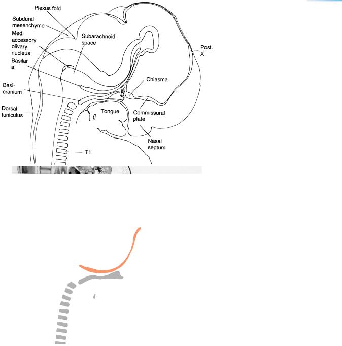
Figure 19–4. Median section of the brain and the related cartilaginous skeleton. The chiasmatic and commissural plates are important landmarks. The choroid fold of the fourth ventricle is evident, and the subdural mesenchyme is a fluid-filled meshwork. Among the injected arteries, the basilar is especially well visible. Fine vessels can be seen in the area of the future falx cerebri. The dorsal funiculus appears as a thick bundle in the spinal cord. The median raphe (His, 1890), known as the septum medullae rhombencephali, consists of sagittally arranged fibers derived from the embryonic floor plate and representing the adult floor plate (Sidman and Rakic, 1982). In the photomicrograph, it is clearly visible above the basilar artery.
The basicranium (reconstructed by O’Rahilly and Muller,¨ 1984b) now consists of an occipital part (developed from sclerotomes 1–4) and a sphenoidal part, which has grown rostrally to reach the adenohypophysis. Although not visible here, the orbital wing of the sphenoid (mainly the postoptic root) is forming.
←−−−−−−−−−−−−−−−−−−−−−−−−−−−−−−−−−−−−−−−−−−−−−−−−−−−−−−−−−−−−−−−−−−−−−−−−−−−−−−−−−−−−−−−
Figure 19–3. (Continued ) The di-telencephalic borderline is indicated by an interrupted line between the velum transversum and the preoptic sulcus. The first optic fibers arrive in the chiasmatic plate but do not yet cross to the opposite side. The commissure shown is formed by supraoptic fibers. The epiphysis cerebri is adjacent to the posterior commissure and contains several follicles. At stages 19–21 it corresponds in general to Stadium IV of Turkewitsch (1933), as pointed out by O’Rahilly (1968). The globus pallidus externus has developed in the subthalamus. The dorsal and ventral thalami are well-separated from each other by the marginal ridge associated with the sulcus medius at the ventricular side and with the zona limitans intrathalamica (Fig. 18–6). The dorsal thalamus possesses an intermediate layer. The epithalamus is well-delimited, and its habenular nucleus (Fig. 19–13) has sent fibers (the habenulo-interpeduncular tract) to the midbrain since stage 17. The fibers arriving in the habenular area constitute the stria medullaris thalami, and arise mostly in the nucleus ovalis of the diencephalon (Figs. 19–9 and 19–23). Medial and lateral ventricular eminences, seen partly by transparency, are shown in the inset.
The isthmic groove between the mesencephalon and the cerebellum is indicated by a short arrow. The lateral recess and choroid fold of the fourth ventricle are shown. The floor of the rhombencephalon is thickened, and this will become more pronounced with the addition of further fibers in subsequent stages. The medial accessory olivary nucleus (Fig. 19–4) is beginning to form in between the roots of the hypoglossal nerve, and its site is indicated schematically by a projection onto the median plane. The lemniscal decussation is visible.
The characteristic hairpin bend of the mesencephalic flexure is formed by the metencephalon and the diencephalon, and indicates the site of the future pontine and interpeduncular cisterns of the subarachnoid space. The apex of the flexure marks the exit of the oculomotor nerves. The pontine flexure is eliminated later, so that the medulla and pons are in a straight line postnatally.
154 C h a p t e r 1 9 : CHOROID PLEXUS OF THE FOURTH VENTRICLE AND THE MEDIAL ACCESSORY OLIVARY NUCLEUS
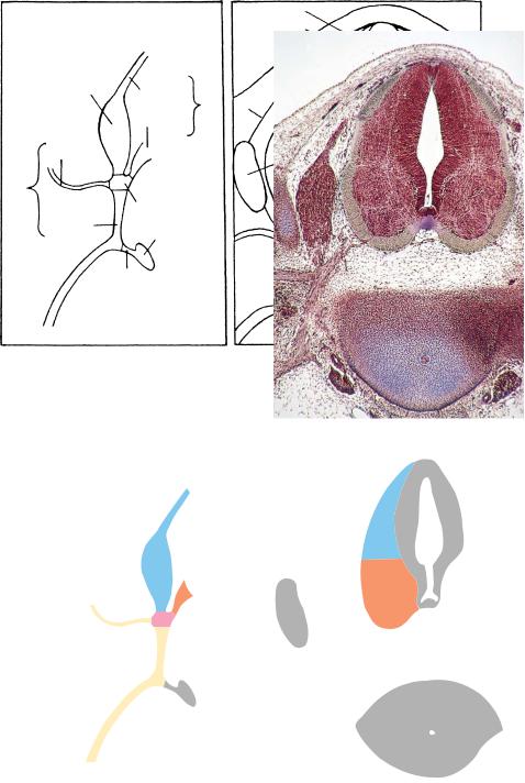
Figure 19–5. Section through the neural tube, clarified in the key below the photomicrograph. The three zones of the neural tube are identified. A spinal
ganglion is visible at the left (medial to the neural arch). The key at the left shows the formation of a spinal nerve.
An atlas of the developing spinal cord is available (Bayer and Altman, 2003).
|
|
Ventricular |
|
|
|
|
Intermediate |
|
|
|
|
Marginal |
|
|
Spinal |
|
|
|
|
ganglion |
Dorsal |
|
|
|
|
Dura |
|
A |
|
|
root |
|
||
|
|
|
|
|
|
Ventral |
|
|
Sulcus |
|
root |
|
|
limitans |
Dorsal |
|
|
Neural |
B |
ramus |
|
|
arch |
|
Spinal n.
Ventral |
|
Dura |
Sympathetic |
|
|
ramus |
|
|
ganglion |
|
|
|
|
|
|
Ramus |
Notochord |
|
|
|
|
communicans |
|
|
|
Centrum |
CHOROID PLEXUS OF THE FOURTH VENTRICLE AND THE MEDIAL ACCESSORY OLIVARY NUCLEUS |
155 |
||||
|
|
|
|
|
|
|
|
|
|
|
|
|
|
|
|
|
|
|
|
|
|
|
|
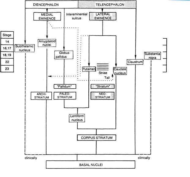
Figure 19–6. Scheme of the basal nuclei showing the development of their diencephalic and telencephalic components. Stages of appearance are also included. Late in the embryonic period, the amygdaloid complex as well as the globus pallidus externus will occupy telencephalic territory. Whether the amygdaloid complex and the claustrum should be included with the other basal nuclei is doubtful. The basal nuclei are best defined as the basal structures affected pathologically in so-called extrapyramidal motor diseases.
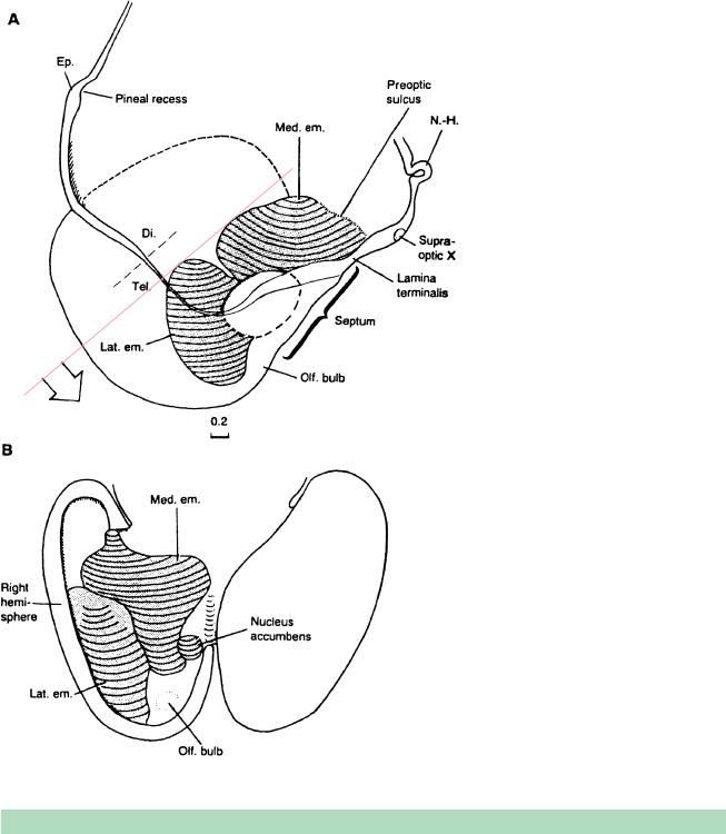
Figure 19–7. Reconstruction of the developing basal nuclei (transitional between stages 18 and 19). (A) Medial view of the right half of the forebrain, in which the medial wall of the right cerebral hemisphere is shown as if transparent. The region of the amygdaloid nuclei is projected as an oval. (B) Dorsal view of the cerebral hemispheres, with the right hemisphere opened to illustrate its basal aspect from the interior. The medial ventricular eminence has almost completely invaded telencephalic territory (cf. Fig. 19–3). The lateral eminence represents the main primordium of the future corpus striatum (Fig. 19–6). From O’Rahilly, Muller,¨ Hutchins, and Moore (1988).
The medial ventricular eminence has been found in the rat to resemble the globus pallidus histochemically far more than it does the lateral eminence, and it is connected to the dopamine pathway that leads from the ventral tegmental region of the midbrain to the forebrain.
The nucleus accumbens develops as a separate entity far earlier than in rodents, and projects as a thickening into the ventricular cavity. It was shown by Hewitt (1958, Fig. 3) in an early human fetus, where it appeared as the elevation known as the “paraterminal body.” It is regarded by various authors as a ventromedial extension of the caudato-putamen complex. Its superficial component includes the nucleus of the diagonal band (Kuhlenbeck, 1973). The nucleus accumbens has been shown in the human to contain dopamine, and it resembles the caudato-putamen complex in its histochemical development (Brana et al., 1995).
TABLE 19–1. Comparison of Medial and Lateral Ventricular Eminences
Medial Ventricular Eminence |
Lateral Ventricular Eminence |
(Archistriatum) |
(Neostriatum) |
|
|
Medial eminence of diencephalic origin gives rise to most of |
Neocortical GABA-expressing interneurons are believed to |
the early amygdaloid nuclei (medial, anterior, basolateral, |
be derived (in the mouse) from the lateral eminence |
and part of lateral) |
Lateral eminence invaded by the cortical nucleus of the |
|
amygdaloid body in and after stage 19. |
Medial eminence with scant striatum-like tissue. Neurochem- |
Lateral eminence characterized by striatum-like tissue, which |
ical resemblance to globus pallidus |
is acetylcholinesterase-positive and dopamine-receptive |
Short proliferative phase |
Long proliferative phase |
|
|
|
|
Medial |
Medial |
|
Anterior |
|
||
|
|
|
forebrain |
|
Baso- |
bundle |
|
|
|
lateral |
|
|
|
Diagonal |
|
|
(septal |
|
|
nucleus) |
Med. em.
Olf. bulb
& tub.
Thal. d.
Thal. v
amygdaloid nuclei
Figures 19–8 to 19–10 show neocortical areas and choroid plexuses. The bars represent 0.1 mm. The inset drawings on this page show the amygdaloid nuclei.
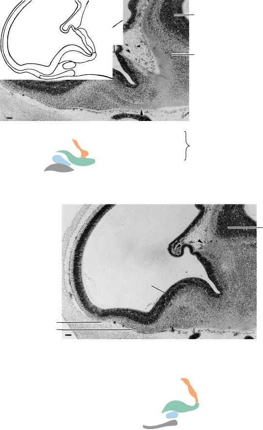
Figure 19–8. Sagittal section showing cerebral hemisphere, choroid plexus (rectangle), medial prosencephalic (forebrain) bundle (arrowhead), and dorsal and ventral thalami. Connections between the amygdaloid nuclei in the medial eminence and the diencephalon belong mostly to the medial prosencephalic bundle, which is the main pathway for longitudinal connections in the hypothalamus (Fig. 19–23). The lateral ventricle is invaded by the choroid fold, which, thicker than that of the fourth ventricle, consists of a multilayered epithelium and a mesenchymal core with thin-walled blood vessels.
Thal. d.
Figure 19–9. Another section, to show the olfactory bulb and tubercle, and the medial ventricular eminence. The stria medullaris thalami (arrowhead at right) arises in the diencephalon from the nucleus taeniae or ovalis (Fig. 19–22), although minor contributions come from the lateral prosencephalic bundle. The choroid fissure is visible (arrowhead at left).