

Книги по МРТ КТ на английском языке / The Embryonic Human Brain An Atlas of Developmental Stages. Third Edition. 2006. By Ronan O'Rahilly
.pdf58 |
|
C h a p t e r 1 1 : CLOSURE OF THE ROSTRAL NEUROPORE |
|
|
|
D |
E |
A |
B |
C |
|
Figure 11–8. Development of the notochord and the floor plate.
(A)After the notochord has developed from the dorsal part of the notochordal process of stages 7 and 8, its cross-sectional shape changes.
(B)It appears as the notochordal plate in stage 9 (Fig. 9–5B). (C, D) It has the form of an inverted U in stages 10 and 11 (Figs. 10–11 and 11–9). (E) Next it appears as a rod in stage 12 (Fig. 10B).
Notochord
Figure 11–9. The notochord in this embryo of stage 11 with 16 pairs of somites still has the form of an inverted U at the level of rhombomere 5. Bar: 0.05 mm.
Figure 11–10. Secondary development of the body. The caudal part of the notochord, caudal to the site of the former neurenteric canal, which is formed from the caudal eminence, in an embryo with 17 pairs of somites. The neural groove above is still open. Bar: 0.04 mm.

CLOSURE OF THE ROSTRAL NEUROPORE |
59 |
A
Aorta
Heart
BCapital v.
RV
LV
Internal carotid a.
C
2
1
Figure 11–11. The early blood vessels. The movement of the blood would be merely ebb and
Otic disc flow. The still open brain would not yet be dependent on a blood supply. (A) At stage 9 (2, 3
somites). (B) At stage 10 (10 somites). (C) At stage
11 (14 somites).
Aorta
Omphalo-
mesenteric v.
Aorta
Umbilical v.
Umbilical
plexus
Cardinal vv.
Cono-
truncus L. & R. ventricles

60 |
C h a p t e r 1 1 : CLOSURE OF THE ROSTRAL NEUROPORE |
NEUROTERATOLOGY:
ENCEPHALO(MENINGO)CELE
Encephalocele and encephalomeningocele may arise, at least in some instances, from a failure in separation of the brain from the surface ectoderm in later stages. This failure in separation would be caused by a disturbance of the mesencephalic neural crest, which is at the height of its formation at stage 10. This mesenchyme is believed to be the main source for the development of the face, and insufficient production of it may lead to a narrow facies, as seen in the fetal alcohol syndrome.
Encephaloceles are, in the Western Hemisphere, most frequently found in the occipital region. They may perhaps be caused by a lack of the basal mesenchyme originally between the neural tube and the notochord; its quantity is accumulating rapidly.
Fronto-ethmoidal encephaloceles may be at or near the situs neuroporicus (as proposed by Hoving from studies
of the rat). It has been speculated (by Hoving, 1993) that insufficient cell death (apoptosis) at the terminal lip of the rostral neuropore (e.g., from retinoic acid deficiency) might result (immediately after fusion of the neural folds) in persistent attachment of the surface ectoderm to the neural ectoderm, producing a subsequent cranial defect and a fronto-ethmoidal encephalocele.
An interesting example of a telencephalic diverticulum in a 35 mm fetus has been recorded (Bossy, 1966). It was situated in the region of the former situs neuroporicus and was considered to represent cerebral dysraphia with secondary encephalocele.
Nasal Glioma. Insufficient cell death in the region of the rostral neuropore causes a persistent “surplus of unintended surviving cells” that “might be represented by heterotopias like nasal gliomas” (Hoving, 1993).

C H A P T E R 12
STAGE 12: CLOSURE OF THE CAUDAL NEUROPORE AND THE BEGINNING OF SECONDARY NEURULATION
Approximately 3–5 mm in Greatest Length;
Approximately 31 Postfertilizational Days
The rostral neuropore is closed, although the situs neuroporicus can frequently be detected and is probably at the future commissural plate in the mid-
dle of the embryonic lamina terminalis. The closure (during stage 11) allows the formation of the telencephalon medium to occur. The mesencephalon consists of two neuromeres (M1 and M2), and the mesencephalic flexure is a right angle. The rhombomeres have important
relationships to the cranial ganglia: Rh.5 is associated with the otic vesicle, and Rh.D is level with the four occipital somites. The first nerve fibers are differentiating, particularly those of the future lateral and ventral longitudinal fasciculi. The hypoglossal nucleus has appeared and four to five intramural hypoglossal roots are present. The caudal neuropore closes during stage 12, and its final prenatal site is at the level of somitic pair 31 (future vertebral level S2).
The Embryonic Human Brain: An Atlas of Developmental Stages, Third Edition. By O’Rahilly and Muller¨ Copyright C 2006 John Wiley & Sons, Inc.
61

62 C h a p t e r 1 2 : CLOSURE OF THE CAUDAL NEUROPORE AND THE BEGINNING OF SECONDARY NEURULATION
Figure 12–1. Right lateral view of an embryo of stage 12 with the brain superimposed. The number of somitic pairs was 28; the range in number at this stage is 21–29.
M
Di.
T
Figure 12–2. Right lateral view of the brain. The features shown are the optic vesicle, the trigeminal and facio-vestibulocochlear ganglia, the otic vesicle, and the glossopharyngeal and vagal-accessory ganglia. The brain now occupies about 40% of the length of the neural tube. Expansion of the brain stretches the surface ectoderm (O’Rahilly and Muller,¨ 1985) so that entrance of mesenchymal cells may be hindered. The prechordal mesenchyme is caudal to the optic vesicles, where the orbital muscles will develop. The optic neural crest (Fig. 12–5) is now at the height of its development. The neural crest contributes to the trigeminal and facio-vestibulocochlear ganglia. Crest cells from the region of ganglia 9–11 migrate, by way of pharyngeal arch 3, towards the aortic sac. Moreover, the surface (epipharyngeal) ectoderm gives rise to cells that may contribute to the facial, inferior glossopharyngeal, and vagal ganglia. The hypoglossal cell cord is forming from the occipital somites and, in the more advanced embryos of this stage, it begins to enter pharyngeal arch 4.

CLOSURE OF THE CAUDAL NEUROPORE AND THE BEGINNING OF SECONDARY NEURULATION |
63 |
Figure 12–3. Reconstruction of the brain. The asterisk indicates the junction with the spinal cord. The rostral neuropore has closed. The site of final closure (the situs neuroporicus) is probably at the future commissural plate in the middle of the embryonic lamina terminalis.
The initial formation of the telencephalon medium normally depends on the closure of the rostral neuropore during stage 11. The prosencephalon now consists of three parts: the telencephalon, D1, and D2. The telencephalon medium, which is rostral to the well-defined neuromere D1, is recognizable externally by its situation between the nasal discs (Fig. 12–7). D1 is still characterized mainly by the optic vesicles. The semilunar opening of the optic ventricle into the third ventricle is indicated by shading. D2 is related to the adenohypophysial primordium and now shows the first sign of the mamillary recess.
The mesencephalon consists of two neuromeres: M1 and M2. The mesencephalic flexure is a right angle between the forebrain and the hindbrain. The sulcus limitans is seen to traverse the midbrain.
The various rhombomeres have distinctive features. For example, the floor is expanded at the level of Rh.2, 4, and 6. Rh.5 is related to the otic vesicle, and the ganglia of the future cranial nerves (which are projected onto the median view) are important relationships of Rh.2, 4, 6, and 7. Rh.D is at the level of somites 1–4. The first nerve fibers are now differentiating. Apart from short fibers in ganglia 5 and 7/8, the fibers are mostly related to cells that will form the nucleus of the lateral longitudinal fasciculus. Moreover, fibers of the future ventral longitudinal fasciculus are discernible in Rh.7. Four intramural hypoglossal roots are present, i.e., the hypoglossal nucleus has developed and some hypoglossal neural crest is still present dorsally. The relative length of the rhombencephalon has been decreasing since stage 9.
Figure 12–4. An almost median section of the brain showing rhombomeres 2–7 and two occipital somites.
64 C h a p t e r 1 2 : CLOSURE OF THE CAUDAL NEUROPORE AND THE BEGINNING OF SECONDARY NEURULATION
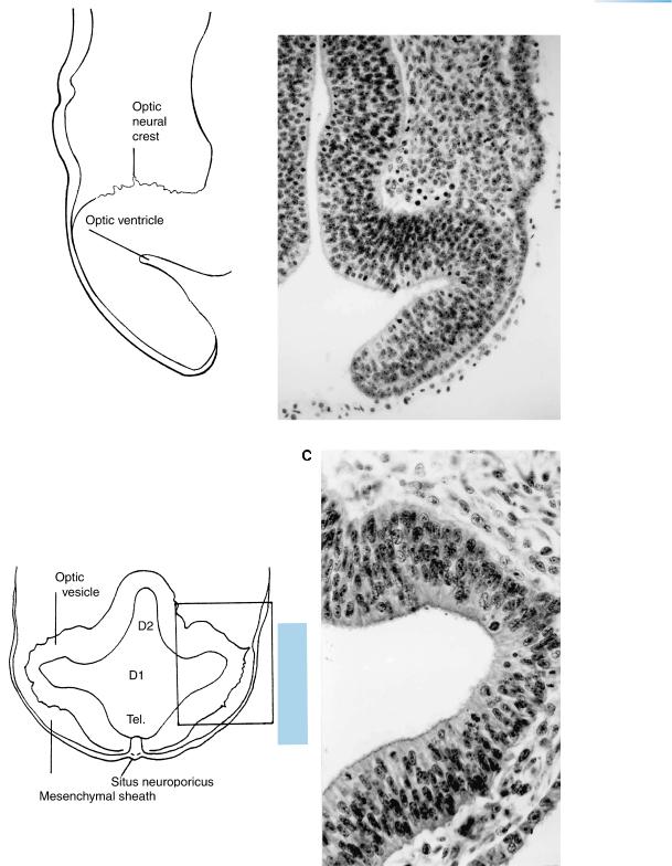
Optic
crest
Opt. D1
Chiasmatic
plate
Oropharyngeal
epithelium
Figure 12–5. Optic vesicles and D1 in a horizontal section. A mesenchymal sheath separates the optic vesicles from the surface ectoderm. Optic neural crest is evident and gives the optic vesicle the appearance of a “frightened hedgehog” (Bartelmez) externally (Fig. 12–2). Little or no mesenchyme is present between the dorsal part of D1 and the surface ectoderm.
Fig. 12 – 5
M
5
7,8v
Fig. 12 – 6
Ot.
Figure 12–6. Rhombencephalon. Several cranial ganglia and the otic pits are visible. The surface ectoderm covering the ventral part of pharyngeal arch 1 (arches 2 and 3 are also visible here) is thicker and gives off cells that join the trigeminal ganglion. The ectoderm is a part of the ectodermal ring (O’Rahilly and Muller,¨ 1985).
CLOSURE OF THE CAUDAL NEUROPORE AND THE BEGINNING OF SECONDARY NEURULATION |
65 |
Situs neuroporicus Tel. medium Nasal disc
Optic
crest
D1
Figure 12–7. Optic vesicles and D1 in a horizontal section of another embryo. Optic neural crest is evident. At this time the diencephalon consists of two neuromeres: D1, related to the optic vesicle, and D2, related to the adenohypophysis. The site of the closed rostral neuropore is conspicuous. The nasal disc becomes bilaminar and becomes shifted laterally. Bar: 0.1 mm.
Sternberg (1927), who placed the rostral neuropore in the region of, and probably dorsal to, the commissural plate, ventured to identify the situs neuroporicus even up to the end of the embryonic period, at which time he showed it between the eyes and at the root of the nose.
D2
Fig. 12 – 8
Fig. 12 – 7
Rh.
2
Rh.
5
5
7,8v
10,11 |
Figure 12–8. Rhombencephalon of still another |
|
embryo. Several cranial ganglia and the otic |
||
|
vesicles are visible. The uppermost part of the |
|
Occipital |
photomicrograph depicts the mesencephalon and |
|
is followed by rhombomeres, which are clearly |
||
somite |
||
delimited by evaginations. The trigeminal |
||
|
||
|
ganglion is related to Rh.2, the facial to Rh.4, the |
|
|
glossopharyngeal to Rh.6, and the vagal and |
|
|
accessory to Rh.7. The part of the brain that is |
|
|
related to the four occipital somites is Rh.D. Bars: |
|
|
0.1 mm. |

66 C h a p t e r 1 2 : CLOSURE OF THE CAUDAL NEUROPORE AND THE BEGINNING OF SECONDARY NEURULATION
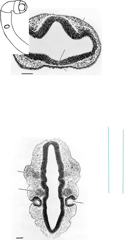
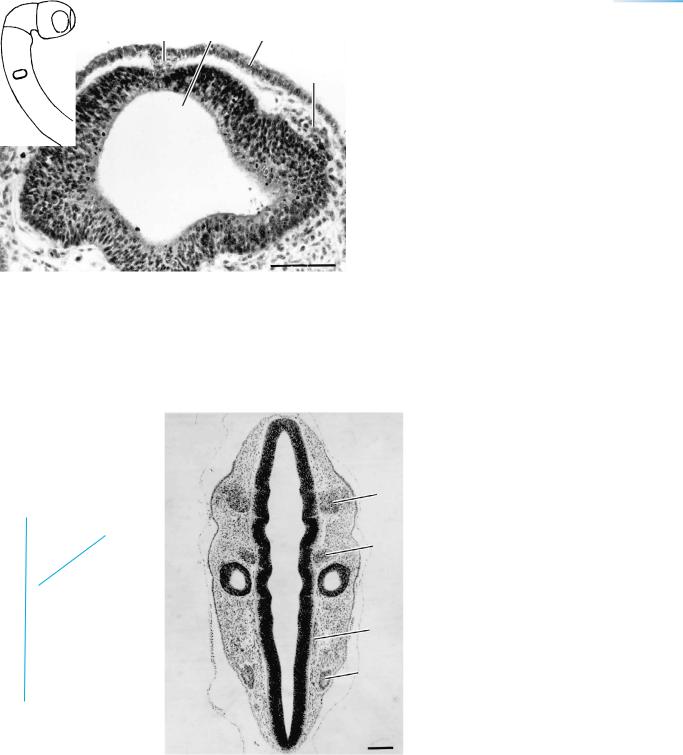
Figure 12–9. The optic vesicles and the optic crest. (A) Transverse section of another embryo showing the optic vesicles on both sides of the diencephalon. In the lower half the first pharyngeal arch and part of the heart are visible. The levels of the sections are indicated in the inset.
(B) In an embryo of stage 11, when the rostral neuropore is still open (arrow), as shown also in Figure 11–7C. The neuropore is covered by amnion, and the ependymal fluid within the future ventricular system is continuous with the amniotic fluid. Mesenchyme is not yet visible between the optic vesicle and the surface ectoderm. Some cells of the optic crest are emerging caudally, but are seen better in B. The vesicle is limited by the caudal limiting sulcus (O’Rahilly, 1966), beyond which blood vessels can be seen. (C) In an embryo of stage 12, when the rostral neuropore has already closed. The striking feature of the optic vesicle here is the emergence from its wall of cells of the optic crest.
CLOSURE OF THE CAUDAL NEUROPORE AND THE BEGINNING OF SECONDARY NEURULATION |
67 |
Di.
Neuropore
Figure 12–9. (Continued )