


64 2.2 Benign Nontraumatic Abnormalities
2.2Benign Nontraumatic Abnormalities
#30 Developmental
Abnormalities (1):
Caroli’s Disease
Related topics: #68 (choledochal cyst),
# 126 (choledochocele)
KEY FACTS: DISEASE
●Communicating cavernous ectasia of intrahepatic ducts with multiple nonobstructive saccular dilations
●Rare, autosomal recessive inheritance
●Age of presentation: children and second/ third decade
●Corresponds to type V bile ducts cysts (Table 30a, b; De Wilde et al. 1991):
●Associations:
–Developmental hepatic fibrosis (Caroli’s syndrome)
–Choledochal cyst
–Polycystic kidney disease
●Symptoms: usually cramplike abdominal pain
●Complications:
–Stone formation (34%) (typically pigmented stones)
–Bile stasis with recurrent cholangitis
–Abscess
–Sepsis
–Cholangiocarcinoma (7%)
–Portal hypertension (in Caroli’s syndrome)
Table 30.1a. Classification of bile duct cysts
KEY FACTS: MRI
●Projective and cross-sectional images:
–Several to multiple saccular dilations of intrahepatic ducts, varying in size (Fig. 30)
–Usually diffuse distribution
●Cross-sectional images:
–Typical feature: portal radicles completely surrounded by dilated bile ducts (central dot sign; Choi et al. 1990) (Fig. 30d)
●Differential diagnosis:
–Multiple liver cysts. This may be difficult on MRCP (unlike ERCP, MRCP cannot prove or exclude the connection between “cysts” and bile ducts with certainty). The diagnosis can be made in most cases, however, by tracing the course of the bile ducts on contiguous cross-sectional images
–Bacterial cholangitis (see #40)
–Oriental cholangitis (see #46)
–Primary sclerosing cholangitis (see #41–44; dilation usually less pronounced)
References
Choi BI, Yeon KM, Kim SH, Han MC (1990) Caroli disease: central dot sign in CT. Radiology 174 : 161–163
De Wilde VG, Elewaut AG, De Vos MP, Hendrix RF, Barbier FE (1991) Choledochal cysts in the adult. Endoscopy 23 : 4–7
Levy AD, Rohrmann CA J., Murakata LA et al. (2002) Caroli’s disease: radiologic spectrum with pathologic correlation. AJR Am J Roentgenol 179 : 1053–1057
Type |
(%) |
Findings |
Type |
(%) |
Findings |
|
|
|
|
|
|
I |
50–80 |
Choledochal cyst |
IV |
18–43 |
Multiple areas of |
|
|
(see #68) |
|
|
cystic dilation |
II |
2 |
Diverticula |
V |
4–13 |
Intrahepatic ducts |
|
|
|
|
|
(e.g., Caroli’s disease) |
III |
1–6 |
Choledochocele |
|
|
|
|
|
(see #126) |
|
|
|
|
|
|
|
|
|
2 Intrahepatic Bile Ducts |
65 |
Table 30.1b. Graphic presentation of bile duct cysts (reprinted with permission from De Wilde et al. 1991)
Type |
Description |
Type |
Description |
|
|
|
|
I |
Solitary fusiform extrahepatic |
IVa |
Fusiform and intrahepatic |
|
|
|
cysts |
II |
Extrahepatic supraduodenal |
IVb |
Multiple extrahepatic cysts |
|
diverticulum |
|
|
III |
Intraduodenal diverticulum; |
V |
Multiple intrahepatic cysts; |
|
choledochocele |
|
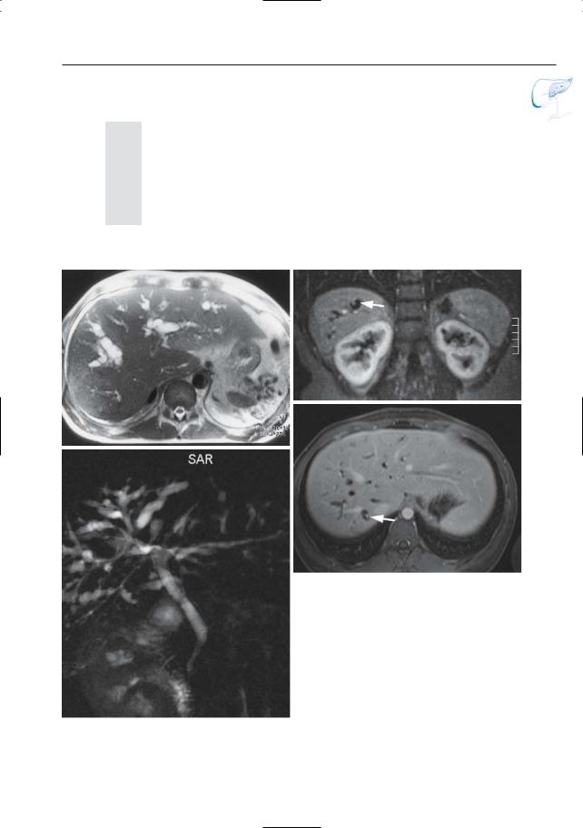
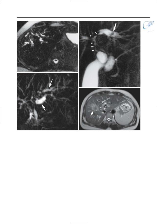
Caroli’s Disease |
|
|
|
|
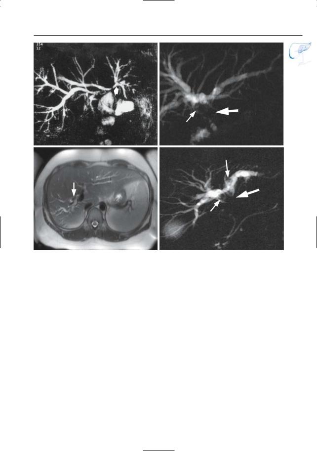
c
a
d
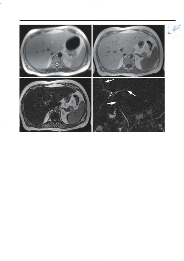
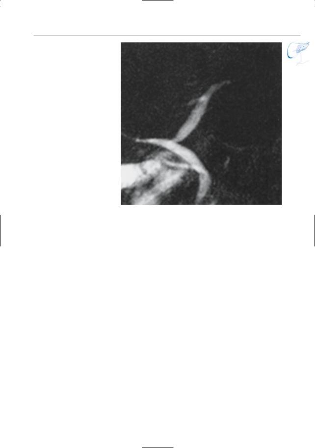
Fig. 30 a–d. Axial T2-weighted image (a) showing marked saccular dilation of intrahepatic ducts in the left and right liver lobe. Projective image (b), coronal (c) and axial (d) post-gadolinium VIBE images in the portovenous phase showing intrahepatic cystic dilatations of the bile ducts (b) with typical ”central-dot” sign on the post-gadolinium
b images (c, d) (arrows). This was a patient with type V bile duct cysts or Caroli’s Disease

66 2.2 Benign Nontraumatic Abnormalities
#31 Developmental
Abnormalities (2):
Hepatic Cysts
Related topic: # 171 (simple true cyst of the pancreas)
KEY FACTS: DISEASE
●Fluid-containing lesions lined by an epithelial layer
●Common
●Types:
–Solitary liver cyst
–Liver cysts in autosomal dominant polycystic kidney disease (ADPKD; most patients with ADPKD have hepatic cysts)
●Symptoms:
–Solitary liver cyst: usually asymptomatic
–Liver cysts in ADPKD: sometimes vague right upper quadrant pain
●Complications (rare in solitary liver cyst):
–Infection
–Hemorrhage
–Bile duct obstruction
KEY FACTS: MRI
●Sharply marginated lesions with a signal intensity typical of fluid
●Easily differentiated from hemangiomas and other lesions on heavily T2weighted images (Bosmans et al. 1997; see #2)
●Septa may be present
●No connection with biliary tree
●Differential diagnosis
(other fluid-containing lesions):
–Biloma: history,evolution, shape, relation to bile ducts
–Abscess: thick wall; irregular contours
–Cystic/necrotic metastases: irregular contours, peripheral soft tissue component on moderately T2-weighted MR images, rim enhancement (see # 193)
–Caroli’s disease (see #30)
–Hydatid cyst (see #47)
–Biliary cystadenoma: thick irregular wall, papillary projections
–Bile duct hamartomas (Fig. 31b, c)
References
Bosmans H, Gryspeerdt S, Van Hoe L et al. (1997) Preliminary experience with a new double echo half-Fourier single-shot turbo spin echo acquisition in the characterisation of liver lesions. MAGMA 5 : 79–84
Levine E, Cook L, Grantham JJ (1985) Liver cysts in autosomal-dominant polycystic kidney disease: clinical and computed tomographic study. AJR Am J Roentgenol 145 : 229–233
Mortelé KJ, Ros PR (2001) Cystic focal liver lesions in the adult: differential CT and MR imaging features. Radiographics 21 : 895–910
2 Intrahepatic Bile Ducts |
67 |
a
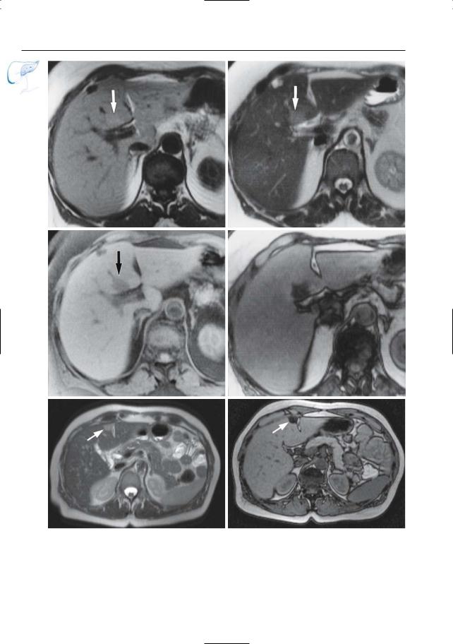
Fig. 31. a Projective image showing multiple cysts. Note the normal size of the bile ducts. b, c Different patient. Projective image (b) and axial T2 HASTE image (TE 360) (c) showing multiple small hepatic cystic structures, compatible with biliary hamartomas
b
c

68 2.2 Benign Nontraumatic Abnormalities
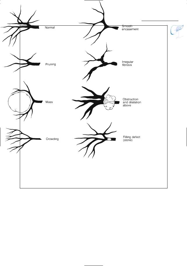
#32 Morphologic Description
of Biliary Abnormalities
in Parenchymal Liver Disease
KEY FACTS
●Use of appropriate terminology is a prerequisite for accurate reporting. Some commonly used terms are the following (Fig. 32):
–Pruning: diminished arborization (implies that peripheral branches are obstructed or obliterated)
–Crowding: decreased distance between structures (usually implies focal or diffuse loss of liver volume)
–Encasement: narrowing caused by circular compression by mural or extrinsic process
–Splaying: spreading apart (suggests parenchymal edema or a space-occu- pying lesion)
References
Rohrmann C, Silverstein F, Templeton F (1997) Intrahepatic conditions of the biliary tree. In: Stewart E,Vennes J, Geenen J (eds) Atlas of endoscopic retrograde cholangiopancreatography. Mosby, St Louis, pp 236–271
2 Intrahepatic Bile Ducts |
69 |
Fig. 32. Intrahepatic ducts as they might appear when affected by various pathologic processes. (Reprinted with permission from Stewart et al. 1977)

70 2.2 Benign Nontraumatic Abnormalities
#33 Bile Duct Lithiasis
Related topic: #72–74 (stones in the common bile duct)
KEY FACTS: DISEASE
●Incidence:
–Intrahepatic bile duct lithiasis is rare as a solitary abnormality
–Common complication of other diseases (see below)
●Predisposing and associated disease:
–Chronic biliary obstruction with or without cholangitis (see #40)
–Biliary ascariasis
–Caroli’s disease (see #30)
–Oriental cholangitis (see #46)
–Primary sclerosing cholangitis (see #44)
●Symptoms: none or signs of cholangitis/obstruction
●Complications: cholangitis, liver abscess, fistula formation
KEY FACTS: MRI
●Stones are easily diagnosed as welldefined intraluminal structures with low signal intensity on T1and T2-weighted images
●Differential diagnosis with aerobilia:
–Small amounts of air in intrahepatic ducts usually not clearly seen at MRI
–If significant aerobilia is present, a fluid–air level may be present (see #67)
–Differentiation may be impossible unless X-ray or CT correlation is obtained
References
Kim JH, Kim MJ, Park SI et al. (2002) Using kinematic MR cholangiopancreatography to evaluate biliary dilatation. AJR Am J Roentgenol. 178 : 909–914
Menu Y, Lorphelin JM, Scherrer A, Grenier P, Nahum H (1985) Sonographic and computed tomographic evaluation of intrahepatic calculi. AJR Am J Roentgenol 145 : 579–583
2 Intrahepatic Bile Ducts |
71 |
a
Fig. 33. a Patient with antecedents of symptomatic cholelithiasis and laparoscopic cholecystectomy. Projective image showing a small intraluminal filling defect in a branch of the left hepatic duct (arrow), corresponding to a small stone. Note the severe cystic dilation of the proximal bile duct (arrowheads) b A 20-year-old patient with mucoviscidosis. Projective image shows a stone in the left hepatic duct with secondary bile duct dilatation (large arrow). Note also the small stone in the distal common bile duct (small arrow). c Patient with antecedents of symptomatic cholelithiasis. Projective image shows dilatation of the posterior right hepatic ducts with multiple intraluminal filling defects (arrows), corresponding to multiple small stones
b
c

72 2.2 Benign Nontraumatic Abnormalities
#34 Acute Hepatitis
KEY FACTS: DISEASE
●Acute inflammation of liver parenchyma
●Pathology: diffuse inflammation with centrilobular necrosis,portal infiltration by lymphocytes, and reactive changes in Kupffer cells
●Causes:
–Viral (hepatitis A–E, G)
–Bacterial
–Fungal
–Toxic agents and drugs
●Symptoms:
–Fatigue, anorexia, nausea, vomiting
–Jaundice usually appears 1–2 weeks after the onset of symptoms
●Complications:
–Massive hepatic necrosis (primarily seen in hepatitis B, D, and E)
–Chronic hepatitis (> 6 months; particularly hepatitis B–D)
–Cirrhosis (viral hepatitis, alcoholic hepatitis; see #36, 37)
–Hepatocellular carcinoma (hepatitis B and C, alcoholic hepatitis; see #58, 59)
KEY FACTS: MRI
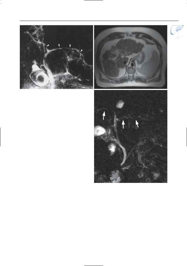
●Cholangiographic images: edema of liver parenchyma may cause separation (splaying) and diffuse narrowing of the bile ducts (Fig. 34a–c; Rohrmann et al. 1977)
●Cross-sectional images:
–Hepatomegaly
–Gallbladder wall thickening (see # 111) (Fig. 34d)
–Periportal cuffing (Itoh et al. 1992)
●Note: Ultrasonography may be more specific than MRI by showing abnormally bright portal venule walls (seen in 60% of cases; Needleman et al. 1986)
●Differential diagnosis:
–Infiltrative diseases (amyloidosis, sarcoidosis, infiltrating tumor)
–Other causes of diffuse hepatocellular edema (e.g., alcohol)
–Severe steatosis (#35)
References
Itoh H, Sakai T, Takahashi N et al. (1992) Periportal high intensity on T2-weighted MR images in acute viral hepatitis. J Comp Assist Tomogr 16 : 564–567
Needleman L, Kurtz AB, Rifkin MD, Cooper HS, Pasto ME, Goldberg BB (1986) Sonography of diffuse benign liver disease: accuracy of pattern recognition and grading. AJR Am J Roentgenol 146 :1011–1015
Rohrmann CA, Ansel HJ, Ayoola EA, Silvis SE, Vennes JA (1977) Endoscopic retrograde intrahepatic cholangiogram: radiographic findings in intrahepatic disease. AJR Am J Roentgenol 128 : 45–52
2 Intrahepatic Bile Ducts |
73 |
a |
b |
c |
d |
Fig. 34 a–c. a Projective MR image and b ERCP obtained in 23-year-old woman, both showing splaying and displacement of the central intrahepatic ducts (arrows) and narrowing of the more peripheral ducts (arrowheads), probably secondary to hepatocellular edema. The ducts in the right lobe are not clearly seen in a because they are not within the section c. Same patient. Heavily T2-weighted axial cross-sectional image showing marked pau-
city of bile ducts in the right liver lobe. Biopsy revealed acute hepatitis. d Projective image (different patient) in acute hepatitis due to graft-versus-host disease, showing splaying and displacement of the intrahepatic ducts (arrowheads) and reactive thickening of the gallbladder wall (arrow)
Note: In a and b, the right side of the image corresponds to the right side of the patient

74 2.2 Benign Nontraumatic Abnormalities
#35 Fatty Metamorphosis
(Steatosis)
KEY FACTS: DISEASE
●Histology: hepatocytes with large cytoplasmic fat vacuoles
●Causes:
–Metabolic disorder
(diabetes, obesity, corticosteroids)
–Hepatotoxins
(e.g., alcohol, chemotherapy)
●Types:
–Diffuse fatty infiltration
–Focal fatty infiltration (predominantly periportal and subcapsular)
●Association: focal fatty infiltration in the posterior edge of the quadrate lobe may be associated with aberrant gastric venous drainage (Kawamori et al. 1996)
KEY FACTS: MRI
Diffuse Fatty Infiltration (Fig. 35 a– d)
●Cross-sectional images:
–Sometimes no abnormalities (MRI less sensitive than ultrasonography and CT)
–Hepatomegaly
–Hyperintensity on T1-weighted images (not always present)
–Variable signal intensity on T2weighted images, depending on the type of sequence used (fat is rendered bright on fast spin echo and HASTE sequences unless fat suppression is applied)
●Projective images:
–Stretching and splaying of the intrahepatic biliary tree may eventually be seen
Focal Fatty Infiltration (Fig. 35 e– l) |
|
● Focal lesion, usually with sharp borders |
|
● Undisplaced course of vessels |
|
● No bulging of liver contour |
! |
● Specific diagnosis can be made by com- |
|
paring images obtained: |
–With and without fat suppression
–“In phase” and “out of phase” (Mitchell et al. 1991)
●May be inhomogeneous (Fig. 35 k, l)
Focal Sparing (Fig. 35 m, n)
●Non-steatotic areas in otherwise steatotic liver
References
Kawamori Y, Matsui O, Takahashi S, Kadoya M, Takashima T, Miyayama S (1996) Focal hepatic fatty infiltration in the posterior edge of the medial segment associated with aberrant gastric venous drainage: CT, US, and MR findings. J Comp Assist Tomogr 20 : 356–359
Mitchell DG, Kim I, Chang TS et al. (1991) Fatty liver. Chemical shift phase-difference and suppression magnetic resonance imaging techniques in animals, phantoms, and humans. Invest Radiol 26 :1041–1052
Van Steenbergen W, Lanckmans S (1995) Liver disturbances in obesity and diabetes mellitus. Int J Obesity 19 [Suppl 3] : 27–36
2 Intrahepatic Bile Ducts |
75 |
a |
b |
c |
d |
Fig. 35 a–d. Diffuse steatosis and mild hepatomegaly. a Axial T2-weighted HASTE image (TE, 60 msec) showing high signal intensity of the hepatic parenchyma (the liver and spleen are nearly isointense), which can be explained by the markedly increased hepatic fat content. b, c T1-weighted images
in phase and out of phase showing a dramatic drop of the signal intensity of the liver on the “out of phase” image due to the diffuse steatosis. d projective image showing the narrowing and paucity of the intrahepatic bile ducts (arrows)
76 2.2 Benign Nontraumatic Abnormalities
e |
f |
g |
h |
i |
j |
Fig. 35 e–j. e Axial T1and f T2-weighted images showing area of discrete hyperintensity posteriorly in the quadrate lobe (arrows) g Frequency-selective fat-suppressed T1-weighted image and h T1-weight- ed image “out of phase” showing the lesion as hypointense, thus confirming the diagnosis of focal fatty infiltration. Axial T2-weighted HASTE image
(TE 60) (i) and T1-weighted image “out of phase” (j) showing a small area of focal fatty infiltration adjacent to the falciform ligament (arrow). This area is hyperintense on the T2-weighted image and shows a loss of signal on the T1-weighted “out of phase” image.
2 Intrahepatic Bile Ducts |
77 |
k |
l |
m |
n |
Fig. 35 k–n. Axial T2-weighted HASTE image (TE 60) (k) and T1-weighted image “out of phase” (l) showing an area of inhomogeneous steatosis in the right lobe of the liver. Note that the areas with increased fat content show a subtle increase in signal intensity on the T2-weighted HASTE image, and a marked drop in signal on the T1-weighted image out of phase (arrows). m, n Different patient. Axial
T2-weighted HASTE image (TE 60) (m) and T1weighted image “out of phase” (n) showing two small areas of focal sparing in de right lobe in a otherwise steatotic liver. Note that the signal intensity of the non-steatotic parenchyma is low on the T2-weighted HASTE image and high on the out- of-phase T1-weighted image (arrows)

78 2.2 Benign Nontraumatic Abnormalities
#36 Cirrhosis, Ductal Changes
Related topics: #38 (primary biliary cirrhosis), #58, 59 (hepatocellular carcinoma)
KEY FACTS: DISEASE
–Hepatic encephalopathy
–Development of hepatocellular carcinoma (cirrhosis is the main risk factor)
KEY FACTS: MRI
●Chronic liver disease characterized by diffuse parenchymal destruction, fibrosis, and nodular regeneration, with abnormal reconstruction of preexisting lobular architecture
●Etiology:
–Toxic agents: alcohol (most common cause in the West, 60%–70%), drugs, iron (hemochromatosis, 5%), copper (Wilson’s disease)
–Inflammation (viral; 10%)
–Biliary obstruction (e.g., cystic fibrosis, primary biliary cirrhosis)
–Other causes (vascular, nutritional, hereditary)
●Symptoms: fatigue, weight loss, …
●Complications:
–Ascites
–Portal hypertension with or without esophageal varices
●Early changes: subtle scattered ductal narrowing, pruning
●Changes occurring in patients with more advanced disease (Fig. 36):
–Ductal tortuosity and crowding (fibrosis, micronodular disease)
–Focal stenoses
–Displacement
(large areas of regeneration)
●Note: Cholangiographic findings are usually nonspecific
References
Altman C,Fabre M,Adrien C et al. (1995) Cholangiographic features in fibrosis and cirrhosis of the liver. Radiological-pathological correlation. Dig Dis Sci 40 : 2128–2133
Legge DA, Carlson HC, Ludwig J (1971) Cholangiographic findings in diseases of the liver: a postmortem study. AJR Am J Roentgenol 113 : 34–40
2 Intrahepatic Bile Ducts |
79 |
a
Fig. 36. a Projective MR image showing relatively large left hepatic duct with caudal displacement. Also note focal changes in caliber (arrowheads) b, c Different patient. Axial T2-weighted HASTE image (TE 60) (b) showing a small liver with diffuse parenchymal nodularity due to the presence of regenerative nodules. Note the secondary enlargement of the spleen suggesting portal hypertension. c Projective image showing diffuse intrahepatic ductal narrowing with ductal tortuosity and displacement (arrows)
b
c

80 2.2 Benign Nontraumatic Abnormalities
#37 Cirrhosis,
Parenchymal Changes
Related topics: #38 (primary biliary cirrhosis), #58, 59 (hepatocellular carcinoma)
KEY FACTS: TYPICAL MR FEATURES
●Volume loss (Fig. 37)
–Segmental, usually quadrate lobe and/or right lobe
–Global (less common)
●Caudate lobe hypertrophy (ratio caudate lobe to right lobe greater than 0.65 on transverse images; most sensitive in patients with post-hepatitis B cirrhosis)
●Periportal cuffing (see #48):
–Presence of fluid, probably lymphedema, around the portal vein tributaries
–Differential diagnosis with biliary dilation: typical location on both sides of venous branches
●Other common findings on cross-sec- tional images:
–Prominence of fissures and porta hepatis
–Surface nodularity
–Signs of portal hypertension (venous collaterals)
–Ascites
–Enlarged hilar lymph nodes
–Thickening of the gallbladder wall (see # 111)
●Differential diagnosis: Budd-Chiari syndrome (obstruction of hepatic vein outflow), which also causes enlargement of the caudate lobe
References
Harbin WP, Robert NJ, Ferruci JT (1980) Diagnosis of cirrhosis based on regional changes in hepatic morphology: a radiological and pathological analysis. Radiology 135 : 273–283
Lafortune M, Matricardi M, Denys A, Favret M, Déry R, Pomier-Layrargues G (1998) Segment 4 (the quadrate lobe): a barometer of cirrhotic liver disease at US. Radiology 206 : 157–160
Wang TF, Hwang SJ, Lee EY et al. (1997) Gallbladder wall thickening in patients with liver cirrhosis. J Gastroenterol Hepatol 12 : 445–449
2 Intrahepatic Bile Ducts |
81 |
a |
b |
c |
d |
Fig. 37 a, b. a Moderately and b heavily T2-weighted images showing irregular contours of hepatic parenchyma, relative atrophy of the quadrate lobe (arrowheads in a), hypertrophy of the caudate lobe (arrows in a), reactive thickening of the gallbladder
wall (arrow in b), and ascites c, d Different patient. Axial T2-weighted HASTE images (TE 60) showing marked volume loss of segment IV (arrow) together with hypertrophy of the caudate lobe (arrowheads) and (to a lesser degree) segment II and III

82 2.2 Benign Nontraumatic Abnormalities
#38 Primary Biliary Cirrhosis
Related topics: #36, 37 (cirrhosis)
KEY FACTS: DISEASE
●Progressive destructive cholangitis of interlobar and septal bile ducts with inflammatory cellular infiltrate, portal fibrosis, nodular regeneration, and shrinkage of hepatic parenchyma
●Presenting age: 35–55 years
●Ratio of women to men: 9 : 1
●Etiology: immune response disorder
●Associated diseases: rheumatoid arthritis, Sjögren’s syndrome, Hashimoto’s thyroiditis, dermatomyositis, systemic lupus erythematosus
●Clinical signs: fatigue, pruritus, keratoconjunctivitis sicca, hyperpigmentation
●Tests for antimitochondrial antibodies usually positive (> 90%)
●Differential diagnosis with primary sclerosing cholangitis (see #41): sex distribution, associated diseases, cholangiographic findings (see below)
●Complications: hepatocellular carcinoma, liver insufficiency
KEY FACTS: MRI
●Early stage: no abnormalities
●Advanced disease (Fig. 38):
–“Tree in winter” appearance: diffuse narrowing or even disappearance (vanishing) of small ducts
–Signs of cirrhosis (tortuosity, narrowing, caliber variation, segmental changes)
●Differential diagnosis with primary sclerosing cholangitis
–Bile duct deformities less common
–Disease initially more limited to small ducts
–No extrahepatic disease
–No diverticular outpouchings
References
Blachar A, Federle MP, Brancatelli G (2001) Primary biliary cirrhosis: clinical, pathologic, and helical CT findings in 53 patients. Radiology 220 : 329
Wiesner RH, LaRusso NF, Ludwig J, Dickson ER (1985) Comparison of the clinicopathologic features of primary sclerosing cholangitis and primary biliary cirrhosis. Gastroenterology 88 : 108–114
2 Intrahepatic Bile Ducts |
83 |
Fig. 38. Projective MR image showing marked discrepancy between the (normal) extrahepatic duct and main right and left hepatic ducts on one hand and the (nearly invisible) more distal branches on the other hand (“pruning”)

84 2.2 Benign Nontraumatic Abnormalities
#39 Vanishing Bile Duct Disease: Differential Diagnosis
KEY FACTS: CAUSES/ASSOCIATED DISEASES
(SHERLOCK 1987; DESMET 1992)
●Immunologically based liver diseases:
–Primary biliary cirrhosis (see #38)
–Graft-versus-host disease
–Chronic liver transplant rejection
–AIDS cholangiopathy
●Sarcoidosis, amyloidosis
●Primary sclerosing cholangitis (see #41–44)
●Bacterial cholangitis (see #40)
●Hepatic artery occlusion with ischemia
●Chemical cholangitis (intra-arterial chemotherapy)
●Developmental disorders (intrahepatic atresia, cystic fibrosis)
●Acute hepatitis (see #34)
●Severe steatosis (see #35)
KEY FACTS: MRI
●Regional or diffuse invisibility of intrahepatic bile ducts (Fig. 39)
References
Desmet VJ (1992) Vanishing bile duct disorders. Prog Liver Dis 10 : 89–121
Sherlock S (1987) The syndrome of disappearing intrahepatic bile ducts. Lancet 29 : 493–496
2 Intrahepatic Bile Ducts |
85 |
a
Fig. 39. a, b Patient with auto immune hepatitis. Projective image showing narrowed and displaced intrahepatic bile ducts (arrowheads). Also note increased signal intensity of background tissue related to presence of ascites. b Same patient. Cross-sec- tional T2-weighted image showing marked parenchymal changes, particularly in the right lobe. Note that no intrahepatic bile ducts are seen,except in the perihilar area (arrowheads) c Projective image in a patient with primary sclerosing cholangitis showing diffuse segmental irregularity, pruning and narrowing of the intrahepatic bile ducts (arrowheads), particularly near the hilar bifurcation (arrow)
b
c

86 2.2 Benign Nontraumatic Abnormalities
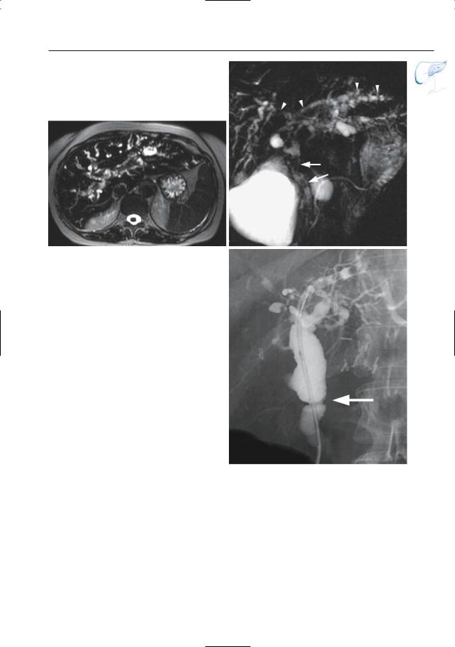
#40 Bacterial Cholangitis
Related topic: #75 (extrahepatic bile duct, bacterial cholangitis)
KEY FACTS: DISEASE
●Bacterial infection of biliary tree
●Synonyms: ascending cholangitis, pyogenic cholangitis, acute cholangitis, obstructive cholangitis
●Organisms: Escherichia coli > Klebsiella > Pseudomonas
●Underlying cause: nearly always obstructive lesion of biliary tree
– Malignant
(e.g., ampullary carcinoma)
– Benign
(e.g., stricture of surgical anastomosis, calculi)
●Symptoms: fever, chills, jaundice
●Complications:
hepatic abscess, septicemia, shock
●Prognosis:
100% mortality without treatment
KEY FACTS: MRI
●Early changes: dilation (may be the only finding)
●Advanced disease:
–Mucosal irregularities
–Intraluminal filling defects (stones, sludge, inflammatory debris)
–Intrahepatic abscess, typically com-
municating with the bile ducts (Fig. 40)
●Differential diagnosis: oriental cholangitis (see #46)
●Note: The primary task of MRCP is not to detect signs of cholangitis, but to detect the underlying obstructive lesion
References
Bader TR, Braga L, Beavers KL, Semelka RC (2001) MR imaging findings of infectious cholangitis. Magn Reson Imaging 19 : 781–788
Mathieu D, Vasile N, Fagniez PL, Segui S, Grably D, Larde D (1985) Dynamic CT features of hepatic abscesses. Radiology 154 : 749–752
Mendez RJ, Schiebler ML, Outwater EK, Kressel HV (1994) Hepatic abscesses: MR imaging findings. Radiology 190 : 431–436
Taylor AJ, Bohorfoush AG (1997) Inflammation of the biliary tract. In: Taylor AJ, Bohorfoush AG (eds) Interpretation of ERCP. Lippincott-Raven, Philadelphia, pp 77–125
2 Intrahepatic Bile Ducts |
87 |
a
c
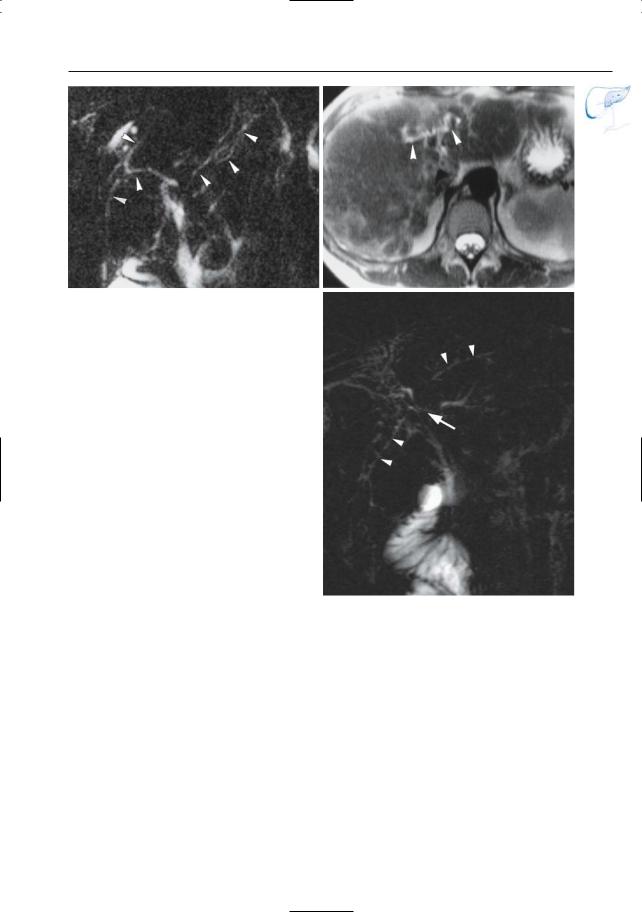
Fig. 40. a Coronal T2-weighted image showing fluid collections in the right liver lobe (arrow). b Heavily T2-weighted axial image showing connection of these fluid collections with the bile duct (arrowheads), pointing to the diagnosis of pyogenic cholangitis with abscess. c–e Patient with a history of hepaticojejunostomy with anastomotic stricture and development of secondary bacterial cholangitis. c, d Axial T2-weighted HASTE images (TE 60 and 360 ms, respectively) showing two fluid collections in the right lobe (arrows) with a connection with the bile duct (arrowhead) due to pyogenic cholangitis with abscess. e RARE image showing the anastomotic stricture of the hepaticojejunostomy (arrow) with secondary dilatation of the bile ducts
b
d
e

88 2.2 Benign Nontraumatic Abnormalities
#41 Primary Sclerosing
Cholangitis, General
Related topic: # 76 (extrahepatic duct, primary sclerosing cholangitis)
KEY FACTS: DISEASE (CHAPMAN 1991)
●Chronic obliterative fibrotic inflammation of bile ducts
●Histology: destructive cholangitis with fibrous thickening of duct walls and with relatively few inflammatory cells
●70% of patients younger than 45 years of age at the time of diagnosis
●Ratio of women to men: 1 : 2
●Etiology: autoimmune
●Localization:
–Both intraand extrahepatic ducts: 72%–96%
–Intrahepatic ducts only: 1%–25%
–Extrahepatic ducts only: 3%
●Cholangiographic classification: see #42, 43
●Associations:
–Inflammatory bowel disease (up to 70% of patients have ulcerative colitis)
–Retroperitoneal fibrosis,Peyronie disease, Riedel thyroiditis, retro-orbital pseudotumor
–Chronic pancreatitis (14%; see also
# 169)
●Symptoms: usually chronic intermittent obstructive jaundice, fatigue, cholangitis, pruritus
●Diagnosis: primarily based on cholangiographic imaging findings
●Complications: see #44
●Prognosis: variable (some patients remain asymptomatic)
●Note: The term “primary” is used even when there is associated disease
References
Brandt DJ, MacCarty RL, Charboneau JW, LaRusso NF,Wiesner RH,Ludwig J (1988) Gallbladder disease in patients with primary sclerosing cholangitis. AJR Am J Roentgenol 150 : 571–574
Fulcher AS,Turner MA,Franklin KJ et al. (2000) Primary sclerosing cholangitis: evaluation with MR cholangiography – a case-control study. Radiology 215 : 71–80
Chapman RW (1991) Aetiology and natural history of primary sclerosing cholangitis – a decade of progress? Gut 32 : 1433–1435
MacCarty RL, LaRusso NF, Wiesner RH, Ludwig J (1983) Primary sclerosing cholangitis: findings on cholangiography and pancreatography. Radiology 149 : 39–44
2 Intrahepatic Bile Ducts |
89 |
#42 Primary Sclerosing
Cholangitis, Early Disease
(Type I)
Related topic: # 76 (extrahepatic duct, primary sclerosing cholangitis)
KEY FACTS: MRI
●Note: Li-Yeng and Goldberg (1984) originally proposed four types of intrahepatic involvement. According to Majoie et al. (1991), a classification into three types is sufficient, since the original types I and II have the same clinical implications
●Cholangiographic description (Fig. 42, see also Fig. 43a; Majoie et al. 1991):
–Segmental irregularity and narrowing, especially at duct bifurcations
–No or only minor dilation
–Pruning
●Cross-sectional images:
–Lack of confluence of dilated intrahepatic ducts toward the hilum (presence of stenoses)
–Direct demonstration of beaded appearance and/or pruning
–T2-weighted images: sometimes presence of peribiliary “cuff” with low signal intensity (see, e.g., #8)
●Differential diagnosis: mainly primary biliary cirrhosis and other types of cirrhosis, diffuse sclerosing cholangiocarcinoma, lymphoma, polycystic liver disease, amyloidosis, liver necrosis (indicative finding: extrahepatic abnormalities)
References
Fulcher AS,Turner MA,Franklin KJ et al. (2000) Primary sclerosing cholangitis: evaluation with MR cholangiography – a case-control study. Radiology 215 : 71–80
Majoie CBLM, Reeders JWAJ, Sanders JB, Huibregtse K, Jansen PLM (1991) Primary sclerosing cholangitis: a modified classification of cholangiographic findings. AJR Am J Roentgenol 157 : 495–497
Li-Yeng C, Goldberg HI (1984) Sclerosing cholangitis: broad spectrum of radiographic findings. Gastrointest Radiol 9 : 39–47
Terada T, Nakanuma Y (1995) Intrahepatic cholangiographic appearance simulating primary sclerosing cholangitis in several hepatobiliary diseases: a postmortem cholangiographic and histopathological study in 154 livers at autopsy. Hepatology 22 : 75–81
Vitellas KM, Keogan MT, Freed KS et al. (2000) Radiologic manifestations of sclerosing cholangitis with emphasis on MR cholangiopancreatography. Radiographics 20 : 959–975
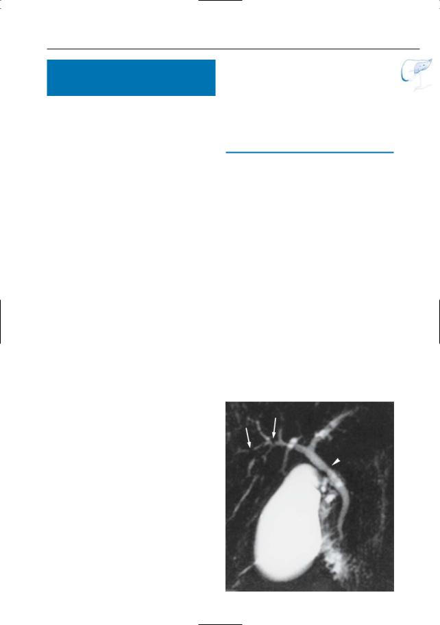
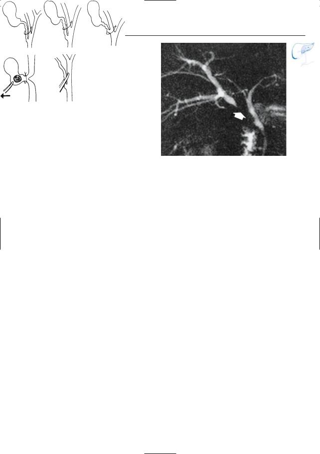
Fig. 42. Projective MR image showing stenoses of intrahepatic ducts (arrows) associated with lowgrade stenosis of the common hepatic duct
(arrowhead)

90 2.2 Benign Nontraumatic Abnormalities
#43 Primary Sclerosing
Cholangitis, Advanced Disease
(Types II and III)
Related topic: # 76 (extrahepatic duct: primary sclerosing cholangitis)
KEY FACTS: MRI
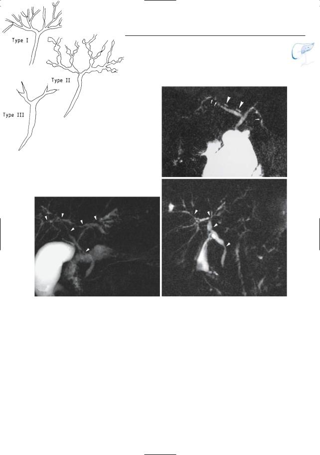
●Cholangiographic classification (Fig. 43a; Li-Yeng and Goldberg 1984; Majoie et al. 1991):
–Type II (marked segmental narrowing and dilation, pruning, beading; sacculations resembling diverticula) (Fig. 43b)
–Type III (peripheral duct obliteration; only narrowed central ducts remain)
●Type II abnormalities: more or less diagnostic
●Differential diagnosis for type III: Klatskin tumor, metastases, diffuse infiltrating hepatic tumors
●Most patients with PSC have associated imaging findings of cirrhosis
!● Note: The key cholangiographic finding is a relative lack of dilation proximal to a stricture (related to diffuse periductal inflammation)
References
Bader TR, Beavers KL, Semelka RC (2003) MR imaging features of primary sclerosing cholangitis: patterns of cirrhosis in relationship to clinical severity of disease. Radiology 226 : 675–685
Fulcher AS, Turner MA, Yelon JA et al. (2000) Magnetic resonance cholangiopancreatography (MRCP) in the assessment of pancreatic duct trauma and its sequelae: preliminary findings. J Trauma 48 : 1001–1007
Li-Yeng C, Goldberg HI (1984) Sclerosing cholangitis: broad spectrum of radiographic findings. Gastrointest Radiol 9 : 39–47
Majoie CBLM, Reeders JWAJ, Sanders JB, Huibregtse K, Jansen PLM (1991) Primary sclerosing cholangitis: a modified classification of cholangiographic findings. AJR Am J Roentgenol 157 : 495–497
Vitellas KM, Keogan MT, Spritzer CE et al. (2000) MR cholangiopancreatography of bile and pancreatic duct abnormalities with emphasis on the single-shot fast spin-echo technique. Radiographics 20 : 939–957
2 Intrahepatic Bile Ducts |
91 |
b
a
c |
d |
Fig. 43. a Type I–III cholangiographic abnormalities. b Projective MR image showing saccular additions (arrowheads) and several stenoses (arrows). c Different patient. Projective image showing multiple intraand extrahepatic bile duct stenoses
(arrowheads) with segmental irregularity and narrowing in this patient with type II PSC. d Projective image (different patient) showing multiple intraand extrahepatic bile duct stenoses (arrowheads) in PSC type II

92 2.2 Benign Nontraumatic Abnormalities
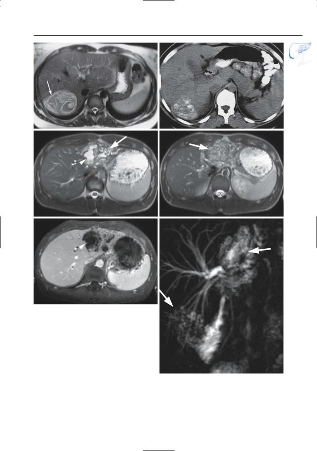
#44 Atypical and Complicated
Primary Sclerosing Cholangitis
Related topic: # 76 (extrahepatic duct, primary sclerosing cholangitis)
KEY FACTS: DISEASE
●Complications:
–Biliary cirrhosis
–Stone disease (8%; Fig. 44c)
–Bacterial cholangitis
–Cholangiocarcinoma (10%)
KEY FACTS: MRI
●Atypical presentation:
–Bile lakes (differential diagnosis with biloma, abscess)
–Segmental disease (differential diagnosis with cholangiocarcinoma may be difficult)
●Cirrhosis: see #36, 37
●Intrahepatic lithiasis: see #33
●Features that should arouse suspicion of cholangiocarcinoma:
–Visualization of a polypoid mass larger than 1 cm
–Rapid progression of stricture disease
–Marked upstream dilation
●Note: In advanced primary sclerosing cholangitis with severe anatomical distortion, detection of stones requires particular attention (differential diagnosis stone versus stricture may be difficult)
References
Dodd GD, Niedzwiecki GA, Campbell WL, Baron RL (1997) Bile duct calculi in patients with primary sclerosing cholangitis. Radiology 203 : 443–447
MacCarty RL,la Russo NF,May GR et al. (1985) Cholangiocarcinoma complicating primary sclerosing cholangitis: cholangiographic appearances. Radiology 156 : 43–46
2 Intrahepatic Bile Ducts |
93 |
a
b
c |
d |
Fig. 44 a–d. a Segmental dilation of intrahepatic bile ducts in primary sclerosing cholangitis. b Projective MR image showing narrowing of the extrahepatic duct and bifurcation (arrowheads) and saccular dilation of the left hepatic duct, with presence of an intraductal stone (arrow). c, d Projective image (different patient) showing multiple bile duct steno-
ses due to known PSC. Note the marked dilatation of several intrahepatic ducts (arrows). This atypical finding should suggests the possibility of cholangiocarcinoma. d T2-weighted HASTE image (TE 60) confirmed the presence of a mass lesion. Final diagnosis was cholangiocarcinoma

94 2.2 Benign Nontraumatic Abnormalities
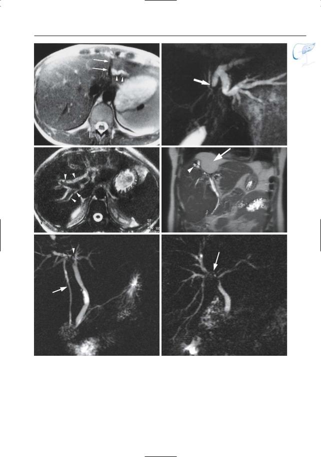
#45 Secondary Sclerosing
Cholangitis
Related topic: #40 (bacterial cholangitis)
KEY FACTS: DISEASE
●Chronic biliary inflammation and fibrosis attributable to local predisposing conditions
●Can be considered as the chronic form of pyogenic (obstructive) cholangitis (see #40)
●Causes: similar to acute cholangitis (e.g., stone disease, postoperative or posttraumatic stricture, papillary stenosis, chronic pancreatitis with bile duct narrowing; Fig. 45)
●Complications:
–Severe duct stricture
–Biliary cirrhosis
KEY FACTS: MRI
●Features of acute cholangitis (see #40)
●Specific findings:
–Duct narrowing, diminished arborization, pruning (fibrosis)
–Intraluminal concretions common
References
Amor A, Chapoutot C, Michel J, Pageaux GP, Larrey D, Michel H (1995) Les cholangites sclerosantes secondaires. Presse Med 24 : 948–952
Park MS, Yu JS, Kim KW, et al. (2001) Recurrent pyogenic cholangitis: comparison between MR cholangiography and direct cholangiography. Radiology 220 : 677–682
Wilson C, Auld CD, Schlinkert R et al. (1989) Hepatobiliary complications in chronic pancreatitis. Gut 30 : 520–527
2 Intrahepatic Bile Ducts |
95 |
a
Fig. 45 a–c. Patient with a history of liver transplantation with presence of a anastomotic stricture on the extrahepatic bile duct with development of secondary sclerosing cholangitis due to chronic bile duct obstruction and dilatation. a T2-weighted HASTE image (TE 360) showing intrahepatic bile duct dilatation. b projective image showing multiple intrahepatic bile duct stenoses and irregularity with beading (arrowheads). Note aerobilia in the common bile duct (arrows). c Corresponding ERCP image showing the anastomotic stricture of the extrahepatic duct (arrow). Note that this stricture is not well seen on the projective image, which may explained either by the presence of air, or by the lack of ductal distension in MRCP (see #23)
b
c

96 |
|
2.2 Benign Nontraumatic Abnormalities |
|
|
|
|
|
||||
|
|
|
|
|
|
|
|
|
|
||
|
|
|
|
|
|
|
KEY FACTS: MRI |
|
|||
|
|
|
|
|
|
|
|
||||
|
|
|
|
#46 Oriental Cholangitis |
|
|
|||||
|
|
|
|
|
|
|
|
|
|
||
|
|
|
KEY FACTS: DISEASE |
● Typical features (Fig. 46): |
|
||||||
|
|
|
|
– |
Marked dilation of large intrahepatic |
|
|||||
|
|
|
|
|
|
|
|
|
|||
|
|
|
|
|
|
|
|
|
|||
|
|
|
● |
Chronic/recurrent infection of bile ducts |
|
|
ducts |
|
|||
|
|
|
|
– Strictures of intrahepatic ducts,mainly |
|
||||||
|
|
|
● Synonyms: recurrent pyogenic cholangi- |
|
|
||||||
|
|
|
|
|
of left duct |
|
|||||
|
|
|
|
tis, Hong Kong disease, intrahepatic pig- |
|
|
|
||||
|
|
|
|
|
– Decreased arborization of smaller |
|
|||||
|
|
|
|
ment stone disease |
|
|
|||||
|
|
|
|
|
|
intrahepatic radicles (pruning) |
|
||||
|
|
|
● |
Pathology: |
|
|
|
||||
|
|
|
|
– Intrahepatic bile ducts filled with pig- |
|
||||||
|
|
|
|
– |
Inflammation of the biliary tree and |
|
|
||||
|
|
|
|
|
|
ment stones and sludge |
|
||||
|
|
|
|
|
surrounding parenchyma |
|
|
|
|||
|
|
|
|
|
|
– |
Marked dilation of common bile duct |
|
|||
|
|
|
|
– |
Duct wall destruction, obliteration of |
|
|
||||
|
|
|
|
|
|
(68%), choledocholithiasis (30%) |
|
||||
|
|
|
|
|
peripheral ducts |
|
|
|
|||
|
|
|
|
|
|
– |
Segmental hepatic atrophy |
|
|||
|
|
|
|
– Fibrosis,especially around portal tracts |
|
|
|||||
|
|
|
|
|
– |
Abscesses |
|
||||
|
|
|
|
– Typically inflammation and fibrosis |
|
|
|||||
|
|
|
|
● |
Note: ERCP may underestimate the |
|
|||||
|
|
|
|
|
of the vaterian sphincter complex |
! |
|||||
|
|
|
|
|
|
abnormalities if contrast medium does |
|||||
|
|
|
|
– Typically formation of pigment stones |
|
||||||
|
|
|
|
|
not pass a severe stenosis (Fig. 46) |
||||||
|
|
|
|
|
(bacterial deconjugation of bilirubin) |
|
|
||||
|
|
|
|
|
● |
Note: Pigment stones characteristically |
|
||||
|
|
|
● |
Incidence: |
|
||||||
|
|
|
|
have a high density on CT |
|
||||||
|
|
|
|
– |
Rare in the West, but common in |
|
|
||||
|
|
|
|
|
|
|
|
|
|||
South-East Asia
–Third most common cause of acute References abdomen in Hong Kong after appen-
|
dicitis and perforated peptic ulcer |
Afagh A, Pancu D (2004) Radiologic findings in |
|
● Location: lateral segment of left lobe and |
recurrent pyogenic cholangitis. J Emerg Med |
||
posterior segment of right lobe most |
26 : 343–346 |
||
commonly involved |
Chau EM, Leong LL, Chan FL (1987) Recurrent pyo- |
||
genic cholangitis: ultrasound evaluation com- |
|||
● Etiology (hypotheses): |
|||
pared with endoscopic retrograde cholan- |
|||
– |
Parasitic infection (Clonorchis sinen- |
giopancreatography. Clin Radiol 38 : 79–85 |
|
|
sis, Ascaris) as initiating factor |
Federle MP, Cello JP, Laing FC, Jeffrey RB Jr (1982) |
|
– |
Bacterial infection (E. coli) related to |
Recurrent pyogenic cholangitis in Asian immi- |
|
grants: use of ultrasonography, computed tomo- |
|||
|
malnutrition |
||
|
graphy, and cholangiography. Radiology 143 : |
||
● Symptoms: recurrent attacks of fever, |
151–156 |
||
chills, abdominal pain, jaundice |
Lim LH (1991) OCH: pathologic, clinical and radio- |
||
● Complications: abscess (18%), segmen- |
logic features. AJR Am J Roentgenol 157 : 1–8 |
||
|
|||
tal/lobar atrophy or even destruction
2 Intrahepatic Bile Ducts |
97 |
a
b
c
d
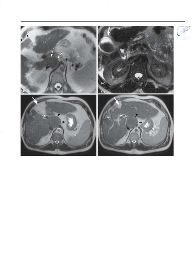
Fig. 46 a–e. Projective MR image (a) showing dilation of the extrahepatic duct and left and right hepatic duct, with pruning of peripheral branches in the right lobe. Also note marked saccular dilation of the bile duct in the left lobe with intraductal lithasis (arrow). The reduced signal intensity of the intrahepatic ducts and common bile duct is explained by the presence of debris. b Axial contrastenhanced T1-weighted MR image showing multiple abscesses in the left liver lobe (arrowheads), located in close proximity to the dilated left hepatic duct (L). c CT image showing typical pigmented stone (arrow). d ERCP images initially failed to reveal the large stone in the left hepatic duct. e After several attempts, successful opacification of the left hepatic duct was obtained and the presence of a large stone
confirmed. Also note tapering of intrahepatic bile e ducts, best seen in d, e

98 2.2 Benign Nontraumatic Abnormalities
#47 Echinococcosis
KEY FACTS: DISEASE
●Infection with Echinococcus granulosus: most common (hydatid disease)
– Histology:
Endocyst (= parasitic component of capsule): (a) inner germinative layer (= brood capsule), (b) cyst membrane (chitin)
Pericyst (= granulation tissue)
–Endemic to many parts of Australia, Africa, and the Middle East
–Organs mainly affected: liver (73%), lung (14%), peritoneum (12%)
–Symptoms: pain, recurrent jaundice (biliary obstruction by membrane fragments)
–Eosinophilia in 20%–50%
–Complications: intrabiliary rupture (66%), intraperitoneal rupture (13%), suppuration (13%), intrathoracic rupture (6%)
●Echinococcus multilocularis: rare
KEY FACTS: MRI
●Typical features of hydatid disease (Fig. 47 a, b):
–Floating membranes within a fluidcontaining and encapsulated lesion
–The floating membranes usually have low signal intensity; this is not an absolute criterion, however
–Signal intensity of fluid: usually high, may be low (hydatid sand)
●Calcifications (seen in 10%–30%) usually not visible on MRI
●If rupture has occurred:
–Interruption of cyst wall
–Filling defects (hydatid sand and/or daughter cyst) in a dilated bile duct
●Echinococcus multilocularis has a more atypical presentation; lesions may mimic either metastases or pyogenic abscesses (Fig. 47c–f)
References
Kumar R, Reddy SN, Thulkar S (2002) Intrabiliary rupture of hydatid cyst: diagnosis with MRI and hepatobiliary isotope study. Br J Radiol 75 : 271–274
Mortelé KJ,Wiesner W, Zou KH et al. (2004) Asymptomatic nonspecific serum hyperamylasemia and hyperlipasemia: spectrum of MRCP findings and clinical implications. Abdom Imaging 29 : 109–114
von Sinner W (1991) New diagnostic signs in hydatid disease: radiography, ultrasound, CT, and MRI correlated to pathology. Eur J Radiol 12 : 150–159
Zargar SA, Khuroo MS, Khan BA, Dar MY, Alai MS, Koul P (1992) Intrabiliary rupture of hepatic hydatid cyst: sonographic and cholangiographic appearances. Gastrointest Radiol 17 : 41–45
2 Intrahepatic Bile Ducts |
99 |
a |
b |
c |
d |
e
Fig. 47 a–f. Hydatid disease. a T2-weighted MR image showing typical aspect of floating membranes (arrow). b Same patient. CT image showing calcifications, which are also typical. In this patient, there were no signs of rupture into the biliary tree. c–f Echinococcus multilocularis c, d T2-weighted HASTE image (TE 60) showing an echinococcus lesion in the left lobe (arrow) with an area of central necrosis (arrowhead) and innumerable small peri-
pheral cysts. e Axial post-gadolinium VIBE image f showing the area of central necrosis. f Projective image showing two echinococcus lesions (arrows), one in the left lobe and a second in the right lobe (not shown on the axial images)

100 2.3 Traumatic, Postoperative, and Iatrogenic Abnormalities
2.3Traumatic, Postoperative, and Iatrogenic Abnormalities
#48 Sequelae
of Direct Liver Trauma
Related topics: # 113 (blunt gallbladder trauma), # 181 (pancreatic duct injury)
KEY FACTS: DISEASE
●Incidence: severe bile duct injury after upper abdominal trauma is rare (< 0.1%; Dawson et al. 1991)
●Mechanism: hepatic laceration extending into a bile duct
●Complications:
–Obstruction with or without stone formation
–Bile leakage
–Biloma formation
–Partial liver atrophy
KEY FACTS: MRI
●MRI may show ductal narrowing or obstruction,bile leakage,etc. (Fig. 48a,b)
●Note: “Periportal cuffing” (Fig. 48c): (= fluid around venous tributaries)
–Synonyms: “periportal halo,” “periportal tracking”
–Commonly seen after blunt hepatic trauma
–May also be seen as a normal finding after hepatic transplantation and in patients with acute hepatitis, cirrhosis,AIDS, veno-occlusive disease, congestive heart failure,etc.(Lawson et al. 1993)
●Some types of injury may present with hilar stenoses, possibly mimicking neoplastic disease (Fig. 48 d–f)
References
Dawson DL, Johansen KH, Jurkovich GJ (1991) Injuries to the portal triad. Am J Surg 161 : 545–551
Fulcher AS, Turner MA (2002) MR cholangiopancreatography. Radiol Clin North Am 40 : 1363–1376
Lawson TL, Thorsen MK, Erickson SJ, Perret RS, Quiroz FA, Foley WD (1993) Periportal halo: a CT sign of liver disease. Abdom Imaging 18 : 42–46
2 Intrahepatic Bile Ducts 101
a |
b |
c |
d |
e |
f |
Fig. 48 a–f. Axial T2-weighted MR image (a) showing a hypointense laceration in the left liver lobe (arrows) and a dilated left hepatic duct (arrowheads). b Projective MR image showing occlusion of the bile duct secondary to the laceration (arrow). c Different patient. Heavily T2-weighted MR image showing “periportal cuffing” (arrowheads). d, e Patient with history of segmentectomy of the liver. d Coronal T2-weighted HASTE image (TE 60) showing the segmentectomy with fat interposition (arrow), together with right intrahepatic bile duct
dilatation (arrow-head). e Projective image reveals a focal stenosis of the right hepatic duct (arrowhead), corresponding to a postoperative cicatricial stenosis. Note the unusually large right segmental posterior bile duct (arrow) related to hypertrophy of the right lobe. f Patient with a history of surgical treatment of a (suicidal) gunshot causing extensive liver damage. Postoperatively, progressive jaundice was observed. Projective image showing a focal hilar cicatricial bile duct stenosis (arrow) with secondary dilatation of the intrahepatic bile ducts

102 2.3 Traumatic, Postoperative, and Iatrogenic Abnormalities
#49 After Hepaticojejunostomy:
Anastomotic Stricture
Related topics: #29 (postoperative anatomy: after hepat(ic)ojejunostomy), #56 (recurrent tumor after hepaticojejunostomy)
KEY FACTS: DISEASE
●Incidence: up to 23%
(Bismuth et al. 1978; Lane et al. 1973)
●Mechanisms:
–Fibrosis
–Recurrent neoplasm (see #56)
●Complications:
–Stone formation
–Cholangitis
KEY FACTS: MRI
●Dilation of intrahepatic bile ducts nearly invariably present
●Typical features of fibrotic strictures (Fig. 49):
–Usually short
–Limited to anastomosis
–Smooth delineation
–No mass lesion on cross-sectional images
●Note: In comparison with PTC, MRCP may overestimate strictures; ERCP is usually impossible
References
Bismuth H, Franco D, Corlette MB, Hepp J (1978) Long-term results of Roux-en-y hepaticojejunostomy. Surg Gynecol Obstet 146 : 161–167
Lane CE, Sawyers JL, Riddel DH, Scott HW Jr (1973) Long-term results of Roux-en-y hepatocholangiojejunostomy. Ann Surg 177 : 714–722
Pavone P, Laghi A, Catalano C et al. (1997) MR cholangiography in the examination of patients with biliary-enteric anastomoses. AJR Am J Roentgenol 169 : 807–811
2 Intrahepatic Bile Ducts 103
a |
b |
c |
d |
Fig. 49. a Projective MR image showing a short stricture of a hepaticojejunal anastomosis (arrow). b, c Patient with history of hepaticojejunostomy. b Projective image showing an anastomotic stricture (large arrow) with a stone (small arrow) and secondary dilatation of the intrahepatic bile ducts.
c T2-weighted HASTE image showing the intrahepatic bile duct dilatation and the stone (arrow) d Different patient. Projective image showing a stricture of the hepaticojejunostomy (large arrow) with secondary intrahepatic bile duct dilatation and stones (small arrows)

104 2.3 Traumatic, Postoperative, and Iatrogenic Abnormalities
#50 After Hepatic
Transplantation (1): Ischemia
Related topics: #80, 81 (extrahepatic bile duct, complications after hepatic transplantation)
KEY FACTS: DISEASE
●Biliary complications after liver transplantation are common (up to 37%; Lerut et al. 1987; Ward et al. 1990)
●Ischemia is an important cause of biliary injury
●Mechanism: bile ducts receive their blood supply from the hepatic artery (not portal vein); any abnormality leading to decreased arterial inflow may cause ischemia
●Common causes:
–Hepatic artery thrombosis (7% of transplants)
–Severe arterial stenosis (1%)
–Prolonged cold ischemic graft perfusion time
●Complications (occurring in up to 50%):
–Stricture formation (multifocal)
–Leakage of bile with or without biloma
–Diffuse dilation
–Intraductal debris, sloughed mucosa, clot
KEY FACTS: MRI (FIG. 50)
●Typical findings:
–Multiple irregularities/strictures typically involving both the intrahepatic ducts and the proximal common hepatic duct
–Bile duct dilation
–Nonanastomotic leakage, biloma formation
–Presence of multiple intraluminal fill-
ing defects
● Differential diagnosis: e.g., primary sclerosing cholangitis, chronic rejection, cholangitis
● See also #81
References
Lerut T, Gordon RD, Iwatsuki S et al. (1987) Biliary tract complications in human orthotopic liver transplantation. Transplantation 43 : 47–51
Ward EM, Kiely MJ, Maus TP, Wiesner RH, Krom RA (1990) Hilar biliary strictures after liver transplantation: cholangiographic and percutaneous treatment. Radiology 177 : 259–263
Zajko AB, Campbell WL, Logsdon GA et al. (1987) Cholangiographic findings in hepatic artery occlusion after liver transplantation. AJR Am J Roentgenol 149 : 485–489
Zoepf T,Maldonado-Lopez EJ,Hilgard P,et al. (2005) Diagnosis of biliary strictures after liver transplantation: which is the best tool? World J Gastroenterol 11 : 2945–2948

2 Intrahepatic Bile Ducts 105
a
Fig. 50 a, b. Projective MR image (a) and ERCP image (b) showing several strictures of bifurcation and the right intrahepatic duct (arrows). There is also an anastomotic stricture. Also shown in a is a small stone in the distal common bile duct (arrowhead). c Different patient. Projective image showing irregular strictures of the common hepatic duct and the left hepatic duct (arrows)
b
c

106 2.3 Traumatic, Postoperative, and Iatrogenic Abnormalities
#51 After Hepatic
Transplantation (2):
Other Complications
Related topics: #80, 81 (extrahepatic bile duct, complications after hepatic transplantation)
KEY FACTS: DISEASE
●Other complications of orthotopic liver transplantation apart from ischemia include:
–Stone formation
–Biloma (may occur secondary to ischemia)
–Hematoma (common in the immediate postoperative period)
–Abscess
–Portal/hepatic vein thrombosis
–Acute rejection
–Chronic rejection
–Viral hepatitis
–Anastomotic stricture/leakage
KEY FACTS: MRI
●Hematomas (Fig. 51) have a variable signal intensity, depending on their age (see Table 51 for a simplified classification). Moreover, the central and peripheral parts of a hematoma may display a different signal intensity
●Bilomas have signal intensity features typical of fluid
●Acute rejection: may give the intrahepatic biliary tree a primary sclerosing cholangitis type of appearance (narrowing, stretching, and separation, reflecting edema and cellular infiltration)
●Chronic rejection: disappearance of small bile ducts (“vanishing bile ducts,” see #39), nonanastomotic strictures,
ductal irregularity, intraductal casts, etc.
● Note: A common finding after liver trans- |
! |
|
|
plantation is the presence of periportal |
|
lymphedema related to surgical disrup- |
|
tion of lymphatic channels. It should not |
|
be erroneously interpreted as biliary dila- |
|
tion (see #37, 48) |
|
References
Bauman J, Campbell WL, Demetris AJ, Zajko AB (1988) Intrahepatic cholangiographic abnormalities in liver transplants: correlation with biopsy evidence of rejection and other disorders. AJR Am J Roentgenol 152 : 275–279
Dominguez R, Cuervas-Mons V, Van Thiel DH, Lecky JW, Starzl TE (1986) Radiographic features of liver allograft rejection. Gastrointest Radiol 11 : 326–329
Moncorge C, Baudin F, Vigouroux C et al. (1989) Transplantation hepatique chez l’adulte: prise en charge postoperatoire et evolution au cours des premiers mois. Ann Fr Anesth Reanim 8 : 497–517
Zoepf T,Maldonado-Lopez EJ,Hilgard P,et al. (2005) Diagnosis of biliary strictures after liver transplantation: which is the best tool? World J Gastroenterol 11 : 2945–2948
|
|
|
2 Intrahepatic Bile Ducts 107 |
|
|
|
|
|
|
Table 51.1 Simplified classification of hematomas |
|
|
|
|
|
|
|
|
|
Age of hematoma |
Composition |
Signal intensity |
Signal intensity |
|
|
|
on T1 |
on T2 |
|
|
|
|
|
|
Hyperacute (<1 h) |
OxyHb |
Low/intermediate |
High |
|
Acute (< 3–4 days) |
Intracellular deoxyHb |
Intermediate |
Low |
|
|
Extracellular deoxyHb (lysis) |
Intermediate |
High |
|
Subacute |
MetHb |
High |
High |
|
(months/years) |
|
|
|
|
Chronic |
MetHb + hemosiderin |
Low at periphery |
Low at periphery |
|
|
(periphery) |
|
|
|
|
|
|
|
|
OxyHb, oxyhemoglobin; deoxyHb, deoxyhemoglobin; MetHb, methemoglobin.
a
Fig. 51 a, b. Images obtained 3 months after hepatic |
|
|
transplantation. a Post-gadolinium axial VIBE |
|
|
image showing a focal area of hepatonecrosis |
|
|
(arrow). b Projective image showing the area of |
b |
|
necrosis filled with fluid and debris (arrow) |
||
|

108 2.3 Traumatic, Postoperative, and Iatrogenic Abnormalities
#52 After Cholecystectomy:
Stricture/Transection
of an Aberrant Bile Duct
Related topics: #27 (variable junction of the posterior right hepatic duct), #82 (after cholecystectomy: stricture of the common bile duct)
KEY FACTS: DISEASE
●Inadvertent transection of an aberrant bile duct (see #27)
●Complications:
–Stenosis
–Occlusion
–Bile leakage
–Infection (cholangitis)
–Partial liver atrophy
!● Note: Other anatomical variations that increase the risk of bile duct injury during laparoscopic cholecystectomy include the following (Fig. 52a):
–Low bifurcation of the common bile duct (see #63)
–Very low or very high insertion of the cystic duct (see #93)
–Short cystic duct
–Parallel course of cystic and hepatic ducts
–Cystic duct entering the common hepatic duct along its medial surface
KEY FACTS: MRI (FIG. 52)
●Depending on the severity of the injury, MR images may show:
–Mild to severe segmental intrahepatic bile duct dilation
–Focal narrowing of an accessory bile duct
–Subhepatic/hilar bile collection
● Note: In comparison with ERCP, MRCP |
! |
|
|
may underor overestimate the severity |
|
of the stenosis (see also Fig. 27c, d) |
|
References
Davidoff AM, Pappas TN, Murray EA et al. (1992) Mechnisms of major bile duct injury during laparoscopic cholecystectomy. Ann Surg 215 : 196–202
Deziel DJ, Millikan KW, Economou SG, Doolas A, Ko ST, Airan MC (1993) Complications of laparoscopic cholecystectomy: a national survey of 4.292 hospitals and 77.604 cases. Am J Surg 165 : 9–14
Vitellas KM, Keogan MT, Spritzer CE et al. (2000) MR cholangiopancreatography of bile and pancreatic duct abnormalities with emphasis on the single-shot fast spin-echo technique. Radiographics 20 : 939–957
2 Intrahepatic Bile Ducts 109
a
Fig. 52. a Some anatomical variations predisposing to bile duct injury. 1, low insertion of the right posterior duct close to the cystic duct; 2, low bifurcation; 3, cystic duct draining into the right hepatic duct; 4, very short cystic duct; 5, long parallel course of the cystic duct and common hepatic duct. (Reprinted with permission from Kune and Sali 1980). b Patient
b
who had had a laparoscopic cholecystectomy now presenting with fever. Projective MR image showing dilated right posterior biliary branches with an aberrant insertion. Note severe narrowing of the distalmost portion of this aberrant duct (arrow), probably related to direct trauma during surgery

110 2.3 Traumatic, Postoperative, and Iatrogenic Abnormalities
#53 Biliary Complications
of Percutaneous Procedures
KEY FACTS: DISEASE (GAZZANIGA ET AL. 1991;
VAN THIEL ET AL. 1993)
●Complications of PTC and other percutaneous interventions:
–Bacteremia
–Sepsis
–Subcapsular hematoma
–Bile leak
–Biloma
–Peritonitis
–Arteriovenous fistula
–Vasculobiliary fistula
●Complication rate:
–PTC using the Chiba needle: 1.8%
–PTC combined with external drainage: 10%–15%
–Liver biopsy: <1%
● MR detection of small arteriovenous fis- |
! |
|
|
||
tulae and vasculobiliary fistulae requires |
|
|
a special technique (preferably three- |
|
|
dimensional breathhold contrast-en- |
|
|
hanced MRI; Gaa et al. 1997; Rofsky |
|
|
et al. 1999; Fig. 53) |
|
|
KEY FACTS: MRI |
|
|
● Biloma, hematoma, etc.: see #51 |
|
|
|
|
|
References |
|
|
Gazzaniga GM, Gaffioni A, Bondanza G, Bararolo C, |
|
|
Filauro M (1991) Percutaneous transhepatic bil- |
|
|
iary drainage: 12 year experience. Hepatogas- |
|
|
troenterology 38 : 154–59 |
|
|
Rofsky, NM, Lee VS, Laub G, et al. (1999) Abdominal |
|
|
MR imaging with a volumetric interpolated |
|
|
breath-hold examination. Radiology 212 : 876– |
|
|
884 |
|
|
Van Thiel DH, Gavaler JS, Wright H, Tzakis A (1993) |
|
|
Liver biopsy: its safety and complications as seen |
|
|
at a liver transplant center. Transplantation 55 : |
|
|
1087–1090 |
|
|

2 Intrahepatic Bile Ducts 111
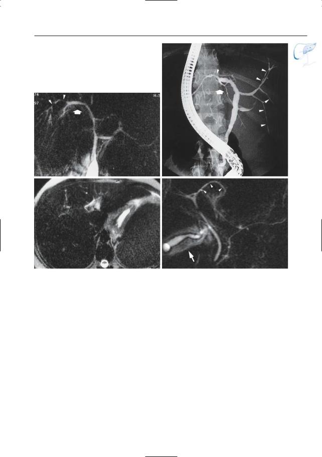
a |
b |
c |
d |
Fig. 53 a–d. Patient who underwent a PTC procedure complicated by a biliary venous fistula. a, b Post-gadolinium axial VIBE images obtained in the venous phase showing the percutaneous drain (arrows) complicated by a thrombus in the right
hepatic vein (arrowheads) due to a biliary venous fistula. c, d Corresponding ERCP images showing the opacification of the right hepatic vein when contrast is injected through the percutaneous drain
(arrows)