


144 3.2 Benign Nontraumatic Abnormalities
3.2Benign Nontraumatic Abnormalities
#68 Developmental
Abnormalities (1):
Choledochal Cyst
Related topics: #30 (Caroli’s disease), # 126 (choledochocele)
KEY FACTS: DISEASE
●Cystic dilation of the extrahepatic bile duct (type I bile duct cyst; see #30)
●50%–80% of bile duct cysts
●Patients present during childhood or adolescence
●Ratio of women to men: 3 :1
●Etiology: anomalous junction of the pancreatic duct and common bile duct (long common channel; see also # 123)
●Mechanism: free reflux of pancreatic enzymes in the biliary tree with weakening of the wall of the common bile duct
●Associations:
–Dilation/stenosis or atresia of other portions of biliary tract
–Gallbladder anomalies
–Polycystic liver disease
–Gallbladder carcinoma
●Complications:
–Stones in the common bile duct (8%–70%)
–Recurrent pancreatitis (1%–34%)
–Cholangitis (20%)
–Malignant transformation (3%–28%; age, > 20 years)
–Biliary cirrhosis (1%–13%)
–Rupture with bile duct peritonitis (1.8%)
–Bleeding
●Note: The choledochal cysts seen in neonates with biliary atresia (#69) are considered to represent a separate entity
KEY FACTS: MRI
●Typical features:
–Markedly dilated extrahepatic bile duct
–The hallmark is a long common channel (Fig. 68a–f)
–No or only mild intrahepatic bile duct
dilation; abrupt changes of caliber |
|
|
– Usually saccular in configuration |
|
|
! |
||
● Irregular ductal contours may herald |
||
malignant degeneration |
|
References
De Wilde V, Elewaut A, De Vos MP, Hendrix R, Barbier F (1991) Choledochal cysts in the adult. Endoscopy 23 : 4–7
Fulcher AS,Turner MA (1999) MR pancreatography: a useful tool for evaluating pancreatic disorders. Radiographics 19 : 5–24
3 Extrahepatic Bile Duct 145
b
a
d
c
e |
f |
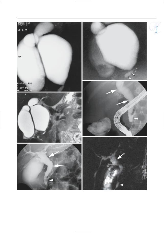
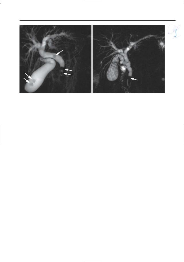
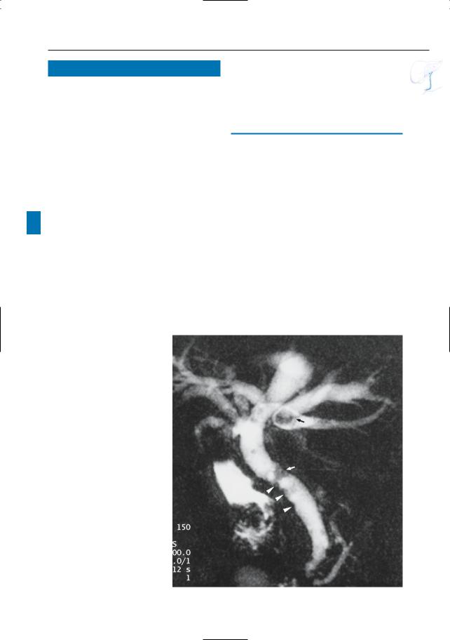
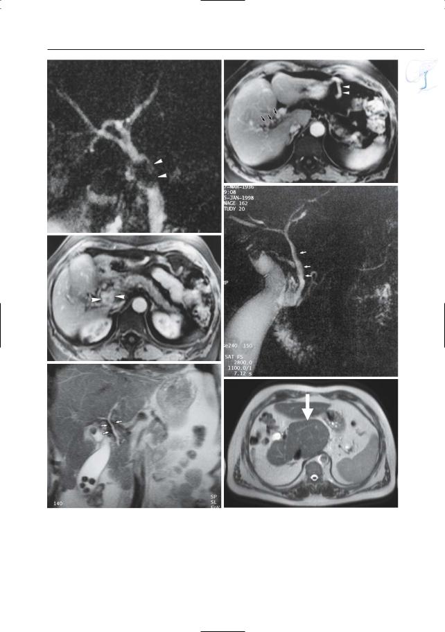
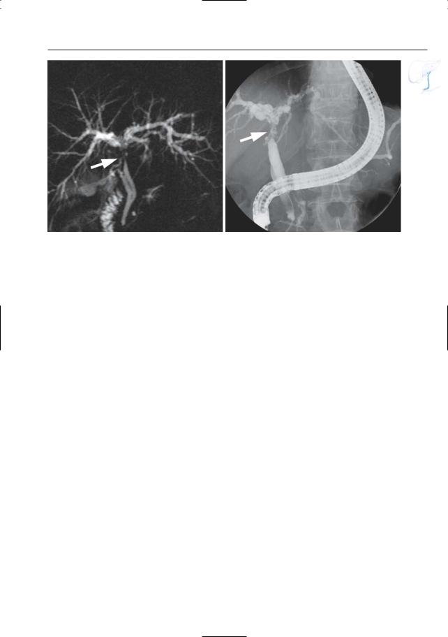
Fig. 68 a–f. a, b Projective MR images showing marked saccular dilation of the common bile duct. The pancreatic duct joins the common bile duct just below the dilated segment (arrows in b). Note the long common channel (arrowheads).Also note the dilated gallbladder containing a stone. c Same patient. Coronal cross-sectional image showing the position of a choledochal cyst in relation to the liver. d–f Different
Patient. ERCP images showing a fusiform dilatation of the common bile duct and the common hepatic duct (arrows) compatible with a type I bile duct cyst. Note long common channel (arrowhead) and aerobilia (arrowhead in e). f Projective image showing dilated common hepatic duct (arrow) and long common channel (arrowhead). Image quality is suboptimal due to aerobilia and respiratory artifact

146 3.2 Benign Nontraumatic Abnormalities
|
|
|
|
|
|
|
|
|
|
|
KEY FACTS: MRI |
|
|
|
|
|
|
#69 Developmental |
|
|
|
|
|
|
|||||||
|
|
|
|
|
|
|
|
|
|
||||||
|
|
|
Abnormalities (2): Atresia |
|
● Typical features: |
|
|||||||||
|
|
|
|
|
|
|
|
|
|
|
|
||||
|
|
KEY FACTS: DISEASE |
|
|
|
|
– |
No visualization of extrahepatic bile |
|
||||||
|
|
|
|
|
|
|
|||||||||
|
|
|
|
|
|
|
duct |
|
|
|
|||||
|
|
|
|
|
|
|
|
|
|
|
|
|
|
|
|
|
|
● Progressive inflammation, fibrosis, and |
– |
“Periportal |
thickening” (correspon- |
|
|||||||||
|
|
|
ding to periportal fibrosis) |
|
|||||||||||
|
|
obliteration of bile ducts |
|
|
|
|
|
||||||||
|
|
|
|
|
● Remarks: |
|
|
|
|||||||
|
|
● Most |
common |
cause |
of |
chronic |
|
|
! |
||||||
|
|
– |
Knee or surface coils should be used |
||||||||||||
|
|
cholestasis in infants and children |
|||||||||||||
|
|
|
as much as possible (high-resolution |
||||||||||||
|
|
● Classification: |
|
|
|
|
|
|
|
||||||
|
|
|
|
|
|
|
|
imaging) |
|
|
|
||||
|
|
– Type I: atresia of the distal part of the |
|
|
|
|
|||||||||
|
|
– |
Atresia can only be ruled out if the |
|
|||||||||||
|
|
|
common bile duct |
|
|
|
|
||||||||
|
|
|
|
|
|
|
entire extrahepatic bile duct is identi- |
|
|||||||
|
|
– |
Type |
II: atresia |
of the |
extrahepatic |
|
|
|||||||
|
|
|
fied |
|
|
|
|||||||||
|
|
|
duct; normal bifurcation and intra- |
|
|
|
|
||||||||
|
|
|
– |
Evaluation |
on the integrity of the |
|
|||||||||
|
|
|
hepatic ducts |
|
|
|
|
|
|||||||
|
|
|
|
|
|
|
|
intrahepatic ducts in infants is diffi- |
|
||||||
|
|
– |
Type |
III |
(most |
common): disease |
|
|
|||||||
|
|
|
cult because normal ducts are barely |
|
|||||||||||
|
|
|
extending proximally into the porta |
|
|
||||||||||
|
|
|
|
visible at MRI |
|
||||||||||
|
|
|
hepatis |
|
|
|
|
|
|
|
|
||||
|
|
|
|
|
|
|
|
|
– |
MRI may beuseful for postoperative |
|
||||
|
|
● Associated disease: developmental chole- |
|
||||||||||||
|
|
|
follow up (Fig. 69 a,b) |
|
|||||||||||
|
|
dochal cyst of the neonate |
|
|
|
|
|
||||||||
|
|
|
|
|
|
|
|
|
|
||||||
|
|
● Symptoms: jaundice, pale stools |
|
|
|
|
|
|
|
||||||
|
|
● Diagnosis: |
hepatobiliary |
scintigraphy |
|
|
|
|
|
||||||
|
|
References |
|
|
|
||||||||||
|
|
(sensitivity up to 97%, lower specificity) |
|
|
|
||||||||||
|
|
|
|
|
|
|
|||||||||
|
|
● Treatment: |
|
|
|
|
|
|
Engelskirchen R,Holschneider AM,Gharib M,Vente |
|
|||||
|
|
– Correctable |
type (type I and II): |
C (1991) Biliary atresia – a 25 year survey. Eur J |
|
||||||||||
|
|
|
hepaticojejunostomy |
|
|
|
Pediatr Surg 1 : 154–160 |
|
|||||||
|
|
– |
Uncorrectable type (type III): usually |
Guibaud L, Lachaud A, Touraine R et al. (1998) MR |
|
||||||||||
|
|
cholangiography in neonates and children: feasi- |
|
||||||||||||
|
|
|
Kasai operation: hepatic portoje- |
|
|||||||||||
|
|
|
bility and preliminary applications. AJR Am J |
|
|||||||||||
|
|
|
junostomy |
(anastomosis |
between |
Roentgenol 170 : 27–31 |
|
||||||||
|
|
|
jejunal limb and cut surface of the |
Kasai M, Suzuki H, Ohashi E, Ohi R, Chiba T, |
|
||||||||||
|
|
|
fibrous mass replacing |
the |
hepatic |
Okamoto A (1978) Technique and results of oper- |
|
||||||||
|
|
|
ative management of biliary atresia.World J Surg |
|
|||||||||||
|
|
|
duct radicles at the porta hepatis) |
|
|||||||||||
|
|
|
2 : 571–580 |
|
|
|
|||||||||
|
|
– |
Liver transplantation |
|
|
|
|
|
|
||||||
|
|
|
|
|
Krause D, Cercueil JP, Dranssart M et al. (2002) MRI |
|
|||||||||
|
|
● Complications (occurring even in pa- |
for evaluating congenital bile duct abnormali- |
|
|||||||||||
|
|
tients |
with |
|
“successful” |
bilioenteric |
ties. J Comput Assist Tomogr 26 : 541–552 |
|
|||||||
anastomoses):
–Chronic cholangitis
–Extensive hepatic fibrosis
–Portal hypertension
●Differential diagnosis (clinical): neonatal cholangitis
●Note: True agenesis of extrahepatic bile ducts is rare
3 Extrahepatic Bile Duct 147
a |
b |
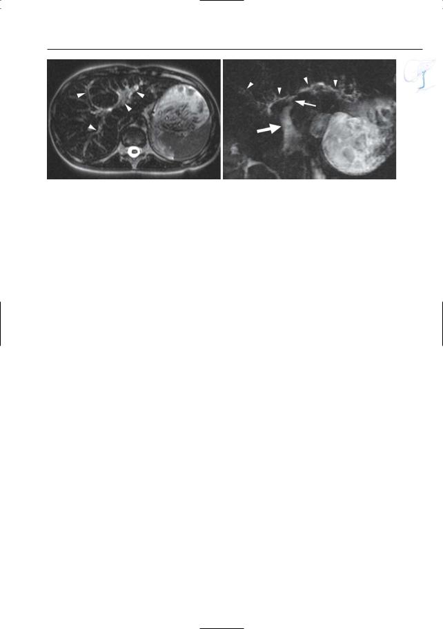
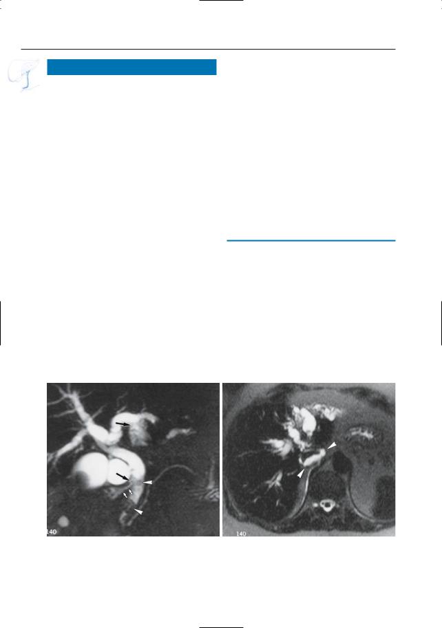
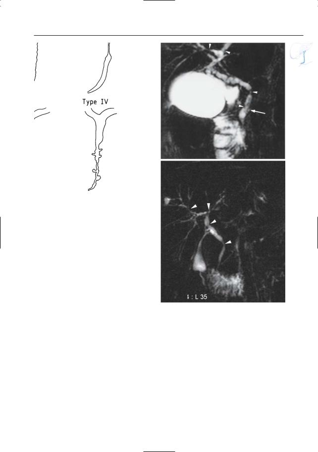
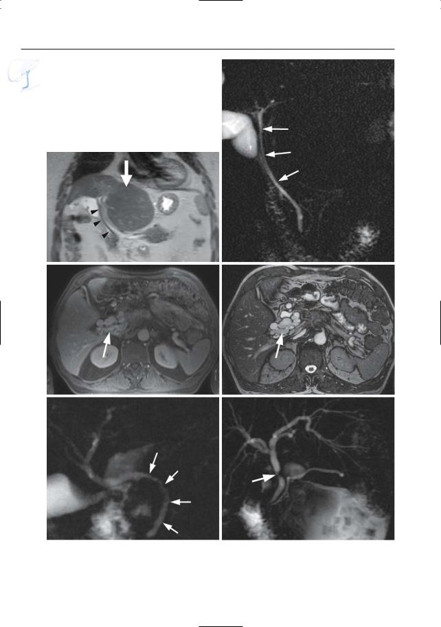
Fig. 69 a, b. Patient with biliary atresia treated by |
tion. The intrahepatic bile ducts are markedly irreg- |
Kasai operation. a Axial T2-weighted HASTE image |
ular (arrowheads) with discrete dilatation of the left |
(TE 360) showing discrete dilatation of the left in- |
intrahepatic bile duct. These findings are due to |
trahepatic bile ducts (arrowheads). b Projective im- |
chronic cholangitis and hepatic fibrosis secondary |
age showing the jejunal limb (large arrow) and the |
to the atresia, and are commonly observed, even |
anastomosis (small arrow) due to the Kasai opera- |
after successful bilioenteric anastomosis |
148 3.2 Benign Nontraumatic Abnormalities
#70 Web
Related topic: # 73 (stones in the common bile duct: pitfalls in diagnosis)
KEY FACTS
●Valvulae in the common bile duct
●Histology: folds or plates containing fibrous and muscular tissue with a redundant epithelial surface
●Rare
KEY FACTS: MRI
●Typical features:
–Less than 1 mm thick
–Elongated aspect
–Sharply marginated
–Not completely surrounded by bile
● Remarks: |
|
|
! |
||
– The MRCP appearance of a web may |
||
differ from its ERCP appearance (ab- |
|
|
sence of distention in MRCP) (Fig. 70) |
|
|
– Occasionally, differentiation between |
|
|
a web and a stone may be difficult (see |
|
|
Fig. 73) |
|
References
Belsito AA, Cramer GG, Dickinson PB (1977) Normal biliary tree. In: Stewart E,Vennes J,Geenen J (eds) Atlas of endoscopic retrograde cholangiopancreatography. Mosby, Saint Louis, pp 88–123
Dolar ME, Ates KB, Dalay AR, Caner ME, Sasmaz N, Sahin B (1993) Congenital stricture of the common hepatic duct due to a web: an unusual case without jaundice. Hepatogastroenterology 40 : 194–195
Papaziogas B, Lazaridis C, Pavlidis T et al. (2002) Congenital web of the common bile duct in association with cholelithiasis. J Hepatobiliary Pancreat Surg 9 : 271–273
3 Extrahepatic Bile Duct 149
a |
b |
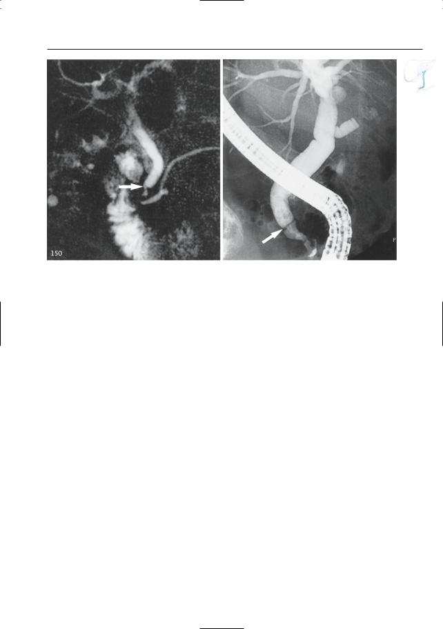
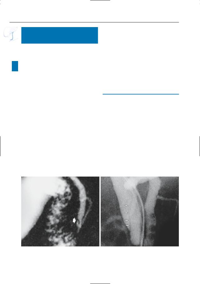
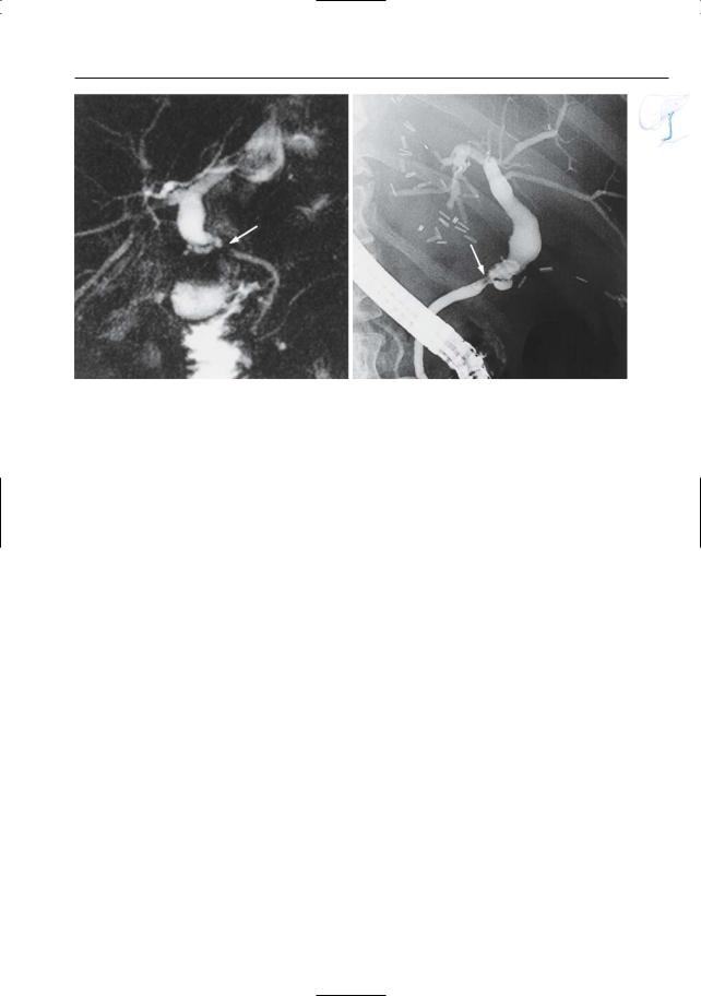
Fig. 70 a, b. a Projective MR image and b ERCP image showing web in the distal common bile duct (arrows). Note collapse of the distal segment of the
common bile duct in a, resulting in a different morphologic appearance of the web

150 3.2 Benign Nontraumatic Abnormalities
#71 Mirizzi Syndrome
KEY FACTS: DISEASE
●Extrinsic right-sided compression of the common bile duct by a large gallstone impacted in the cystic duct, gallbladder neck, or cystic duct remnant, accompanied by chronic inflammatory reaction
●Types:
–Type 1: simple type
–Type 2: fistula between the gallbladder and common hepatic duct
●More common in patients with anomalous low insertion of the cystic duct into the common hepatic duct with a parallel course of the ducts
●Associated disease: gallbladder carcinoma (see #22; Miller and Sica 1996)
KEY FACTS: MRI
●Typical features:
–Type 1: smooth, focal, laterally scalloped narrowing of the common bile duct caused by a stone in the gallbladder neck/cystic duct (Fig. 71a, b)
–Type 2: stone located at the junction of the cystic duct and common hepatic duct, partially located within the bile duct; no smooth lateral compression (Fig. 71c–f)
–In both types, infiltration of fat surrounding the junction may be seen (inflammation)
●The presence of a soft tissue mass ! around the cystic duct and/or in the hepatic portal should arouse suspicion
of associated neoplastic disease (usually gallbladder carcinoma; see #22)
●Differential diagnosis of smooth extrinsic bile duct narrowing: extrinsic inflammatory process, acute cholecystitis with gallbladder enlargement, pancreatic pseudocysts, choledochal varices
References
Binmoeller KF, Thonke F, Soehendra N (1993) Endoscopic treatment of Mirizzi’s syndrome. Gastrointest Endoscop 39 : 532–536
Koehler RE, Melson GL, Lee JK, Long J (1979) Common hepatic duct obstruction by cystic duct stone: Mirizzi syndrome. AJR Am J Roentgenol 132 : 1007–1009
Miller FH, Sica GT (1996) Mirizzi syndrome associated with gallbladder cancer and biliary-enteric fistulas. Case report. AJR Am J Roentgenol 167 : 95–97
Redaelli CA, Buchler MW, Schilling MK et al. (1997) High coincidence of Mirizzi syndrome and gallbladder carcinoma. Surgery 121 : 58–63
3 Extrahepatic Bile Duct 151
a |
b |
c
d
e |
f |
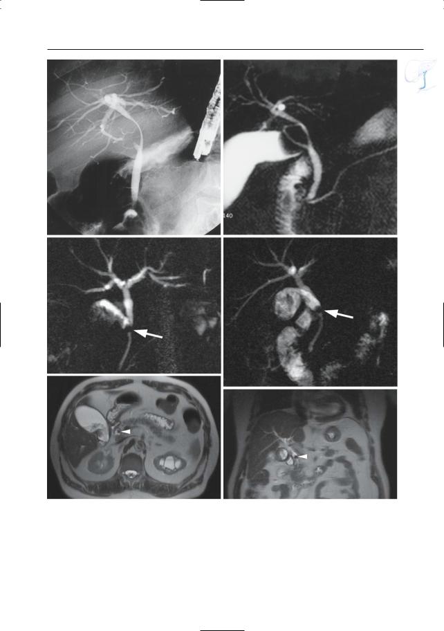
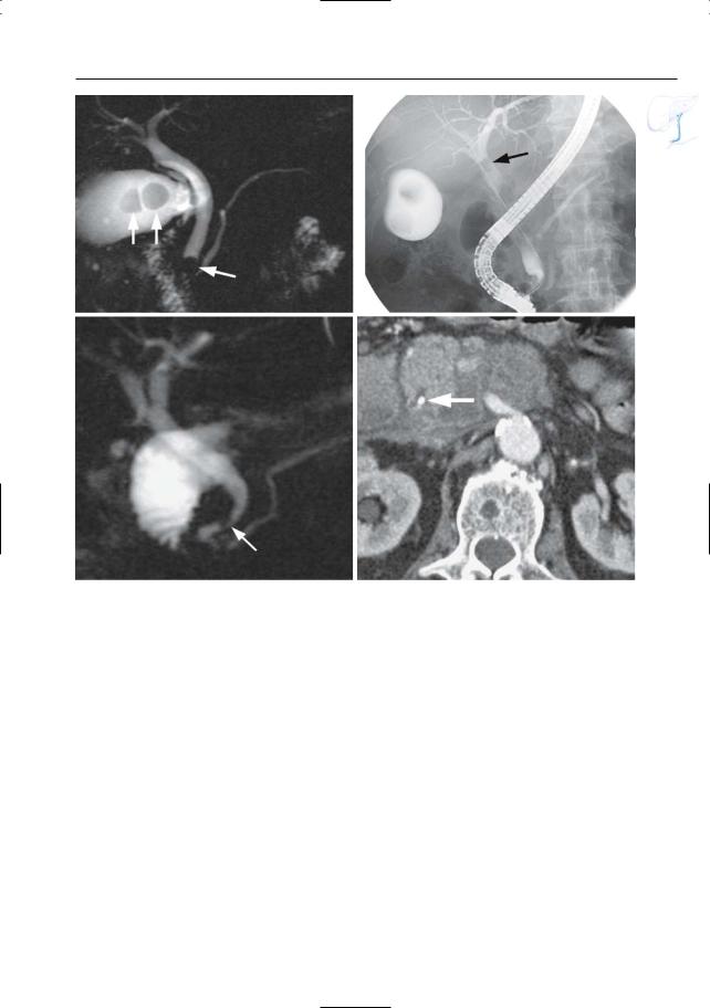
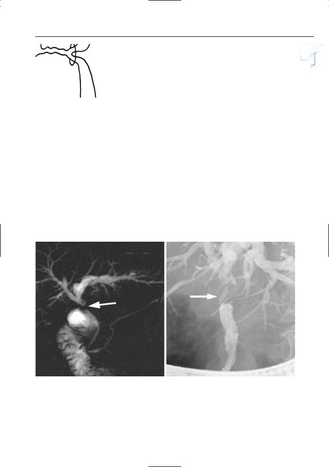
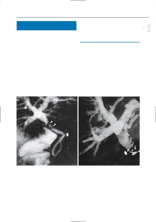
Fig. 71 a–f. Type I Mirizzi syndrome. a ERCP image showing smooth lateral impression on the extrahepatic bile duct. b Projective MR image showing an impacted stone in the gallbladder neck c–f. Type II Mirizzi syndrome. c, d Projective image showing an impacted stone at the junction of the cystic duct and
common hepatic duct (arrow) with secondary obstruction and intrahepatic bile duct dilatation. e, f Axial (e) and coronal (f) T2-weighted HASTE image (TE 60) showing the impacted stone at the junction (arrowhead)

152 3.2 Benign Nontraumatic Abnormalities
|
|
|
|
|
|
|
KEY FACTS: MRI |
|
|
|
|
|
|
#72 Stones in the Common |
|
|
|
|
|
|
|||
|
|
|
|
|
|
|
|
|
|||
|
|
|
Bile Duct |
|
|
● MR is highly accurate in the diagnosis |
|
||||
|
|
|
|
|
|
|
|
||||
|
|
Related topics: #33 (intrahepatic bile duct |
of bile duct lithiasis (Topal et al. 2003; |
|
|||||||
|
|
|
|||||||||
|
|
Tayler et al. 2002; Kim et al. 2002) |
|
||||||||
|
|
lithiasis), # 125 |
(vaterian sphincter |
com- |
|
||||||
|
|
● The large majority of stones are seen as |
|
||||||||
|
|
plex, impacted stone) |
|
|
|
||||||
|
|
|
|
T1and T2-hypointense structures sur- |
|
||||||
|
|
|
|
|
|
|
|
||||
|
|
KEY FACTS: DISEASE |
|
|
rounded by bile |
|
|
|
|
||
|
|
|
|
● Differential diagnosis: aerobilia (see #67) |
|
||||||
|
|
|
|
|
|
|
|
||||
|
|
● Most common cause of bile duct obstruc- |
● Pitfalls: see # 73 |
|
|
|
|
||||
|
|
● Note: 20%–35% of |
stones |
occur in |
|
||||||
|
|
tion |
|
|
|
! |
|||||
|
|
|
|
|
nondilated ducts (Reinhold |
and Bret |
|||||
|
|
● Found in 12%–15% of patients undergo- |
|||||||||
|
|
1996) |
|
|
|
||||||
|
|
ing cholecystectomy |
|
|
|
|
|
|
|||
|
|
|
|
|
|
|
|
|
|||
|
|
● Types: |
|
|
|
|
|
|
|
|
|
|
|
– |
In Western countries, most (95% of) |
|
|
|
|
|
|||
|
|
References |
|
|
|
|
|||||
|
|
|
patients with choledocholithiasis have |
|
|
|
|
||||
|
|
|
|
|
|
|
|
||||
|
|
|
secondary |
choledocholithiasis, |
i.e., |
Kim TK, Kim BS, Kim JH et al. (2002) Diagnosis of |
|
||||
|
|
|
the stones have migrated from the |
intrahepatic stones: superiority of MR cholan- |
|
||||||
|
|
|
gallbladder into the common |
bile |
giopancreatography over endoscopic retrograde |
|
|||||
|
|
|
duct |
|
|
|
cholangiopancreatography. AJR Am J Roent- |
|
|||
|
|
|
|
|
|
genol. 179 : 429–434Reinhold C, Bret PM (1996) |
|
||||
|
|
– |
Primary choledocholithiasis is found |
|
|||||||
|
|
Current status of MR cholangiopancreatogra- |
|
||||||||
|
|
|
in individuals of Asian or Eastern cul- |
phy. AJR Am J Roentgenol 1285–1295 |
|
||||||
|
|
|
tures and is associated with parasitic |
Taylor AC, Little AF, Hennessy OF et al. (2002) |
|
||||||
|
|
|
infection |
|
|
|
Prospective assessment of magnetic resonance |
|
|||
|
|
|
|
|
|
cholangiopancreatography |
for |
noninvasive |
|
||
|
|
● Predisposing factors: |
|
|
|
||||||
|
|
|
|
imaging of the biliary tree. Gastrointest Endosc |
|
||||||
|
|
– |
Cholecystolithiasis |
|
|
55 : 17–22 |
|
|
|
|
|
|
|
– |
Bile duct stenosis |
|
|
Topal B,Van de Moortel M,Fieuws S et al. (2003) The |
|
||||
|
|
– |
Dysfunction of the vaterian sphincter |
value of magnetic resonance cholangiopancre- |
|
||||||
|
|
|
complex (see # 127) |
|
|
atography in predicting |
common bile duct |
|
|||
|
|
|
|
|
stones in patients with gallstone disease. Br J |
|
|||||
|
|
● Associated disease: cholangitis |
|
|
|
||||||
|
|
|
|
Surg 90 : 42–47 |
|
|
|
|
|||
●Symptoms (75%): jaundice, chills, fever
●Complications:
–Acute pancreatitis (see # 151)
–Duct stricture
–Fistula (e.g., bilioenteric; see # 74)
–Bacterial cholangitis (see # 75)
–Liver abscess (see #40)
–Secondary sclerosing cholangitis (see #45)
3 Extrahepatic Bile Duct 153
a |
b |
Fig. 72. a Projective image showing several stones in the gallbladder and in the common bile duct (arrows) b Different patient. Projective image showing
multiple small stones in the gallbladder and an impacted stone in the distal common bile duct (arrow), with secondary bile duct dilatation
154 3.2 Benign Nontraumatic Abnormalities
#73 Stones in the Common Bile Duct (2): Pitfalls in Diagnosis with MRCP/ERCP
Related topics: #67 (aerobilia), #70 (web),
# 125 (impacted stone), # 122 (vaterian sphincter complex, pseudocalculus sign)
! KEY FACTS
● False-negative diagnoses on MRCP are usually caused by a lack of fluid surrounding the stone:
– Lack of ductal distention (e.g., primary sclerosing cholangitis, intermittent obstruction)
– Stone impacted in sphincteric segment (see # 125)
– Very large stone; multiple stones with or without sludge
● Causes of a false-positive diagnosis on MRCP:
– Bile duct web (rare) (see # 70 and Fig. 73a, b)
–Inverted sphincteric segment (“pseudocalculus sign”; see # 122)
a
–Aerobilia (see #67)
–Flow artifact (Sugita et al. 2003; Van Hoe et al. 2004)
●Causes of a false-negative diagnosis on ERCP:
–Small stone invisible due to overfilling/superimposition of contrast medium (intrinsic limitation of projective technique)
–Proximal displacement of stones during filling with contrast medium (Fig. 73c–d)
References
Irie H, Honda H, Kuroiwa T et al. (2001) Pitfalls in MR cholangiopancreatographic interpretation. Radiographics 21 : 23–37
Sugita R, Sugimura E, Itoh M et al. (2003) Pseudolesion of the bile duct caused by flow effect: a diagnostic pitfall of MR cholangiopancreatography. AJR Am J Roentgenol. 180 : 467–471
Van Hoe L, Mermuys K, Vanhoenacker P (2004) MRCP pitfalls. Abdom Imaging 29 : 360–387 Watanabe Y, Dohke M, Ishimori T et al. (1999) Diag-
nostic pitfalls of MR cholangiopancreatography in the evaluation of the biliary tract and gallbladder. Radiographics 19 : 415–429
b
Fig. 73 a–f. a Projective MR image showing hypointense elongated structure in the distal common bile duct (arrow), which was initially interpreted
as lithiasis. b ERCP showing that this lesion represented a web (arrows).
3 Extrahepatic Bile Duct 155
c |
d |
e |
f |
Fig. 73. (continued) c, d Different patient. Projective image showing two large stones in the gallbladder and an impacted stone in the distal common bile duct (arrows). d ERCP image shows no stone in the distal common bile duct; the stone now has a more
proximal position (arrow). e, f Different patient. Projective image showing a hypointense structure along the medial wall of the distal common bile duct (arrow), not quite typical for a stone. f CT image shows heavily calcified bile duct stone (arrow)
156 3.2 Benign Nontraumatic Abnormalities
#74 Stones Complicated by Fistula
Related topic: # 166 (complications of chronic pancreatitis)
KEY FACTS: DISEASE
●Fistulas occur in up to 7% of patients with common bile duct stones
●Most common types (decreasing order of frequency):
–Choledochoduodenal fistula
–Choledochogastric fistula
–Choledochojejunal fistula
–Choledochocolonic fistula
–Choledochopleural fistula
●Other causes of fistula with the common bile duct:
–Peptic ulcer disease
–Hepatic hydatid disease (Ponchon et al. 1989)
–Trauma (usually surgical)
–Gallbladder disease (see # 103)
a
KEY FACTS: MRI
●In typical cases, fistulas are seen as bandlike T2-hyperintense structures communicating with the common bile duct (Fig. 74)
●Small fistulas containing air rather than fluid are probably more difficult to visualize
●Note: If significant periductal inflammation is present, fistulas may be difficult to assess with projective MRCP; in such cases, heavily T2-weighted cross-sec- tional images are quite useful to avoid superimposition of fluid/edema (Fig. 74)
References
Ikeda S, Okada Y (1975) Classification of choledochoduodenal fistula and its etiological significance. Gastroenterology 69 : 130–137
Ponchon T,Gallez JF,Valette PJ,Chavaillon A,Bory R (1989) Endoscopic treatment of biliary tract fistulas. Gastrointest Endoscop 35 : 490–498
b
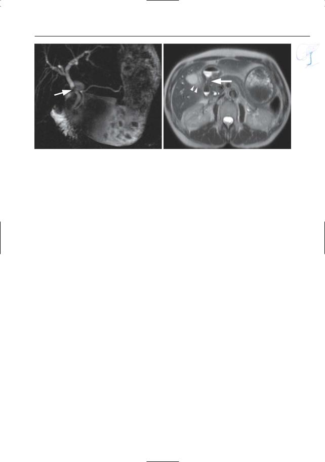
Fig. 74 a, b. a Projective MR image showing stones in the distal common bile duct (arrowheads) and rupture of both the left intrahepatic bile duct and the common bile duct (arrows). b Heavily T2-
weighted axial image showing presence of fluid around the vena cava and in the proximity of the right diaphragmatic crus (arrowheads)
3 Extrahepatic Bile Duct 157
#75 Bacterial Cholangitis
Related topic: #40 (intrahepatic bile ducts: bacterial cholangitis)
KEY FACTS: DISEASE
●Differential diagnosis:
–Primary sclerosing cholangitis (see
# 76)
–Bile duct varices (may cause narrowing with an undulated aspect; see # 79)
● See #40
KEY FACTS: MRI
●Typical feature: mucosal irregularities (Fig. 75)
!● Note: MRCP is less sensitive than ERCP in the detection of subtle abnormalities (lack of distention)
References
Bader TR, Braga L, Beavers KL, Semelka RC (2001) MR imaging findings of infectious cholangitis. Magn Reson Imaging 19 : 781–788
Sinanan MN (1992) Acute cholangitis. Infect Dis Clin North Am 6 : 571–599
Taylor AJ, Bohorfoush AG (1997) Inflammation of the biliary tract. In: Taylor AJ, Bohorfoush AG (eds) Interpretation of ERCP. Lippincott-Raven, Philadelphia, pp 77–125
Fig. 75. Projective image showing irregular contours of the common bile duct (arrowheads). Also note the
presence of two stones (arrows)
158 3.2 Benign Nontraumatic Abnormalities
#76 Primary Sclerosing Cholangitis
Related topics: #41–44 (intrahepatic bile ducts, primary sclerosing cholangitis)
KEY FACTS: DISEASE
● See #41–44
KEY FACTS: MRI
●Typical findings in different stages of disease (Fig. 76a; Majoie et al. 1991):
–Type I: marginal irregularities (differential diagnosis with cholangitis)
–Type II: focal stenosis (if solitary, differential diagnosis with cholangiocarcinoma, metastasis, traumatic stricture)
–Type III: diffuse, irregular narrowing (differential diagnosis with sclerosing carcinoma, metastases)
–Type IV: diverticular outpouchings, sacculations (typical)
● Key features: |
|
||
! |
|||
– |
Usually multiple strictures |
||
– Relative lack of dilation proximal to a |
|
||
|
stricture |
|
|
– |
Involvement of intraand extrahep- |
|
|
|
atic ducts |
|
|
References
Chapman RW (1991) Aetiology and natural history of primary sclerosing cholangitis – a decade of progress? Gut 32 : 1433–1435
Fulcher AS,Turner MA,Franklin KJ et al. (2000) Primary sclerosing cholangitis: evaluation with MR cholangiography – a case-control study. Radiology 215 : 71–80
MacCarty RL, LaRusso NF, Wiesner RH, Ludwig J (1983) Primary sclerosing cholangitis: findings on cholangiography and pancreatography. Radiology 149 : 39–44
Majoie CB, Reeders JWAJ, Sanders JB, Huibregtse K, Jansen PLM (1991) Primary sclerosing cholangitis: a modified classification of cholangiographic findings. AJR Am J Roentgenol 157 : 495–497
Vitellas KM, Keogan MT, Freed KS et al. (2000) Radiologic manifestations of sclerosing cholangitis with emphasis on MR cholangiopancreatography. Radiographics 20 : 959–975
3 Extrahepatic Bile Duct 159
a
Fig. 76. a Type I–IV cholangiographic abnormalities. b Projective MR image showing multiple focal stenoses of intraand extrahepatic bile ducts (arrowheads). Saccular outpouchings may also be present (arrow). c Different patient. Projective image showing multiple focal stenoses of the intraand extrahepatic bile ducts (arrowheads)
b
c

160 3.2 Benign Nontraumatic Abnormalities
#77 Common Bile Duct Stenosis
in Acute Pancreatitis
Related topics: # 151–157 (acute pancreatitis)
KEY FACTS: DISEASE
●A variety of diseases may cause hyperbilirubinemia in patients with acute pancreatitis
●In a minority of these patients, a stenosis of the common bile duct can be demonstrated
●Causes of common bile duct stenosis:
–Pancreatic edema (usually transient)
–Complications of pancreatitis (e.g., pseudocysts)
–Coexistent or underlying disease (e.g., common bile duct stones)
KEY FACTS: MRI (FIG. 77)
●MR appearance of the common bile duct in edematous pancreatitis:
–Commonly normal
–Smooth, gradual tapering may be observed
●Differential diagnosis: chronic pancreatitis, pancreatic cancer (see clinical data/ cross-sectional images/aspect of the pancreatic duct)
References
Frieden JH (1965) The significance of jaundice in acute pancreatitis. Arch Surg 90 : 435–441
Rohrmann CA, Baron RL (1989) Biliary complications of pancreatitis. Radiol Clin North Am 27 : 93–104
3 Extrahepatic Bile Duct 161
b
a |
c |
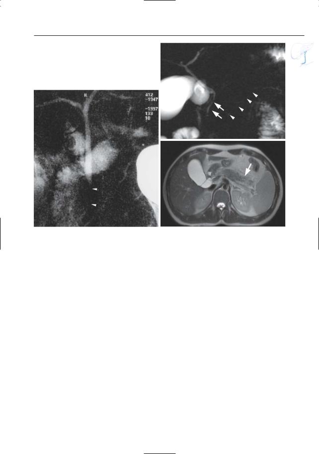
Fig. 77 . a Patient with acute pancreatitis. Projective MR image showing severe narrowing of the distal (intrapancreatic) part of the common bile duct (arrowheads). Also note increased background signal intensity due to peripancreatic fluid/edema. b, c Different patient. Projective image showing dis-
crete narrowing of the distal common bile duct (arrows) and apparent absence of the main pancreatic duct due to pancreatic oedema (arrowheads). c Axial T2-weighted HASTE image (TE 60) showing increased signal intensity of the pancreatic tissue, particularly in the tail (arrow)

162 3.2 Benign Nontraumatic Abnormalities
|
|
|
|
|
|
|
KEY FACTS: MRI |
|
|
||
|
|
|
#78 Common Bile Duct Stenosis |
|
|
|
|||||
|
|
|
|
|
|
|
|
|
|||
|
|
|
|
in Chronic Pancreatitis |
|
|
Typical MR features (Fig. 78): |
|
|||
|
|
|
|
|
|
|
● |
! |
|||
|
|
|
|
|
|
|
|||||
|
|
Related topics: # 159–167 (chronic pancre- |
|
– |
Long,smooth,gentle,progressive nar- |
||||||
|
|
|
|||||||||
|
|
|
|
rowing of entire pancreatic portion of |
|||||||
|
|
atitis) |
|
|
|
|
|
||||
|
|
|
|
|
|
the common bile duct (differential |
|
||||
|
|
|
|
|
|
|
|
|
|
||
|
|
KEY FACTS: DISEASE |
|
|
diagnosis |
with carcinoma: usually |
|
||||
|
|
|
|
abrupt narrowing) |
|
||||||
|
|
|
|
|
|
|
|
|
|
||
|
|
● |
Incidence: |
|
|
|
– |
Signs of |
attraction/distortion not |
|
|
|
|
|
|
|
|
uncommon (Fig. 78b) |
|
||||
|
|
|
– |
Prevalence of clinically relevant com- |
|
|
|
||||
|
|
|
|
– |
Displacement if direct compression |
|
|||||
|
|
|
|
mon bile |
duct stenosis in chronic |
|
|
||||
|
|
|
|
|
|
by pseudocyst is present |
|
||||
|
|
|
|
alcoholic pancreatitis: ± 9% |
|
|
|
||||
|
|
|
|
● |
The distalmost segment of the intrapan- |
|
|||||
|
|
● |
Causes: |
|
|
|
|||||
|
|
|
|
|
creatic common bile duct may widen |
|
|||||
|
|
|
– |
Fibrosis |
|
|
|
|
|||
|
|
|
|
|
|
slightly again before it narrows to enter |
|
||||
|
|
|
– |
Pseudocyst(s) |
|
|
|||||
|
|
|
|
the sphincter (“hourglass” appearance), |
|
||||||
|
|
|
– |
Inflammatory mass |
|
|
|||||
|
|
|
|
thus reflecting the extrapancreatic loca- |
|
||||||
|
|
● |
Symptoms/signs: |
|
|
||||||
|
|
|
tion of the distalmost part of the com- |
|
|||||||
|
|
|
– Abdominal pain (not specific) |
|
|
||||||
|
|
|
|
mon bile duct (see Fig. 163b) |
|
||||||
|
|
|
– Jaundice (may also be related to hepa- |
|
|
||||||
|
|
|
● Note: Atypical patterns of common bile |
|
|||||||
|
|
|
|
tocellular damage) |
|
||||||
|
|
|
|
|
duct narrowing are not uncommon (see |
|
|||||
|
|
● |
Complication: secondary sclerosing chol- |
|
|
||||||
|
|
|
# 164,“double duct sign” in chronic pan- |
|
|||||||
|
|
|
angitis with |
or without cirrhosis (see |
|
|
|||||
|
|
|
|
creatitis) |
|
|
|||||
|
|
|
#45) |
|
|
|
|
|
|||
|
|
|
|
|
|
|
|
|
|
||
References
Rohrmann CA, Baron RL (1989) Biliary complications of pancreatitis. Radiol Clin North Am 27 : 93–104
Stahl TJ, Allen MO, Ansel HJ, Vennes JA (1988) Partial biliary obstruction caused by chronic pancreatitis. An appraisal of indications for surgical biliary drainage. Ann Surg 207 : 26–32
3 Extrahepatic Bile Duct 163
a
b |
c |
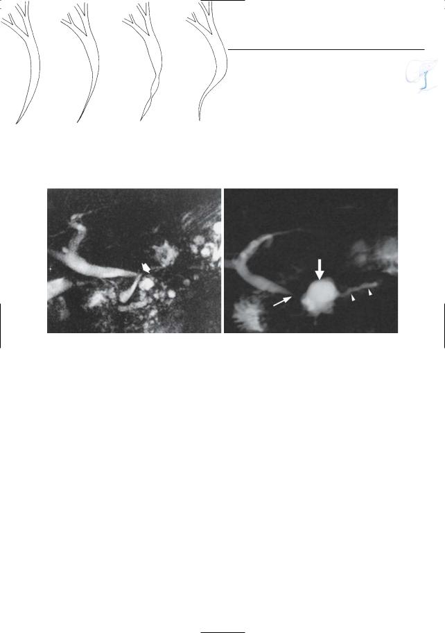
Fig. 78. a Typical configurations of the common bile duct in chronic pancreatitis. From left to right: normal; gradual tapering; alternating stenosis and dilation; focal medial or lateral deflection, usually due to pseudocyst and/or fibrosis. (From Rohrman and Baron 1989, with permission). b Projective MR image showing displacement and marked angulation of the common bile duct (arrow), caused by
extensive parenchymal fibrosis in a patient with severe chronic pancreatitis. c Different patient. Attraction and smooth progressive stenosis of the distal common bile duct (small arrow) caused by fibrosis. Note pancreatic pseudocyst (large arrow) and dilatation of pancreatic duct (arrowheads) in this patient with severe chronic pancreatitis

164 3.2 Benign Nontraumatic Abnormalities
#79 Other Benign Causes
of Bile Duct Narrowing
KEY FACTS: DISEASE
●Other benign causes of bile duct narrowing include:
–Retroperitoneal fibrosis
–Reactive lymph node enlargement
–Ectopic pancreas (see also # 145)
–Benign tumors (adenoma, hamartoma; Allaire et al. 1988)
–Bile duct varices resulting from portal vein occlusion (Fig. 79; see also # 112; Kim and Chew 1988)
–Duplication cyst of the duodenum
–Other cysts (post-traumatic, postinfection, idiopathic)
KEY FACTS: MRI
●Findings depend on type of underlying disease
●Patients with cavernous transformation of portal vein typically show “pseudocholangiocarcinoma sign” (irregular, undulating narrowing and nodular extrinsic defects; Fig. 79)
References
Allaire GS, Rabin L, Ishak KG, Sesterhenn IA (1988) Bile duct adenoma: a study of 152 cases. Am J Surg Pathol 12 : 708–715
Bayraktar Y, Balkanci F, Ozenc A et al. (1995) The “pseudo-cholangiocarcinoma sign” in patients with cavernous transformation of the portal vein and its effect on the serum alkaline phosphatase and bilirubin levels. Am J Gastroenterol 90 : 2015–2019
Kim S, Chew FS (1988) Choledochal varices.AJR Am J Roentgenol 150 : 578–580
3 Extrahepatic Bile Duct 165
b
a
c
d
e |
f |
Fig. 79 a–c. a Projective MR image showing severe narrowing of the proximal extrahepatic duct (arrowheads), possibly an impacted stone or a polypoid tumor. Axial contrast-enhanced T1-weighted images showing the absence of a normal portal vein with the formation of multiple collaterals (cavernous transformation) (arrows in b) and extrahepatic venous collaterals (arrowheads in b). The focal
narrowing of the bile duct is explained by the presence of large peribiliary venous collaterals (arrowheads in c). d–e Another patient with long-standing thrombosis of the portal vein. d Projective MR image showing undulated aspect of the extrahepatic bile duct (arrows). e Cross-sectional image obtained in the coronal plane showing flow void in peribiliary varices (arrows). f–h Patient with cirrhosis
166 3.2 Benign Nontraumatic Abnormalities
g |
h |
i |
j |
k |
l |
Fig. 79 a – n. (continued)
3 Extrahepatic Bile Duct 167
m |
n |
Fig. 79 f– h. (continued) Axial (f) and coronal (g) T2-weighted HASTE images showing marked hypertrophy of the caudate lobe (arrows) with secondary displacement of common hepatic duct and common bile duct (arrowheads). h Projective image showing lateral deviation and narrowing of the common hepatic and common bile duct (arrows). i–k Different patient with long-standing portal vein thrombosis. Axial contrast-enhanced T1-weighted VIBE image (i) obtained in the venous phase, and axial True-FISP image (j) showing the absence of a normal portal vein with cavernous transformation and large venous collaterals
(arrows). k Projective image showing nodging and focal narrowing of the common bile duct due to the presence of venous collaterals (arrows). l–n Patient with a large bulbar ulcer with secondary inflammatory changes in the liver hilum and proximal common bile duct. l, m Projective images showing focal narrowing of the proximal common bile duct (arrow). Note the large stomach due to gastric outlet obstruction. n Axial T2-weighted HASTE image showing wall thickening of the pylorus and bulbar area (arrow) due to the ulcer. Note the secondary infiltration in the liver hilum and around the gallbladder (arrowhead)
168 3.3 Traumatic, Postoperative, and Iatrogenic Abnormalities
3.3Traumatic, Postoperative, and Iatrogenic Abnormalities
#80 After Hepatic
Transplantation (1):
Anastomotic Stricture
Related topics: #50, 51 (intrahepatic bile ducts, complications after hepatic transplantation), #65 (anatomy after hepatic transplantation)
KEY FACTS: DISEASE
●Incidence:
–Choledocho-choledochal anastomoses: at least 5%
–Choledochojejunal anastomoses: up to 27%
●Causes:
–Excessively tight anastomotic suturing
–Scar formation (fibrosis)
●Differential diagnosis with other causes of obstruction:
–Ischemic stricture (see #81)
–Strictures at the level of T tube insertion
–Stones, blood clot, sloughed mucosa
–Extrinsic compression (e.g., biloma, hematoma)
●Other complications of extrahepatic bile ducts after hepatic transplantation:
–Anastomotic leakage
–Nonanastomotic leakage (usually secondary to ischemia or from T tube insertion site)
KEY FACTS: MRI (FIG. 80)
●Typical features:
–Usually short
–Only involves the point of anastomosis
–Smoothly marginated
References
Holbert BL, Campbell WL, Skolnick ML (1995) Evaluation of the transplanted liver and postoperative complications. Radiol Clin North Am 33 : 521–540
Sheng R, Zaiko AB, Campbell WL, Abu-Elmagd K (1993) Biliary strictures in hepatic transplant patients: prevalence and types in patients with primary sclerosing cholangitis vs those with other liver diseases. AJR Am J Roentgenol 161 : 297–300
Ward J, Sheridan MB, Guthrie JA et al. (2004) Bile duct strictures after hepatobiliary surgery: assessment with MR cholangiography. Radiology 231 : 101–108
3 Extrahepatic Bile Duct 169
a
Fig. 80 a, b. a Projective MR image suggesting the presence of an anastomotic stricture (arrow). b ERCP confirms the presence of a stricture
b
(arrow). Balloon dilation was performed. Note that the severity of the stricture is overestimated in a

170 3.3 Traumatic, Postoperative, and Iatrogenic Abnormalities
#81 After Hepatic
Transplantation (2):
Ischemic Stricture
Related topics: #50, 51 (intrahepatic bile ducts, complications after hepatic transplantation), #65 (extrahepatic bile duct: anatomy after hepatic transplantation)
KEY FACTS: DISEASE
●Pathophysiology: in addition to the hepatic artery, the gastroduodenal artery and branches from the celiac trunk and superior mesenteric artery help supply the native biliary tree. Following liver transplantation, all but the hepatic artery blood supply is lost
●Complications of ischemia:
–Stricture
–Nonanastomotic leakage of bile with or without biloma
–Diffuse dilation/stasis/cholangitis
–Intraductal debris, sloughed mucosa, clot, stone formation (found in 6%– 29% of patients that have undergone transplantations)
KEY FACTS: MRI
● Typical findings (Fig. 81): |
|
! |
|
|
|
||
– |
Multiple irregularities/strictures typi- |
|
|
|
cally involving both |
intrahepatic |
|
|
ducts and proximal common hepatic |
|
|
|
duct |
|
|
– Minimal irregularity |
usually first |
|
|
|
develops in the donor common |
|
|
|
hepatic duct |
|
|
– |
An irregular cast of sloughed mucosa |
|
|
|
may develop and cause proximal duct |
|
|
|
dilation |
|
|
– |
Strictures are usually relatively short |
|
|
|
and multifocal |
|
|
● |
Differential diagnosis: e.g., primary scle- |
|
rosing cholangitis, bacterial cholangitis |
● |
See also #50 |
References
Holbert BL, Campbell WL, Skolnick ML (1995) Evaluation of the transplanted liver and postoperative complications. Radiol Clin North Am 33 : 521–540
Lerut T, Gordon RD, Iwatsuki S et al. (1987) Biliary tract complications in human orthotopic liver transplantation. Transplantation 43 : 47–51
Ward EM, Kiely MJ, Maus TP,Wiesner RH, Krom RA (1990) Hilar biliary strictures after liver transplantation: cholangiographic and percutaneous treatment. Radiology 177 : 259–263
Zoepf T,Maldonado-Lopez EJ,Hilgard P,et al.(2005) Diagnosis of biliary strictures after liver transplantation: which is the best tool? World J Gastroenterol 11 : 2945–2948
3 Extrahepatic Bile Duct 171
a |
b |
Fig. 81 a, b. Patient after liver transplantation. a Projective image showing a stricture of the proximal extrahepatic bile duct (arrow). b Corresponding
ERCP image confirming the stricture with irregular aspect (arrow)

172 3.3 Traumatic, Postoperative, and Iatrogenic Abnormalities
#82 After Cholecystectomy (1):
Stricture of the Common
Bile Duct
Related topics: #27 (variable junction of the posterior right hepatic duct), #52 (after cholecystectomy: stricture/transection of an aberrant bile duct), #63 (variant anatomy: location of bifurcation), #83 (after cholecystectomy: bile leak), #84 (postcholecystectomy syndrome), #93 (normal and variant anatomy of cystic duct), # 114 (complications with cystic duct remnant)
KEY FACTS: DISEASE
●Incidence:
–After conventional cholecystectomy: 0.1% (Andren-Sandberg et al. 1985)
–After laparoscopic cholecystectomy: at least 0.6% (Deziel et al. 1993)
●Tends to occur when the cystic duct is clamped and ligated close to its junction with the common hepatic duct (Fig. 82a)
●Anatomic variations that increase the risk of bile duct injury during laparoscopic cholecystectomy: see #52 (Davidoff et al. 1992)
●Mechanism: failure to recognize these variants may lead to mistaking the common hepatic duct for the cystic duct and to inadvertent ligation or resection of a portion of the common hepatic duct
●Classification: see Fig. 82 b
KEY FACTS: MRI
●Stenoses are usually seen near the insertion of the cystic duct or at the bifurcation (Fig. 82 c– e)
●Morphologic features: usually short and regular
●Note: In some patients with severe stenoses, ERCP only shows a cutoff. MRCP may give more relevant preoperative information by revealing the distance between the proximal end of the lesion and the bifurcation
References
Andren-Sandberg A, Alinder G, Bengmark S (1985) Accidental lesions of the bile ducts at cholecystectomy: preand perioperative factors of importance. Ann Surg 201 : 328–332
Davidoff AM, Pappas TN, Murray EA et al. (1992) Mechanisms of major biliary injury during laparoscopic cholecystectomy. Ann Surg 215 : 196–202
Deziel DJ, Millikan KW, Economou SG, Dodas A, Ko ST,Airan MC (1993) Complications of laparoscopic cholecystectomy: a national survey of 4.292 hospitals and analysis of 77.604 cases. Am J Surg 165 : 9–14
Khalid TR, Casillas VJ, Montalvo BM et al. (2001) Using MR cholangiopancreatography to evaluate iatrogenic bile duct injury. AJR Am J Roentgenol 177 : 1347–1352
3 Extrahepatic Bile Duct 173
a |
b |
c |
d |
Fig. 82. a Possible mechanism of common bile duct injury. Traction on the common bile duct during cholecystectomy can cause disruption or inclusion in a clamp or ligature (see also Fig. 52a). (Reprinted with permission from Kune and Sali 1980). b Classification of benign bile duct injuries (Bismuth). Type I: remaining common hepatic duct greater than 2 cm; type II: common hepatic duct less than 2 cm; type III: injuries extending to the bifurcation; type IV: injuries exten-
ding to the distal portion of the right and left hepatic duct, with lack of communication between the two ductal systems. c Patient with signs of biliary obstruction after laparoscopic cholecystectomy. Projective images showing a focal narrowing/absence of the proximal extrahepatic bile duct with secondary intrahepatic bile duct dilatation (arrows). d Corresponding ERCP image showing severe narrowing of proximal extrahepatic bile duct and presence of clips (arrow)

174 3.3 Traumatic, Postoperative, and Iatrogenic Abnormalities
#83 After Cholecystectomy (2):
Bile Leak
Related topics: #27 (variable junction of the posterior right hepatic duct), #52 (after cholecystectomy: stricture/transection of an aberrant bile duct), #63 (variant anatomy: location of the bifurcation), #82 (after cholecystectomy: stricture of the common bile duct), #84 (post-cholecystec- tomy syndrome), #93 (normal and variant anatomy of the cystic duct), # 114 (complications with cystic duct remnant)
KEY FACTS: DISEASE
●Causes:
–Leakage at the end of the remaining cystic duct (see # 114)
–Leakage from damaged common bile duct or right hepatic duct
●Predisposing factors: anatomic variants
(see #82)
–Complications:
–Bile peritonitis
–Biloma
–Abscess
KEY FACTS: MRI (FIG. 83)
●Bile collection in close proximity of the common bile duct
●The actual connection with the common bile duct can usually be demonstrated or at least suspected
●Differential diagnosis:
–Hemorrhage (e.g., after inadequate ligation of cystic artery; typical signal intensity)
–“Normal” postoperative collection (seen in up to 20% of patients): a mixture of blood and bile is often seen in the first week after surgery and resolves gradually
References
Davidoff AM, Pappas TN, Murray EA et al. (1992) Mechanisms of major biliary injury during laparoscopic cholecystectomy. Ann Surg 215 : 196–202
Deziel DJ, Millikan KW, Economou SG, Dodas A, Ko ST, Airan MC (1993) Complications of laparoscopic cholecystectomy: a national survey of 4.292 hospitals and analysis of 77.604 cases. Am J Surg 165 : 9–14
Neff CC, Simeone JF, Ferruci JT Jr, Mueller PR, Wittenberg J (1983) The occurrence of fluid collections following routine abdominal surgical procedures: sonographic survey in asymptomatic postoperative patients. Radiology 146 : 463–466
3 Extrahepatic Bile Duct 175
a |
b |
c |
d |
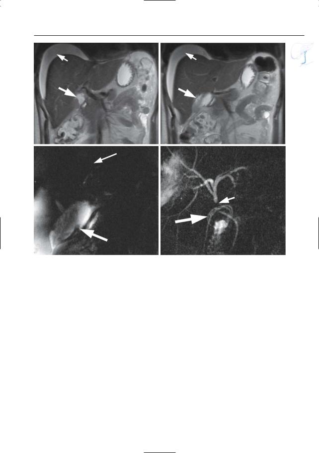
Fig. 83 a–d. Patient complaining of pain after laparoscopic cholecystectomy. a, b Coronal T2weighted HASTE image (TE 60) showing ascites (small arrow) and a fluid collection located in the liver hilum (large arrow). c Projective image showing the fluid collection (biloma) (large arrow); the absence of intrahepatic bile duct dilatation (small arrow) is related to the presence of a persisting bil-
iary leak. The patient was treated with percutaneous drainage of the biloma. d Follow-up projective image 1 month later showing the drain, no residual collection visible (large arrow). The intrahepatic bile ducts are visible again. Note the development of a secondary fibrotic stricture on the proximal extrahepatic bile duct (small arrow)
176 3.3 Traumatic, Postoperative, and Iatrogenic Abnormalities
#84 Post-cholecystectomy
Syndrome
Related topics: #52 (after cholecystectomy: stricture/transection of an aberrant bile duct), #82 (after cholecystectomy: stricture of the common bile duct), #83 (after cholecystectomy: bile leak), #93 (normal and variant anatomy of the cystic duct), # 114 (complications with cystic duct remnant)
KEY FACTS
●Definition: symptoms recurring/persisting after cholecystectomy
●Incidence of severe symptoms: 2.6%– 32%
●Causes:
–Biliary: incomplete surgery (retained stone in cystic duct remnant; overlooked common bile duct stone); operative trauma (bile duct stricture; leakage of bile with or without peritonitis); bile duct pathology (fibrosis of vaterian sphincter complex; biliary dyskinesia; biliary fistula)
–Extrabiliary: disease overlooked during preoperative evaluation (e.g., pancreatitis, hepatitis); new disease
● In symptomatic patients, stones in the |
|
|
! |
||
cystic or common bile duct may be |
||
found in up to 34% (Fig. 84; Marotta et |
|
|
al. 1989) |
|
●Note: Delayed remote abscess formation is another (probably rare) complication that may occur after peritoneal spillage of gallstones during (laparoscopic) cholecystectomy
References
Marotta F, Hada R, Morello P et al. (1989) ERCP in the assessment of patients with postcholecystectomy syndrome: benefits and limitations. Nether J Med 35 : 232–240
McGahan JP, Stein M (1995) Complications of laparoscopic cholecystectomy: imaging and intervention. AJR Am J Roentgenol 165 : 1089– 1097
Moossa AR, Easter DW, vanSonnenberg E, Casola G, D’Agostino H (1993) Laparoscopic injuries to the bile duct. A cause of concern. Ann Surg 218 : 215–216
Rubini G, Dimonte M (1999) Postcholecystectomy syndrome: evaluation by biliary cholescintigraphy and MR cholangiopancreatography. Clin Nucl Med 24 : 784–788
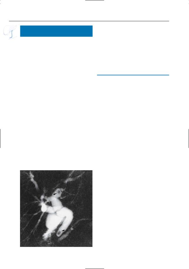
Fig. 84. Projective MR image obtained in patient with persistent complaints after cholecystectomy showing stones in the distal common bile duct and left hepatic duct (arrowheads)
3 Extrahepatic Bile Duct 177
#85 Bile Duct Trauma Related
to Nonbiliary Surgery
KEY FACTS: DISEASE
●Bile duct injury may occur during gastrectomy, pancreatectomy, partial hepatectomy, portocaval shunting, etc.
●Mechanisms:
–Direct injury
–Vascular injury (the blood supply of the middle part of the common bile duct depends entirely on two paracholedochal arteries; injury to these arteries can lead to ischemia and stricture formation)
a
KEY FACTS: MRI
●Most common finding: stricture and/or bile leak (Fig. 85)
References
Khalid TR, Casillas VJ, Montalvo BM et al. (2001) Using MR cholangiopancreatography to evaluate iatrogenic bile duct injury. AJR Am J Roentgenol 177 : 1347–1352
Lillemoe KD, Pitt HA, Cameron JL (1990) Postoperative bile duct strictures. Surg Clin North Am 70 : 1355–1380
b
Fig. 85 a, b. Patient who underwent gastrectomy 5 years earlier. a Projective MR image showing a relatively short stricture of the common hepatic duct (arrowheads). Also note the attraction of the more distal extrahepatic duct due to fibrosis
(arrow). b PTC image showing occlusion of the common hepatic duct (arrow). Despite the short length of the stricture, percutaneous dilation was not possible and the patient was referred for surgery
178 3.3 Traumatic, Postoperative, and Iatrogenic Abnormalities
#86 Choledochoduodenostomy
with Sump Syndrome
KEY FACTS: DISEASE
●“Sump”: reservoir located at the lowest point of a drainage system, collecting fluid and debris
●“Sump syndrome”: cholangitis and biliary obstruction caused by the accumulation of debris and ingested material in the bile ducts
●Incidence of sump syndrome: 0.14%–1.3% of choledochoduodenostomies (Hawes et al. 1992)
●Mechanism: reflux of gas, intestinal contents, and contrast medium into the intrahepatic bile ducts (Fig. 86a)
●Note on choledochoduodenostomy:
–Indications: multiple or unremovable common bile duct stones, chronic pancreatitis, postoperative stricture, ampullary stenosis
–Considered by some as a valuable alternative to endoscopic papillotomy and biliary lithotripsy (Berlatzky and Freund 1990)
KEY FACTS: MRI (FIG. 86 b)
●The common bile duct has a large diameter
●Debris and stones are usually clearly seen as hypointense structures within the lumen
●If the bile duct contains a large amount of air (which is not unusual), the interpretation may become difficult
References
Berlatzky Y, Freund HR (1990) Primary choledochoduodenostomy for benign obstructive biliary tract disease. J Clin Gastroenterol 12 : 420–422
Escudero-Fabre A, Escallon A, Sack J, Halpern NB, Aldrete JS (1991) Choledochoduodenostomy. Analysis of 71 cases followed for 5 to 15 years.Ann Surg 213 : 643–644
Hawes DR, Pelsang RE, Janda RC, Lu CC (1992) Imaging of the biliary sump syndrome.AJR Am J Roentgenol 158 : 315–319
Silvis S, Rohrmann C, Ansel H (eds) (1995) Endoscopic retrograde cholangiopancreatography. Igaku-Shoin, New York, p 287
3 Extrahepatic Bile Duct 179
a
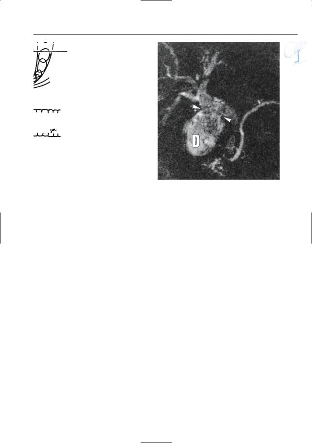
Fig. 86. a Choledochoduodenostomy (arrow). Stones, debris, and sludge may collect in the excluded segment of the distal common bile duct. (Reprinted with permission from Silvis et al. 1995). b Patient who underwent choledochoduodenostomy and
b
presented with fever. Projective MR image showing connection (arrowheads) between the extrahepatic bile duct and the first part of the duodenum (D). Note low overall signal intensity of the bile ducts, related to presence of air and debris