


32 1.3 Use of Contrast Media and Drugs
1.3 Use of Contrast Media and Drugs
#14 Oral Contrast Media
KEY FACTS
T 1-Weighted MRI
Positive Contrast Media
●Examples: manganese or gadolinium compounds
●Advantages: improved distention of stomach, duodenum and jejunum, improved evaluation of mural and intraluminal abnormalities
●Potential applications: evaluation of mucosal and mural abnormalities, assessment of contractility, etc. (Geitung and Gjesdal 1997)
T 2-Weighted MRI
Positive Contrast Media
●Examples: tap water
●Advantages: improved distention of stomach, duodenum, jejunum
●Applications: prerequisite for adequate evaluation of bilioenteric anastomoses (Pavone et al. 1997); also useful in the evaluation of ampullary tumors and tumors of pancreatic head
Negative Contrast Media
●Examples: iron-containing oral suspension
●Advantages: elimination of overlapping fluid-containing organs
●Applications: evaluation of pancreatic fluid outflow (Matos et al. 1997)
●Limitations: exact length and location of the vaterian sphincter complex more difficult to evaluate
Note: We only administer oral contrast media in a minority of patients (see indications for use of tap water). Patients do not fast prior to MR study
References
Chan JH, Tsui EY,Yuen MK et al. (2000) Gadopentetate dimeglumine as an oral negative gastrointestinal contrast agent for MRCP. Abdom Imaging 25 : 405–408
Geitung JT, Gjesdal KI (1997) Dynamic imaging of the gastric ventricle using MRI. MAGMA 5 [Suppl] : 39
Matos C, Metens T, Devière J et al. (1997) Pancreatic duct: morphologic and functional evaluation with dynamic MR pancreatography after secretin stimulation. Radiology 203 : 435–441
Papanikolaou N, Karantanas A, Maris T et al. (2000) MR cholangiopancreatography before and after oral blueberry juice administration. J Comput Assist Tomogr 24 : 229–234
1 MRCP Technique |
33 |
a
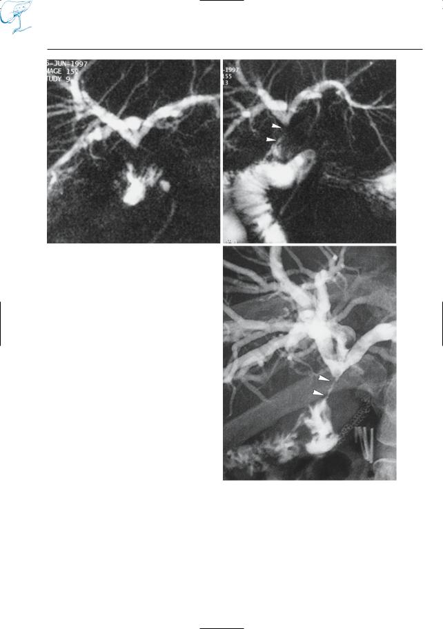
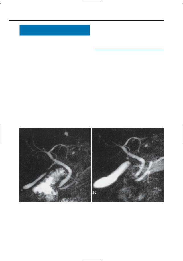
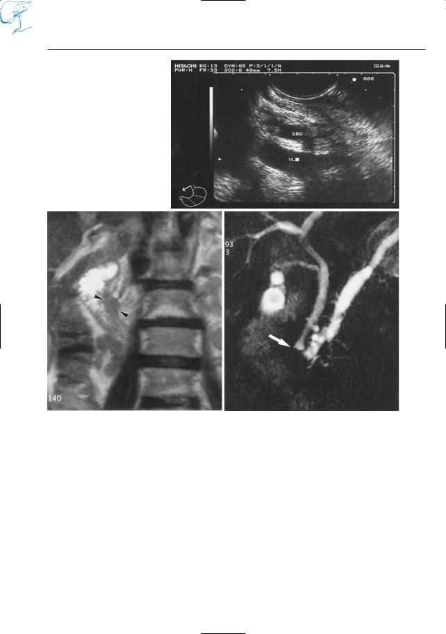
Fig. 14 a–c. Patient with stenosis of hepaticojejunal anastomosis. a, b Before and after oral administration of tap water, respectively. In b, the valvulae conniventes of the anastomosed jejunal loop are clearly seen and the length of the stenosis can easily be determined (arrowheads). c Corresponding image obtained by percutaneous transhepatic cholangiography (PTC) confirms the presence of a relatively long stenosis (arrowheads)
b
c

34 1.3 Use of Contrast Media and Drugs
#15 Nonspecific Intravenous
Contrast Media (1):
Parenchymal Organs
and Lymph Nodes
KEY FACTS
●Type: gadolinium chelates
●May be used routinely or in function of clinical information and/or findings at non-contrast imaging (see # 1)
●Formal indications
–Characterization of focal liver lesions displaying indeterminate features on double-echo HASTE (± 10%–20% of lesions)
–Evaluation of pancreatic perfusion (detection of necrotizing pancreatitis; see # 155)
–Detection of pancreatic carcinoma in patients with associated pancreatitis (see # 194; Gabata et al. 1994)
–Detection/staging of endocrine pancreatic tumors (see # 177, # 197)
–Preoperative evaluation of pancreatic tumors (e.g., detection of lymph nodes; Fig. 15a, b; see also # 16)
●Note: Many abnormalities are clearly displayed on non-contrast-enhanced images (Fig. 15c, d). Moreover, some tumors (particularly endocrine pancreatic tumors) may temporarily become less conspicuous or even invisible after administration of intravenous contrast agents (see # 177, # 197).
●Timing: biphasic imaging at 15 and 45 seconds or later after arrival of contrast material in the abdominal aorta is a practical method for acquisition of highquality dynamic T1-weighted MR images of pancreatic parenchyma (Kanematusu
et al. 2000)
● For dynamic imaging, a fat-saturat- ed volumetric interpolated breath-hold (VIBE) sequence is preferred (Rofsky et al. 1999)
References
Gabata T, Matsui O, Kadoya M et al. (1994) Small pancreatic adenocarcinomas: efficacy of MR imaging with fat suppression and gadolinium enhancement. Radiology 193 : 683–688
Hamm B, Mahfouz A-E, Taupitz M et al. (1997) Liver metastases: improved detection with dynamic gadolinium-enhanced MR imaging? Radiology 202 : 677–682
Kanematsu M, Shiratori Y, Hoshi H, et al. (2000) Pancreas and peripancreatic vessels: effect of imaging delay on gadolinium enhancement at dynamic gradient-recalled-echo MR imaging. Radiology 215 : 95–102
Rofsky, NM, Lee VS, Laub G, et al. (1999) Abdominal MR imaging with a volumetric interpolated breath-hold examination. Radiology 212 : 876– 884
1 MRCP Technique |
35 |
a |
b |
c |
d |
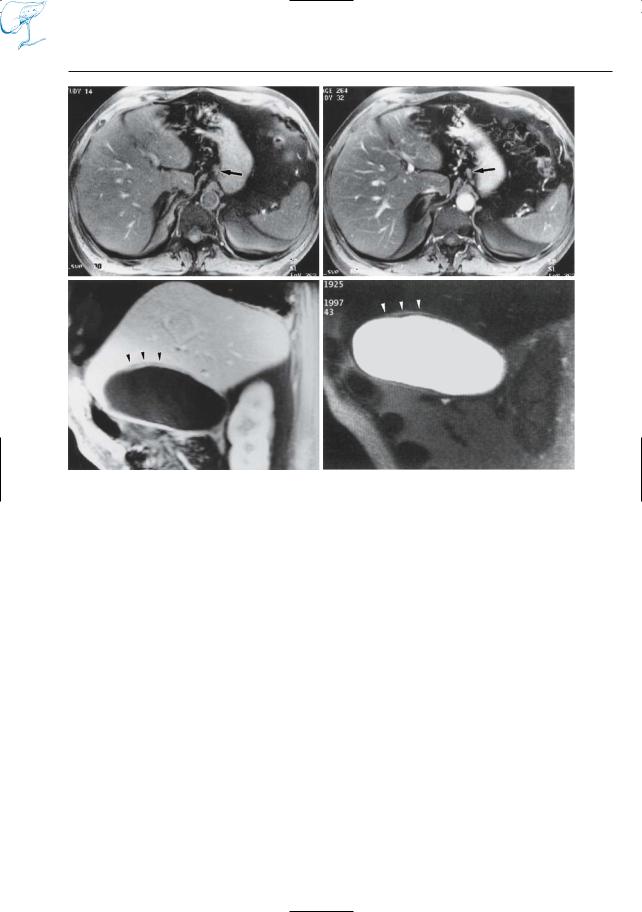
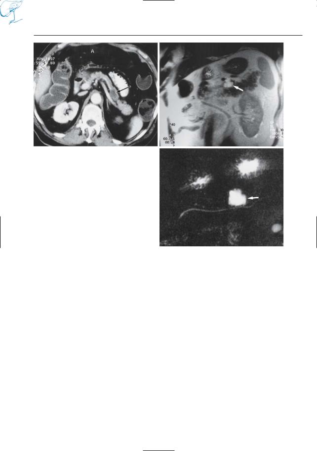
Fig. 15 a–d. Patient with pancreatic cancer and enlarged lymph nodes. a Non-contrast-enhanced and b contrast-enhanced fat-suppressed T1-weighted images. Slightly enlarged lymph node is better seen in b (arrows). c, d Patient with acute cholecystitis. c Contrast-enhanced T1-weighted image obtained
in the sagittal plane showing thin hypoenhancing zone between gallbladder wall and liver (arrowheads), corresponding to fluid. d Heavily T2-weighted snapshot image also nicely shows the small amount of fluid
36 1.3 Use of Contrast Media and Drugs
#16 Nonspecific Intravenous
Contrast Media (2):
Vascular “Roadmapping”
KEY FACTS
●Breathhold three-dimensional contrastenhanced MRI/MRA has emerged as a highly promising method for the evaluation of abdominal vascular disease (Prince et al. 1995)
●Using high-power gradient systems, the arteries of the entire upper abdomen can be imaged during one single breathhold (Earls et al. 1997)
●Critical elements of successful MRA examination (Earls et al. 1997; Prince et al. 1995):
–Timing examination (test dose)
–Patient coaching with or without oxygen administration
–Fast injection of contrast medium
–Optimization of voxel size in function of vessel size
●Indications:
–Preoperative evaluation of (suspected) pancreatic cancer (“all-in- one” approach; see Fig. 191c, d; Gaa et al. 1997)
–Detection of anatomic variants prior to pancreatic or hepatobiliary surgery (Fig. 16)
–Clinical suspicion of pseudoaneurysm, arteriovenous fistula, arterial occlusion
–Suspicion of venous involvement in patients with neoplastic disease (e.g., hepatocellular carcinoma)
●In practice, vascular roadmapping and dynamic contrast-enhanced parenchymal imaging can be combined by using VIBE or similar sequences (Rofsky et al. 1999, Keogan et al. 2000)
References
Earls JP, Rofsky NM, DeCorato DR, Krinsky GA, Weinreb JC (1997) Breath-hold single-dose gadolinium-enhanced three-dimensional MR aortography: usefulness of a timing examination and an MR power injector. Radiology 202 : 268–273
Gaa J, Wendl K, Trede M, Georgi M (1997) New concepts in MR imaging of pancreatic carcinoma: the all-in-one approach. In: Oudkerk M, Edelman R (eds) High-power gradient MR-imaging. Blackwell Science, Berlin, pp 425–430
Keogan MT, Edelman RR (2001) Technologic advances in abdominal MR imaging. Radiology 220 : 310–320
Prince MR, Narasimham DL, Stanley JC, Chenevert TL, Williams DM, Marx MV, Cho KJ (1995) Breath-hold gadolinium-enhanced MR angiography of the abdominal aorta and its major branches. Radiology 197 : 785–792
Rofsky, NM, Lee VS, Laub G, et al. (1999) Abdominal MR imaging with a volumetric interpolated breath-hold examination. Radiology 212 : 876– 884

1 MRCP Technique |
37 |
a
Fig. 16 a–c. Maximum-intensity projection images calculated from images obtained in a the arterial and b the portal vein phases of perfusion showing an anatomic variant in a (early takeoff of left hepatic artery, arrows) and a normal portal vein in b (arrows). c In another patient, the common hepatic artery (arrow) originates from the superior mesenteric artery, representing another anatomic variant. In both cases, MRA was performed as part of a preoperative MRCP–MRI–MRA investigation in patients with pancreatic carcinoma
b
c

38 1.3 Use of Contrast Media and Drugs
#17 Specific Intravenous
Contrast Media (1):
Hepatocyte-Directed Agents
KEY FACTS
●Molecules with large lipophilic component
●Uptake by hepatocytes and excretion in bile; sometimes uptake in other parenchymal organs (see below)
●Influence on tissue contrast: increase in signal intensity on T1-weighted images (paramagnetic agents)
●Examples (Van Beers et al. 1997):
–Manganese-dipyridoxyl diphosphate (manganese-DPDP; uptake also in pancreas and adrenals)
–Gd-ethoxybenzyl-diethylenetri- aminopentoacetic acid (Gd-EOB- DTPA)
–Gd-benzyloxyproprionic-tetraacetic acid (Gd-BOPTA)
●Main advantage:
–Improved visualization of secondary hepatic tumors (e.g., metastases of pancreatic carcinoma) (Fig. 17a, c)
–Evalution of bile duct integrity (Vitellas et al. 2002, Fayad et al. 2005)
●Other advantages/potential applications:
–Evaluation of biliary excretory function (the biliary excretion fraction of Gd-EOB-DTPA, for instance, is 50%) (Fig. 17e)
–Detection of pancreatic tumors (man- ganese-DPDP; Gehl et al. 1991; Kettritz et al. 1996) (Fig. 17b,d)
References
Fayad LM, Kamel IR, Mitchell DG, Bluemke DA (2005) Functional MR cholangiography: diagnosis of functional abnormalities of the gallbladder and biliary tree.Am. J. Roentgenol 184 : 1563–1571 Gehl HB,Vorwerk D, Klose KC, Guenther RW (1991) Pancreatic enhancement after low-dose infusion
of Mn-DPDP. Radiology 180 : 337–339
Kettritz U, Warshauer D, Brown E, Schlund J, Eisenberg L,Semelka R (1996) Enhancement of the normal pancreas: comparison of manganese-DPDP and gadolinium chelate. Eur Radiol 6 : 14–18
Van Beers B, Gallez B, Pringot J (1997) Contrastenhanced MR imaging of the liver. Radiology 203 : 297–306
Vitellas KM, El-Dieb A, Vaswani KK, et al. (2002) Using contrast-enhanced MR cholangiography with IV mangafodipir trisodium (Teslascan) to evaluate bile duct leaks after cholecystectomy: a prospective study of 11 patients. AJR Am J Roentgenol 179 : 409–416
1 MRCP Technique |
39 |
a
c
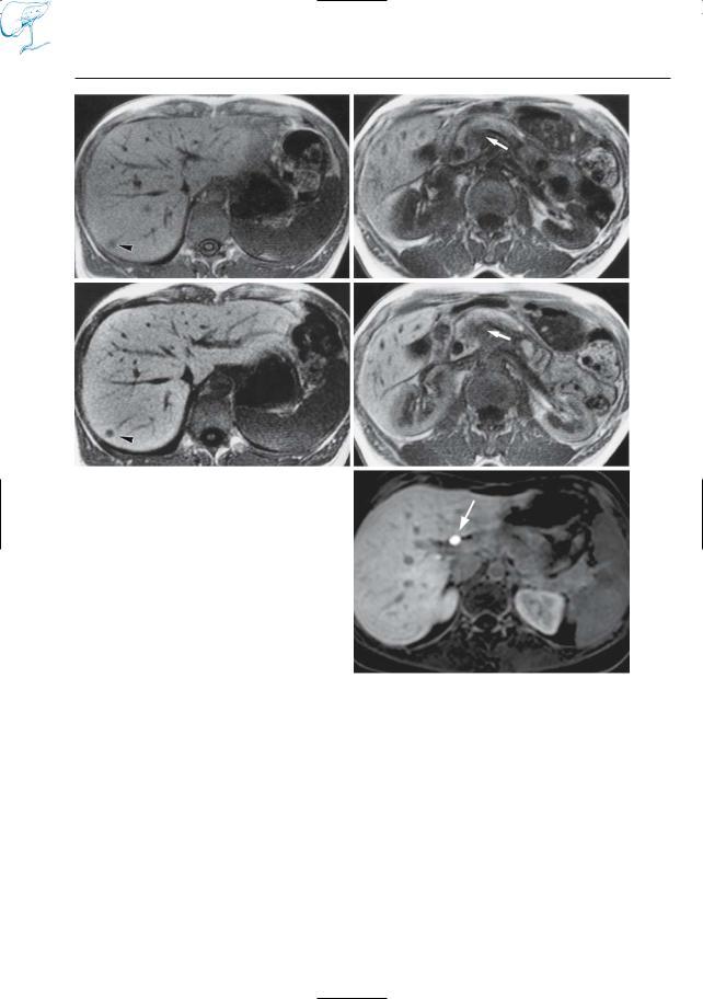
Fig. 17 a–e. T1-weighted images obtained a, b before and c, d after intravenous administration of manganese-DPDP. Both the primary pancreatic tumor (arrows) and the metastatic hepatic tumor (arrowheads) are better seen on the contrast-enhan- ced scans, which is related to uptake of manganese by both liver and pancreas. e Different patient.Axial T1-weighted VIBE image 90 min after administration of a hepatospecific contrast agent, gadobenate dimeglumine (Gd-BOPTA), shows the common hepatic duct filled with dense contrast material
(arrow)
b
d
e

40 1.3 Use of Contrast Media and Drugs
#18 Specific Intravenous
Contrast Media (2):
Reticuloendothelial Agents
KEY FACTS
●Iron-containing particles
●Specific uptake by elements of the reticuloendothelial system in liver, spleen, bone marrow, and lymph nodes
●Influence on contrast of superparamagnetic agents: cause dephasing and focal signal loss,mainly on T2-weighted images (the effect on T1-weighted images is variable)
Small Iron Oxide Particles
●80% uptake by hepatic Kupffer cells
●Improve the detection of hepatic metastases (Fig. 18; Senéterre et al. 1996)
●Useful for characterization of primary liver tumors (uptake by focal nodular hyperplasia and adenoma; Vogl et al. 1996)
Ultrasmall particles
●Accumulation mainly in bone marrow and lymph nodes
●Blood pool contrast agent
●Potential application: characterization of (enlarged) lymph nodes (Anzai et al. 1994; Rogers et al. 1994)
References
Anzai Y, Blackwell KE, Hirschowitz SL et al. (1994) Initial clinical experience with dextran-coated superparamagnetic iron oxide for detection of lymph node metastases in patients with head and neck cancer. Radiology 192 : 709–715
Rogers JM, Lewis J, Josephson L (1994) Visualization of superior mesenteric lymph nodes by the combined oral and intravenous administration of the ultrasmall superparamagnetic iron oxide AMI 227. Magn Reson Imaging 12 : 1161–1165
Senéterre E, Taourel P, Bouvier Y et al. (1996) Detection of hepatic metastases: ferumoxides-en- hanced MR imaging versus unenhanced MR imaging and CT during arterial portography. Radiology 200 : 785–792
Vogl TJ, Hammerstingl R, Schwarz W et al. (1996) Superparamagnetic iron oxide-enhanced versus gadolinium-enhanced MR imaging for differential diagnosis of focal liver lesions. Radiology 198 : 881–887
1 MRCP Technique |
41 |
a |
b |
c |
d |
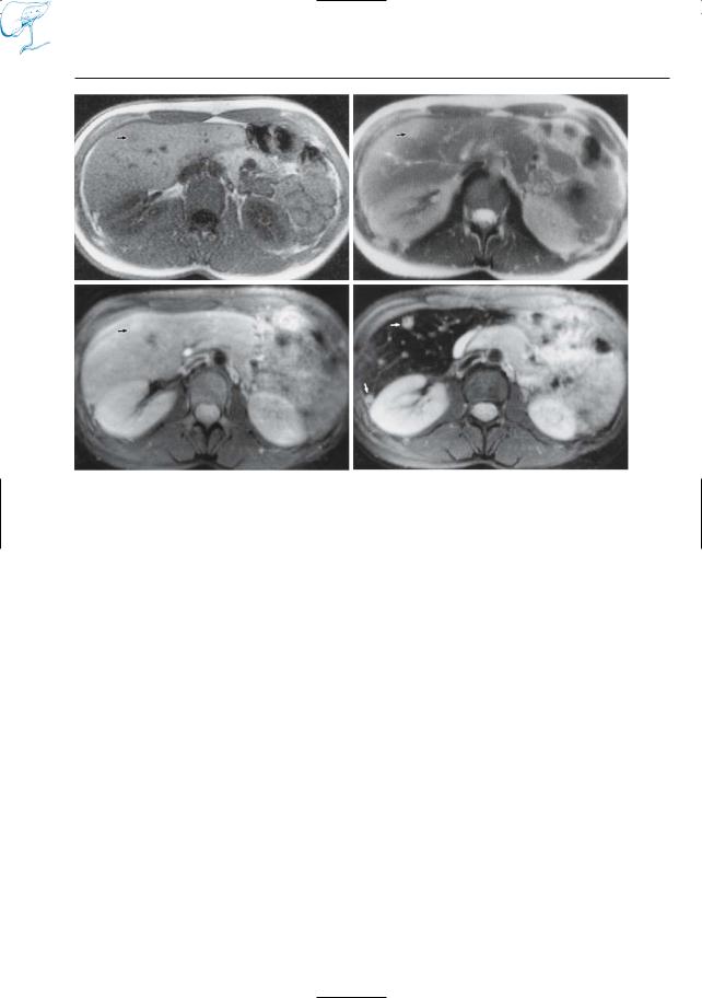
Fig. 18 a–d. Patient with hepatic metastases of cholangiocarcinoma. a, b T1and T2-weighted snapshot images and c T2-weighted fat-suppressed fast spin echo image, all obtained before administration of contrast agent. d T2-weighted fat-suppressed fast
spin echo image obtained after administration of small iron oxide particles. Note the much better visibility of two small metastases (arrows) in d, which is explained by signal loss of normal liver tissue (uptake by Kupffer cells)

42 1.3 Use of Contrast Media and Drugs
#19 Drugs Stimulating
Excretory Function
KEY FACTS
Pancreatic Ducts: Secretin
●Stimulates the secretion of fluid and bicarbonate by the exocrine pancreas
●Potential applications (Matos et al. 1997; Warschaw et al. 1985):
–Assessment of the anatomy of the pancreatic duct (Fig. 19)
–Evaluation of pancreatic excretory function
–Detection of pancreatic outlet obstruction (e.g., papillary obstruction; see # 127)
–Demonstration of pancreatico-biliary reflux
●Limitations:
–Does not allow visualization of normal side branches
Biliary Tree: Cholecystokinin
●Cholecystokinin produces:
–Powerful contraction of the gallbladder
–Decreased resistance of the sphincter of Oddi
–Increased hepatic secretion of bile
●Can be used in conjunction with imaging studies (Krishnamurthy and Krishnamurthy (1996) to detect ampullary obstruction (e.g., dysfunction of vaterian sphincter complex)
●A similar effect can be obtained by administration of a fatty meal (results in endogenous release of cholecystokinin; Darweesh et al. 1988)
References
Czako L, Endes J, Takacs T et al. (2001) Evaluation of pancreatic exocrine function by secretinenhanced magnetic resonance cholangiopancreatography. Pancreas 23 : 323–328
Darweesh R, Dodds W, Hogan W et al. (1988) Efficacy of quantitative hepatobiliary scintigraphy and fatty-meal sonography for evaluating patients with suspected partial common duct obstruction. Gastroenterology 94 : 779–786
Hosoki T,Hasuike Y,Takeda Y et al. (2004) Visualization of pancreaticobiliary reflux in anomalous pancreaticobiliary junction by secretin-stimu- lated dynamic magnetic resonance cholangiopancreatography. Acta Radiol 45 : 375–382
Hellerhoff KJ, Helmberger H, Rösch T, et al. (2002) Dynamic MR pancreatography after secretin administration: image quality and diagnostic accuracy. AJR Am J Roentgenol 179 : 121–129
Krishnamurthy S, Krishnamurthy G (1996) Cholecystokin and morphine pharmacological intervention during 99mTc-HIDA cholescintigraphy: a rational approach. Semin Nucl Med 26 : 16–24
Matos C, Metens T, Devière J et al. (1997) Pancreatic duct: morphologic and functional evaluation with dynamic MR pancreatography after secretin stimulation. Radiology 203 : 435–441

1 MRCP Technique |
43 |
a |
b |
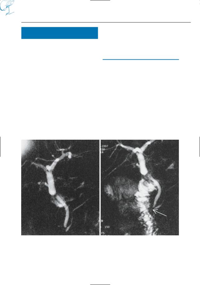
Fig. 19 a, b. Images obtained a before and b after administration of secretin in a patient with chronic pancreatitis. Both images show signs of chronic pancreatitis and a large intraductal stone (arrows).
In b, the degree of filling of the distal pancreatic duct is slightly improved and some additional side branches are shown (arrowheads)
44 1.3 Use of Contrast Media and Drugs
#20 Spasmolytic Drugs (1):
Buscopan
KEY FACTS
●Active component: butylhyoscin
●Spasmolytic drug causing relaxation of smooth muscles in the gastrointestinal tract, pancreatobiliary tract, urogenital tract, and gallbladder (Forssmann and
Singer 1994)
● Half-life after intravenous injection:
± 15 min
●Possible indications:
– Evaluation of stomach, duodenum, ampullary region, pancreatic head
a
Fig. 20 a, b. Projective images obtained a before and b after intravenous administration of Buscopan. Note significantly increased size of the gall-
–Contracted gallbladder: differentiation of organic abnormalities (e.g., chronic cholecystitis) and functional spasm (Fig. 20)
References
Chopra K,Westaby D,Murray-Lyon I (1996) Why use buscopan during diagnostic upper gastrointestinal endoscopy? Gut 38 : 473
Forssmann K, Singer MV (1994) Akute Cholezystitis
– konservative Therapie. Schweiz Rundsch Med Prax 83 : 877–879
b
bladder in b, suggesting (normal) smooth muscle relaxation
1 MRCP Technique |
45 |
#21 Spasmolytic Drugs (2):
Glucagon
KEY FACTS
●Smooth muscle spasmolytic agent
●Relatively short half-life (a few minutes)
●Effects:
–Effective inhibitor of the baseline pressure of the vaterian sphincter (Staritz 1988)
–Effective inhibitor of motility and tonicity of the esophagus, stomach, duodenum, and colon
–Increases the amount of fluid in the duodenum (Fig. 21)
●Applications:
–Evaluation of stomach, duodenum, ampullary region, and pancreatic head
a
–Potentially useful in the evaluation of the distal common bile duct and vaterian sphincter complex (hypotonic cholangiography; Ferruci et al. 1976; Dalal et al. 2004)
References
Dalal PU, Howlett DC, Sallomi DF et al. (2004) Does intravenous glucagon improve common bile duct visualisation during magnetic resonance cholangiopancreatography? Results in 42 patients. Eur J Radiol 49 : 258–261
Ferruci J, Wittenberg J, Stone L, Dreyfuss J (1976) Hypotonic cholangiography with glucagon. Radiology 118 : 466–467
Staritz M (1988) Pharmacology of the sphincter of Oddi. Endoscopy 20 :171–174
b
Fig. 21 a, b. Patient who underwent hepatic transplantation. Images obtained a before and b after intravenous administration of glucagon. Note the significantly increased amount of fluid in the duo-
denum in b, which is a common finding. The arrow in b shows the intramural portion of the common bile duct. Note that no oral contrast medium was given
46 1.4 Comparison with Other Techniques
1.4Comparison
with Other Techniques
#22 MRCP Compared with ERCP (1):
Limitations of ERCP
KEY FACTS
–No opacification of ducts proximal to a complete obstruction (Fig. 22f–h)
–Less accurate MRCP for detection of intrahepatic stones (Kim et al. 2002)
–Less accurate than MRCP for characterization of pancreatic duct stenoses
!● The limitations of endoscopic retrograde cholangiopancreatography (ERCP) can be summarized as follows:
–Invasive nature (complication rate between 4.6% and 12%, depending on the type of procedure, e.g., diagnostic, manometry, sphincterotomy; Cotton and Chong 1995)
–Highly operator dependent
–Unsuccessful cannulation of the common bile duct or pancreatic ducts in 3%–9% of cases (failure rate of MRCP, 1%–4%)
–Sedation usually required
–No direct information on extraductal abnormalities (Fig. 22a–e)
References
Cotton P, Chong W. Complications of ERCP and therapy. In: Silvis S, Rohrmann C, Ansel H (eds) Endoscopic retrograde cholangiopancreatography.IgakuShoin,New York,pp 446–469
Kim JH, Kim MJ, Park SI et al. (2002) Using kinematic MR cholangiopancreatography to evaluate biliary dilatation. AJR Am J Roentgenol. 178 : 909–914
Reinhold C, Bret PM (1996) Current status of MR cholangiopancreatography. AJR Am J Roentgenol 166 : 1285–1295
Soto JA,Yucel EK, Barish MA, Chuttani R, Ferruci JT (1996) MR cholangiography after unsuccessful or incomplete ERCP. Radiology 199 : 91–98
a |
b |
Fig. 22 a–e. ERCP image (a) showing smooth extrinsic compression on distal common bile duct,suggesting Mirizzi syndrome. b Projective MR image confirms gradual narrowing of the common bile duct.

1 MRCP Technique |
47 |
c |
d |
e |
f |
g |
h |
Fig. 22. c, d Cross-sectional T2-weighted images obtained in the coronal plane showing large stone impacted in the cystic duct (arrowheads in c), thus confirming the initial diagnosis, and a soft tissue mass centered on the gallbladder and invading the liver parenchyma (arrowheads in d). e Axial T1weighted image also shows large soft tissue mass in the liver hilum, surrounding the stone (arrows).
Final diagnosis: Mirizzi syndrome associated with gallbladder carcinoma (see also # 71). f–h Other patient. ERCP image (f) showing obstruction of pancreatic duct (arrow): possibly a stone or a tumor. g Cross-sectional axial T2-weighted image showing dilation of the pancreatic duct in the pancreatic tail. h Projective MR image confirms the dilation and shows its cause: a small intraductal stone (arrow)
48 1.4 Comparison with Other Techniques
#23 MRCP Compared with ERCP (2):
Limitations of MRCP
KEY FACTS
●The disadvantages of MRCP can be summarized as follows:
–Artifacts related to the presence of surgical clips, metallic stents (see #57), and pneumobilia (see #67)
–More limited spatial resolution (this is not the most important limitation)
!– Ducts are examined in their physiologic state (in ERCP, ducts are distended due to filling); this may lead to both underand overestimation of stenoses (Figs. 23, 27c, d, 179) and to poor visualization of intraductal lesions (Figs. 23, 125d)
–The normally high signal intensity (and consequently the conspicuity) of biliary ducts on T2-weighted images may be lost in the case of hemobilia, casting of necrotic debris, etc.
–Superimposition of fluid-containing structures (e.g., cysts, exudates) may prevent adequate visualization of biliary and pancreatic ducts and may degrade the quality of projective images (see Fig. 35)
–Currently not suited for guidance of interventional procedures or biopsy
References
Reinhold C, Bret PM (1996) Current status of MR cholangiopancreatography. AJR Am J Roentgenol 166 : 1285–1295
Van Hoe L, Mermuys K, Vanhoenacker P (2004) MRCP pitfalls. Abdom Imaging 29 : 360–387
1 MRCP Technique |
49 |
a |
b |
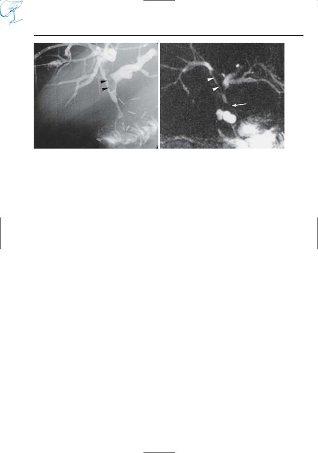
Fig. 23 a, b. Patient who underwent hepatojejunostomy. a PTC image showing stenosis at anastomosis and presence of an intrabiliary structure (arrowheads), perhaps a blood clot or sludge. b Projective MR image overestimates the severity of the
stenosis (arrow). Moreover, the intraductal filling defect (arrowheads) is less conspicuous because it is not completely surrounded by fluid. This case typically illustrates the disadvantage of imaging the biliary tree in its physiologic state

50 1.4 Comparison with Other Techniques
#24 MRCP Compared
with Ultrasonography
KEY FACTS
Transabdominal Ultrasonography
●Advantages of MRCP:
–Higher sensitivity in the detection of common bile duct stones (reported values for sensitivity: ultrasonography, 13%–75%; MRI, > 90%; Guibaud et al. 1995; O’Conner et al. 1986)
–More constant visualization of biliary, pancreatic,and retroperitoneal abnormalities
–Better characterization of focal liver lesions
●Limitations:
–Cost
–Availability
Endoscopic
and Intraductal Ultrasonography
●Relative value largely dependent on equipment/experience
●Theoretical advantages of MRCP:
–Less invasive
–Much larger field of view
–More powerfull tool for tissue characterization
–More versatile and comprehensive technique
●Theoretical limitations of MRCP:
–Spatial resolution
References
Guibaud L, Bret P, Reinhold C, Atri M, Barkun AL (1995) Bile duct obstruction and choledocholithiasis: diagnosis with MR cholangiography. Radiology 197 : 109–115
O’Conner HJ, Hamilton I, Ellis WR,Watters J, Lintott DJ, Axon AT (1986) Ultrasound detection of choledocholithiasis: prospective comparison with ERCP in the postcholecystectomy patient. Gastrointest Radiol 11 : 161–164
Rosch T, Lightdale CJ, Botet JF et al. (1992) Localization of pancreatic endocrine tumors by endoscopic ultrasonography. N Engl J Med 326 : 1721–1726
Zerbey AL, Lee MJ, Brugge WR, Mueller PR (1996) Endoscopic sonography of the upper gastrointestinal tract and pancreas.AJR Am J Roentgenol 166 : 45–50
1 MRCP Technique |
51 |
a
b |
c |
Fig. 24. a In this patient, endoscopic sonography showed no abnormalities (VL, splenic vein; CBD, common bile duct). Axial T1and T2-weighted MR images also revealed no focal lesions. b However, the coronal T2-weighted image suggests the presence of a small mass in the papillary area (arrowheads).
c Projective MR image showing abrupt narrowing of the common bile duct and pancreatic duct (arrow), together with some dilated side branches. This case illustrates the advantages of a comprehensive MR approach: projective and cross-sectional MR images clearly provide complementary information

52 1.4 Comparison with Other Techniques
#25 MRCP Compared
with Multislice Computed
Tomography
KEY FACTS
●General advantages of MRI:
–Absence of ionizing radiation
–Higher intrinsic soft tissue contrast (Fig. 25)
–Possible to obtain projective (e.g., cholangiographic) images in any desired plane
–Direct multiplanar imaging capability
●General advantages of CT:
–Spatial resolution (pixel size typically four to eight times smaller in CT)
–Physical basis and artifact behavior easier to understand
–Cost
–Larger examination volume: possible to investigate the entire abdomen or even thorax and abdomen in one session
–Better visualization of calcifications (see Fig. 47)
●Value in clinical practice:
–In most institutions multislice CT is the first imaging modality in patients referred for evaluation of upper abdominal disease
–MRC/MRCP commonly used for problem solving and for specific indications
References
Cahir JG, Freeman AH, Courtney HM (2004) Multislice CT of the abdomen. Br J Radiol 77 Spec No 1: S64–73
Ichikawa T, Haradome H, Hachiya J et al. (1997) Pancreatic ductal adenocarcinoma: preoperative assessment with helical CT versus dynamic MR imaging. Radiology 202 : 655–662
Semelka RC, Kelekis NL, Molina PL, Sharp TJ, Calvo B (1996) Pancreatic masses with inconclusive findings on spiral CT: is there a role for MRI? J Magn Reson Imaging 6 : 585–588
1 MRCP Technique |
53 |
a
Fig. 25 a–c. CT image (a) obtained in patient with malignant melanoma showing pancreatic mass with a density of 35 HU (arrow) perhaps metastasis, cyst, pseudocyst, or cystic tumor. b Cross-sectional and c projective MR images showing the mass as a fluidcontaining lesion with sharp borders (arrows); metastasis can be excluded. The normal morphology of the pancreatic duct makes chronic pancreatitis unlikely. Final diagnosis (follow-up): pseudocyst after previous attack of acute pancreatitis
b
c