


Vaterian Sphincter Complex

246
5 Vaterian Sphincter Complex
5.1 Normal Anatomy and Variants
# 119 Normal Anatomy
KEY FACTS: ANATOMY
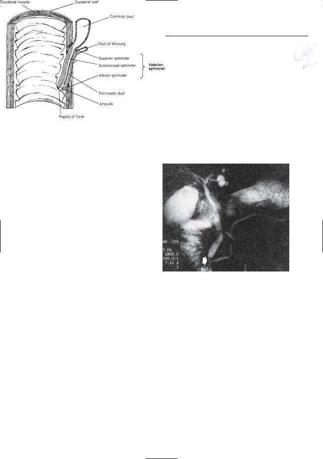
●The vaterian sphincter complex includes the distal (intramural) part of the common bile duct and pancreatic duct, the papilla, and the surrounding smooth muscle (sphincter of Oddi) (Fig. 119a)
●Length of vaterian sphincter complex: 10–15 mm
●Course: oblique in a laterocaudal direction
●Diameter of intramural duct: a few millimeters
●In 60%–80% of individuals,the pancreatic and bile ducts unite to form a common channel (length, 2–15 mm; average, 5 mm; Misra and Dwivedi 1990; see # 123)
●The slightly dilated distal segment of the common channel is also called the ampulla
●The papilla forms an endoluminal protrusion at the medial wall of the duode-
num and contains the orifice
● Size of papilla: usually less than 10–
15 mm
●Shape of papilla: variable (may be polypoid)
●Location of papilla:
– Usually (92%) located at the posteromedial wall of the mid-descending duodenum
– Can be anywhere between the pylorus and the ligament of Treitz
●Location of minor papilla: 1–2 cm proximal to the major papilla
KEY FACTS: MRI
●If MRI is performed during relaxation of the sphincter, images show the distal portion of the pancreatic duct and common bile duct as thin, fluid-containing structures connected with the duodenum (Fig. 119b)
●Nonvisualization is a normal variant if
only single images are obtained (see
# 120; Van Hoe et al. 1998)
●The papilla may be seen as an endoluminal protrusion at the medial wall of the duodenum
References
Misra SP, Dwivedi M (1990) Pancreaticobiliary ductal union. Gut 31 : 1144–1149
Stewart E, Vennes J, Geenen J (1977) Atlas of endoscopic retrograde cholangiopancreatography. Mosby, St Louis, p 41
Van Hoe L, Gryspeerdt S, Vanbeckevoort D et al. (1998) Normal Vaterian sphincter complex: evaluation of morphology and contractility with dynamic single-shot MR cholangiography. AJR 170 : 1497–1500
5 Vaterian Sphincter Complex 247
a
Fig. 119. a Anatomy of the vaterian sphincter complex. The common bile duct and pancreatic duct become invested by smooth muscle fibers as they approach the duodenal wall. This is reflected by a marked decrease in internal diameter. (Reprinted with permission from Stewart et al. 1977). b Projective MR image showing the distal common bile duct and pancreatic duct joining to form a common channel. Note the (normal) abrupt change in diame-
ter of the distalmost part of the common bile duct b
(arrow)

248 5.1 Normal Anatomy and Variants
|
|
|
|
|
|
|
|
|
|
|
|
KEY FACTS: DYNAMIC MRI |
|
|
|
|
#120 |
Normal Contractile Activity |
|
|
|||||||||
|
|
|
|
|
|
|||||||||
|
|
Related |
topics: # 13 (dynamic evaluation |
● Serial breathhold MRI using a single- |
|
|||||||||
|
|
|
shot technique (kinematic MRCP) allows |
|
||||||||||
|
|
of |
vaterian |
sphincter complex), |
# 128 |
|
|
|
||||||
|
|
|
|
|
||||||||||
|
|
|
|
visualization of the vaterian sphincter |
|
|||||||||
|
|
(sphincter |
dysfunction, |
|
features |
on |
|
|
||||||
|
|
|
|
complex in 95% of normal patients (Van |
|
|||||||||
|
|
dynamic MRCP) |
|
|
|
|
|
|
|
|
||||
|
|
|
|
|
|
|
|
|
Hoe et al. 1998; Kim et al. 2002) |
|
||||
|
|
|
|
|
|
|
|
|
|
|
|
|
|
|
|
|
KEY FACTS: PHYSIOLOGY |
|
|
|
|
|
● Normal features: |
|
|||||
|
|
|
|
|
|
|
|
– On serial images, the aspect of the |
|
|||||
|
|
|
|
|
|
|
|
|
|
|
|
|
|
|
|
|
● |
The |
vaterian sphincter |
complex |
func- |
|
vaterian sphincter complex shows |
|
|||||
|
|
|
temporal variability: it is either not |
|
||||||||||
|
|
|
tions as an independent motor unit that |
|
|
|||||||||
|
|
|
|
shown or is shown as a narrow ductal |
|
|||||||||
|
|
|
regulates the flow of bile and pancreatic |
|
|
|||||||||
|
|
|
|
segment |
|
|||||||||
|
|
|
secretions |
|
|
|
|
|
|
|
|
|||
|
|
|
|
|
|
|
|
|
|
– The distal aspect of the presphincteric |
|
|||
|
|
● |
Following a meal, the gallbladder con- |
|
|
|||||||||
|
|
|
segment of the common bile duct also |
|
||||||||||
|
|
|
tracts and the sphincter relaxes,allowing |
|
|
|||||||||
|
|
|
|
shows temporal variability in shape, |
|
|||||||||
|
|
|
free flow of bile into the duodenum. This |
|
|
|||||||||
|
|
|
|
which is related to the contractile |
|
|||||||||
|
|
|
response is mediated by cholecystokinin |
|
|
|||||||||
|
|
|
|
activity of the sphincter (Fig. 120a) |
|
|||||||||
|
|
● |
On manometric studies, the sphincter |
|
|
|||||||||
|
|
● |
Note: Forceful contraction of the sphinc- |
|
||||||||||
|
|
|
exhibits a basal pressure of 15 ± 5 mmHg. |
! |
||||||||||
|
|
|
|
ter is not uncommonly asociated with |
||||||||||
|
|
|
Superimposed |
on the |
basal |
tone are |
|
|||||||
|
|
|
|
disappearance of fluid in the adjacent |
||||||||||
|
|
|
high-amplitude |
phasic |
wave |
contrac- |
|
|
||||||
|
|
|
|
portion of the duodenum. This may be a |
|
|||||||||
|
|
|
tions occurring at a mean frequency of |
|
|
|||||||||
|
|
|
|
helpful feature in the interpretation of |
|
|||||||||
|
|
|
4 ± 0.5 per min. These |
contractions |
|
|
||||||||
|
|
|
|
dynamic studies of the sphincter (see |
|
|||||||||
|
|
|
measure |
150 ± 16 mmHg |
in amplitude |
|
|
|||||||
|
|
|
|
Figs. 122b, c, 125) |
|
|||||||||
|
|
|
and have a duration of 4.3 ± 0.5 s |
|
|
|
|
|||||||
|
|
|
|
|
|
|
|
|||||||
References
Staritz M (1988) Pharmacology of the sphincter of Oddi. Endoscopy 20 : 171–174
Van Hoe L, Gryspeerdt S, Vanbeckevoort D et al. (1998) Normal Vaterian sphincter complex: evaluation of morphology and contractility with dynamic single-shot MR cholangiography. AJR 170 : 1497–1500

5 Vaterian Sphincter Complex 249
b
a
d
c
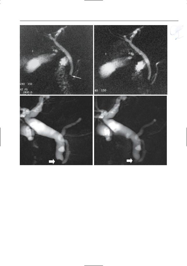
Fig. 120. a Functional activity of the sphincter and |
|
|
its influence on the appearance of the distalmost por- |
|
|
tions of the bile duct (B) and pancreatic duct (P).Left, |
|
|
the sphincter is relaxed. Right, contraction of the |
|
|
sphincter causes obliteration of the lumina of both |
|
|
ducts.The distal aspect of the presphincteric segment |
|
|
of the bile duct may be flat, slightly concave (bottom |
|
|
right, arrow), or slightly convex (top right, arrow). b, c |
|
|
Projective images obtained at 30-s intervals. The |
|
|
images show the variable appearance of the intramu- |
|
|
ral portion of the common bile duct, which is invisi- |
|
|
ble in (b) and can clearly been seen in (c) (arrow). d, e |
|
|
Different patient. Projective images with an interval |
|
|
of 130-s. These images also show the variable appea- |
|
|
rance of the intramural portions of the pancreatic |
|
|
duct and bile duct: they are clearly visible in (d) (rela- |
e |
|
xation) and invisible in (e) (contraction) (arrow) |
||
|

250 5.1 Normal Anatomy and Variants
#121 Variant Anatomy (1):
Type of Junction
KEY FACTS: ANATOMY
●The junction between the distal common bile duct and the pancreatic duct can be classified as follows:
–Y-type junction (± 70%)
–V-type junction (± 20%)
–U-type junction (± 10%)
–Other types (rare)
●In less than 0.2% of patients, the common bile duct and pancreatic ducts have separate orifices
References
Bocker V, Stolte M (1979) Functionelle Morphologie. In: Ottenjann R, Classen M (eds) Gastroenterologische Endoskopie. Enke, Stuttgart, pp 142–149 Wiedmeyer DA, Sewart ET, Taylor AJ (1993) Radiologic evaluation of structure and function of the sphincter of Oddi. Gastrointest Endoscop Clin
North Am 3 : 13–40
5 Vaterian Sphincter Complex 251
a
b
Fig. 121. a Y-type (left), V-type (center), and U- type junction (right). CBD, common bile duct; PD, pancreatic duct. (Reprinted with permission from Wiedmeyer et al. 1993). b–d Projective images showing junction with b Y-type, c V-type, and d U-type morphology (arrows)
c
d

252 5.1 Normal Anatomy and Variants
#122 Variant Anatomy (2):
“Pseudocalculus” Sign
Related topics: # 73 (stones in common bile duct: pitfalls in diagnosis), # 125 (impacted stone)
KEY FACTS
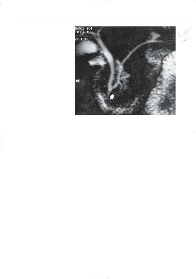
●Pseudocalculus sign: concave aspect of distal border of suprasphincteric part of the common bile duct, mimicking a stone (Fig. 122)
●Cause: forceful contraction of the sphincter with retrograde “invagination”
●Normal variant
●Potential cause of false-positive MR ! diagnosis of common bile duct lithiasis
(if dynamic imaging is not performed)
References
Ferruci J, Wittenberg J, Stone L, Dreyfuss J (1976) Hypotonic cholangiography with glucagon. Radiology 118 : 466–467
Van Hoe L, Gryspeerdt S, Vanbeckevoort D et al. (1998) Normal Vaterian sphincter complex: evaluation of morphology and contractility with dynamic single-shot MR cholangiography. AJR 170 : 1497–1500
Van Hoe L, Mermuys K, Vanhoenacker P (2004) MRCP pitfalls. Abdom Imaging 29 : 360–387
Fig. 122 a – g. a Pseudocalculus sign. Left, the sphincter is relaxed. Right, forceful contraction of the sphincter leads to retrograde invagination (arrow). b, c Projective image obtained during contraction showing pseudocalculus sign (arrow). c Projective image obtained 10 s later showing normal sphincter area (arrows). Note the absence of fluid in the second portion of the duodenum in b
a
b |
c |
5 Vaterian Sphincter Complex 253
d |
e |
f |
g |
Fig. 122 a – g. (continued) d, e Other patient. Projective images obtained within a 15-s interval.Again, the aspect of the distal common bile duct in d could erroneously be interpreted as a small stone (arrow). f , g. Different patient. Projective image during for-
ceful sphincter contraction (f) showing retrograde invagination resulting in the pseudocalculus sign (arrow). Projective image obtained 20-s later (g) showing a normal sphincter area (arrow)

254 5.1 Normal Anatomy and Variants
#123 Variant Anatomy (3):
Length of Common Channel
KEY FACTS
●In 60%–80%, the pancreatic and bile ducts unite to form a common channel (length, 2–15 mm; average, 5 mm; Kochhar et al. 1989)
●Presence of an anomalous pancreaticobiliary duct union (common channel greater than 15 mm in length) may predispose to intrabiliary reflux of pancreatic juice and formation of a choledochal cyst (see #68)
●Note: A key factor in the evaluation of the length of the common channel is adequate visualization of the exact location of the papilla. In MRI, sufficient filling of the duodenum with fluid is an essential prerequisite
●Note: Techniques to ensure that the duodenum is filled with fluid during an MRCP study:
–Dynamic (repetitive) scanning (mechanism: intermittent passage of fluid)
–Injection of a spasmolytic agent (possible mechanism: increased outflow of bile and/or gastric content; see #21)
–Oral administration of water
References
Hand BH (1962) An anatomical study of the choledochoduodenal area. Br J Surg 50 : 486
Hosoki T,Hasuike Y,Takeda Y et al. (2004) Visualization of pancreaticobiliary reflux in anomalous pancreaticobiliary junction by secretin-stimu- lated dynamic magnetic resonance cholangiopancreatography. Acta Radiol 45 : 375–382
Kochhar R, Nagi B, Chawla S et al (198) The clinical spectrum of anomalous pancreaticobiliary junction. Surg Endoscop 3 : 83–86
5 Vaterian Sphincter Complex 255
Fig. 123. Projective image showing a relatively long common channel
(± 16 mm; arrow)

256 5.1 Normal Anatomy and Variants
#124 Variant Anatomy (4): Papilla in or Adjacent
to a Duodenal Diverticulum
KEY FACTS: ANATOMY
● Incidence of duodenal diverticula: 5%–10% (Shemesh et al. 1987)
●Anatomic relationship to papilla:
–Papilla on the rim of the diverticulum (most common)
–Papilla in the diverticulum (may be a cause of failed ERCP)
●Associations:
–Increased incidence of common bile duct stones
–Possibly also increased incidence of pancreatitis (Osnes et al. 1981)
KEY FACTS: MRI (FIG. 124)
●Appearance of diverticula: paraduodenal structures containing:
–Fluid
–Air
–Air and fluid (with air–fluid level)
● The terminal ducts may be narrowed as they pass around the diverticulum
● If the papilla is within the diverticulum, ! intermittent reflux of air may be seen (may be interpreted as a stone if dynamic imaging is not performed)
●Duodenal diverticula may mimic cystic neoplasms of the pancreas when filled only with fluid (Macari et al. 2003)
References
Balci NC, Noone T, Akun E et al. (2003) Juxtapapillary diverticulum: findings on MRI. J Magn Reson Imaging 17 : 487–492
Macari M, Lazarus D, Israel G, Megibow A (2003) Duodenal diverticula mimicking cystic neoplasms of the pancreas: CT and MR imaging findings in seven patients. AJR Am J Roentgenol 180 : 195–199
Osnes M, Lootveit T, Larsen S, Aune S (1981) Diverticula and their relationship to age, sex, and biliary calculi. Scand J Gastroenterol 16 : 103–107
Shemesh E, Friedman E, Czesniak A, Bat L (1987) The association of biliary and pancreatic anomalies with periampullary duodenal diverticula. Correlation with clinical presentations. Arch Surg 122 : 1055–1057
5 Vaterian Sphincter Complex 257
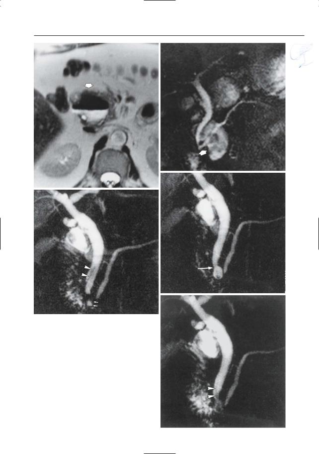
a
c
Fig. 124 a, b. a Axial T2-weighted image showing a large diverticulum at the medial border of the duodenum (arrow). Note the presence of an air-fluid level. b Projective image showing that the papilla is located adjacent to (not in) the diverticulum (arrow). c–e Other patient. Three consecutively obtained projective images. c Weblike low-intensity structure in the distal common bile duct (arrowheads), representing reflux of air. Note the small duodenal diverticulum (arrows). d The air has disappeared. In addition, the connection between the distal common bile duct and the diverticulum is clearly demonstrated (arrow). e Air refluxing into the bile duct (arrowheads). This case illustrates that small diverticula embedded in the duodenal wall may show temporal changes in size and shape caused by smooth muscle contractions
b
d
e