Добавил:
Опубликованный материал нарушает ваши авторские права? Сообщите нам.
Вуз: Предмет: Файл:
Книги по МРТ КТ на английском языке / MR Cholangiopancreatography Atlas with Cross-Sectional Imaging Correlation - L Van Hoe Dirk Vanbeckevoort Koen Mermuys We.pdf
Скачиваний:
1
Добавлен:
05.10.2023
Размер:
37.07 Mб
Скачать

6 Pancreatic Ducts 375

6.4Malignant Tumors and Tumors with Malignant Potential

#185 Adenocarcinoma, General

KEY FACTS: DISEASE

Fourth to fifth leading cause of cancer death in the United States

Incidence in autopsy series: 2%

Age peak: seventh decade

Resectable at presentation: 8%–15%

CA 19-9 increased in 80%–90%

Origin:

Exocrine ductal epithelium: 99%

Acinar portion of the pancreatic gland: 1%

Extent (Cubilla et al. 1979)

65% of patients have advanced local disease or distant metastases

21% have local disease with spread to regional lymph nodes

14% have tumor confined to the pancreas

Metastases

Liver: 30%–60%

Regional lymph nodes: 15%–28%

Peritoneum: 7%–10%

Other sites: e.g., lung, pleura, bone

Symptoms:

Epigastric pain radiating to the back

Obstructive jaundice

New-onset diabetes, steatorrhea

Five-year survival rate: 1%–5%

References

Cubilla AL, Fitzgerald PJ (1979) Cancer of the pancreas (nonendocrine): a suggested morphological classification. Semin Oncol 6 : 285–297

Warshaw AL, Fernandez-del Castillo C (1992) Pancreatic carcinoma. New Engl J Med 326 : 455–465

376 6.4 Malignant Tumors and Tumors with Malignant Potential

#186 Adenocarcinoma,

Signal Intensity

KEY FACTS: MRI

Signal intensity:

Nearly invariably hypointense on T1-weighted magnetization-prepared snapshot gradient echo images (see #9)

May be isointense, however, if the surrounding pancreatic tissue is abnormal (pancreatitis)

Isoor hyperintense on T2-weighted images

Usually hypointense on contrast-en- hanced scans

Technical remarks about contrast-en- hanced scans:

Rapid bolus injection is mandatory

Timing is important: imaging should

be done in the pancreatic phase (± 40 s after injection)

Note: Value of sequences:


!

T1 much better than T2 for tumor


detection


Magnetization-prepared

snapshot


gradient echo particularly reliable

– Contrast-enhanced scans

often re-


quired for preoperative evaluation (as-


sessment of vascular anatomy, detec-


tion of lymph node metastases)

References

Gabata T,Matsui O,Kadoya M et al. (1994) Small pancreatic adenocarcinomas: efficacy of MR imaging with fat suppression and gadolinium enhancement. Radiology 193 : 683–688

Ichikawa T, Haradome H, Hachiya J et al. (1997) Pancreatic ductal adenocarcinoma: preoperative assessment with helical CT versus dynamic MR imaging. Radiology 202 : 655–662

Kelekis NL, Semelka RC (1997) MRI of pancreatic tumors. Eur Radiol 7 : 875–886

6 Pancreatic Ducts 377

a

b

c

d

e

f

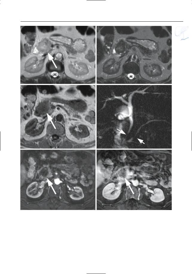
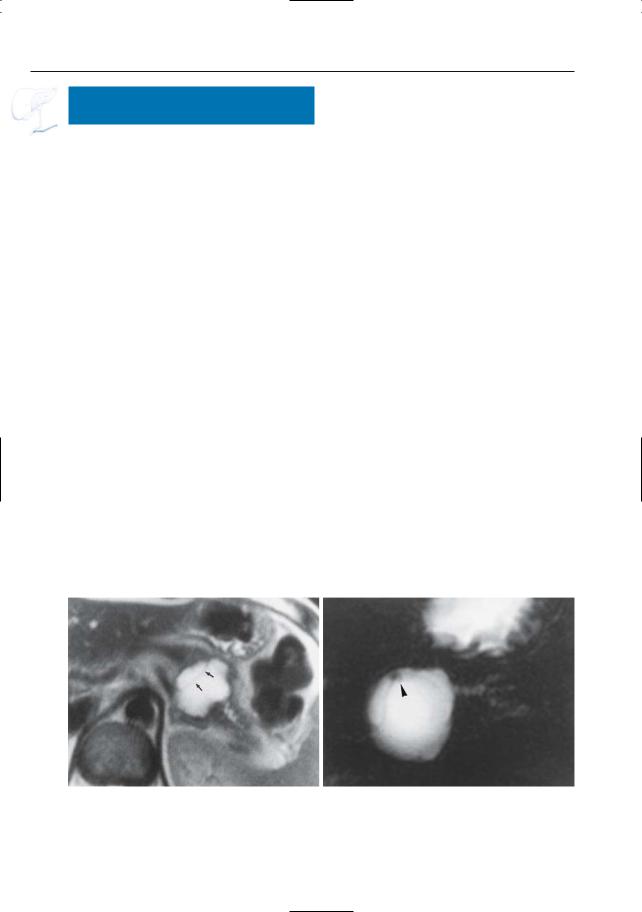
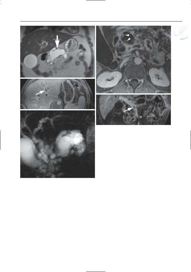
Fig. 186 a–f. Axial T2-weighted HASTE images (TE 60 and 360) (a, b) and (c) axial T1-weighted image showing a focal lesion in pancreatic head (arrows): adenocarcinoma. The lesion is markedly hypointense to normal pancreatic tissue on the T1weighted image. d Projective image showing focal narrowing of the distal common bile duct and pancreatic duct,approximately at the same level (double

duct sign) (arrows). Note nearly normal size of proximal pancreatic duct, which is an unusual finding. e, f Axial contrast-enhanced T1-weighted VIBE images obtained in the pancreatic and late venous phase showing no enhancement of the tumor in the pancreatic phase and only minimal enhancement of the tumor in the late venous phase (arrow)

378 6.4 Malignant Tumors and Tumors with Malignant Potential

#187 Adenocarcinoma,

Ductal Changes

KEY FACTS: MRI

Classical patterns of involvement of the main duct (Fig. 187a):

(Apparent) occlusion (> 50% on MRCP)

Stenosis, usually irregular with abrupt termination

Dilation of the proximal main duct

Double duct sign (Freeny et al. 1978; e.g., see #90, Fig. 187b–e, 188a, b); sometimes “four-segment sign” (Kim et al. 2002)

!Side branches may be:

Invisible (normal or obliterated)

Dilated (obstruction; may appear like bizarre cystic pockets located adjacent to the tumor)

Distorted/displaced

Comments:

Long strictures of the pancreatic duct, especially in the body or tail, are most commonly malignant (see, however,

# 164, # 167 and # 169); short strictures are more common in chronic alcoholic pancreatitis

Projective images should be obtained routinely: subtle ductal changes may herald the presence of small tumors

References

Freeny PC, Bilbao MK, Katon RM (1976) “Blind” evaluation of endoscopic retrograde cholangiopancreatography (ERCP) in the diagnosis of pancreatic carcinoma: the “double duct” and other signs. Radiology 119 : 271–274

Kim JH, Kim MJ, Chung JJ et al. (2002) Differential diagnosis of periampullary carcinomas at MR imaging. Radiographics 22 : 1335–1352

Stewart E, Vennes J, Geenen J (1977) Atlas of endoscopic retrograde cholangiopancreatography. Mosby, St Louis, p 182

6 Pancreatic Ducts 379

a

b


c

d

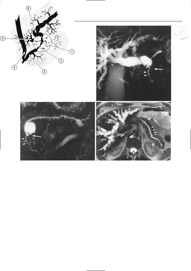
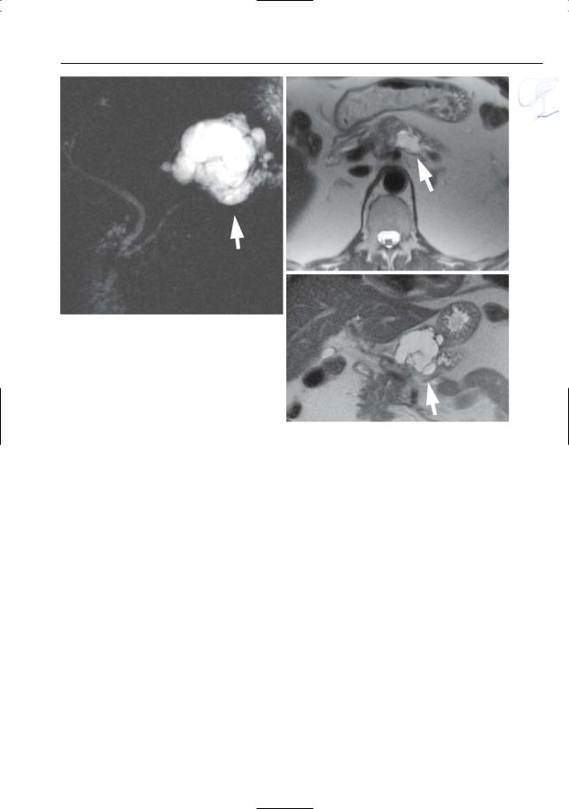
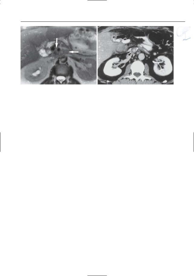
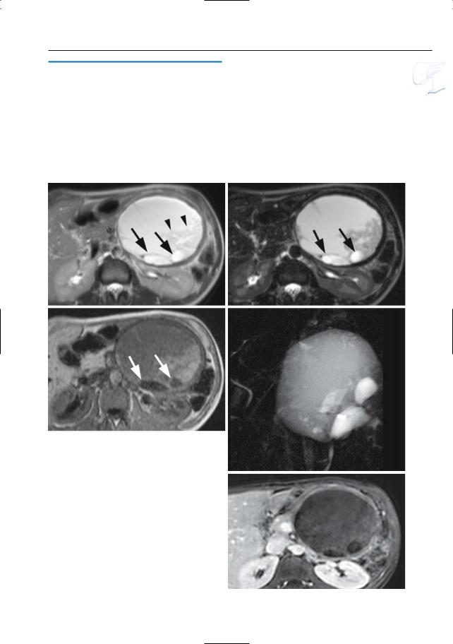
Fig. 187. a Ductal changes in pancreatic cancer. The tumor (1) causes abrupt narrowing of the pancreatic duct (2). Note the irregular contours of the stricture (3). The common bile duct (4) may be normal or stenotic at the same level.Dilated side branches are seen adjacent to the tumor mass (5). The proximal pancreatic duct shows dilation (6). Note that necrotic areas within the tumor may appear as small “cysts.” In chronic pancreatitis, dilated side branches may be seen within an inflammatory mass, rather than adjacent to the mass (see # 167). (Reprinted with permission from Stewart et al. 1997). b–d Patient with cancer of pancreatic head. Projective images showing

marked dilation of intrahepatic bile ducts with abrupt termination (arrow in b) and slightly dilated pancreatic duct that exhibits narrowing at the same level (arrow in c). Note the rather homogeneous dilation of side branches in c and the presence of a dilated side branch in the pancreatic head that is displaced by the tumor (arrowheads in b,c). The cystic lesion in the pancreatic neck proved to be a benign mucinous neoplasm (incidental finding). d Same patient. Heavily T2-weighted axial image also showing rather uniform dilation of the side branches (arrows). The final diagnosis was infiltrating adenocarcinoma of the pancreatic neck

380 6.4 Malignant Tumors and Tumors with Malignant Potential

e

f

g

h

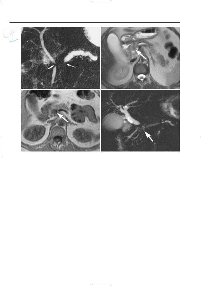
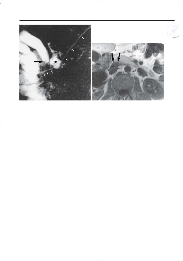
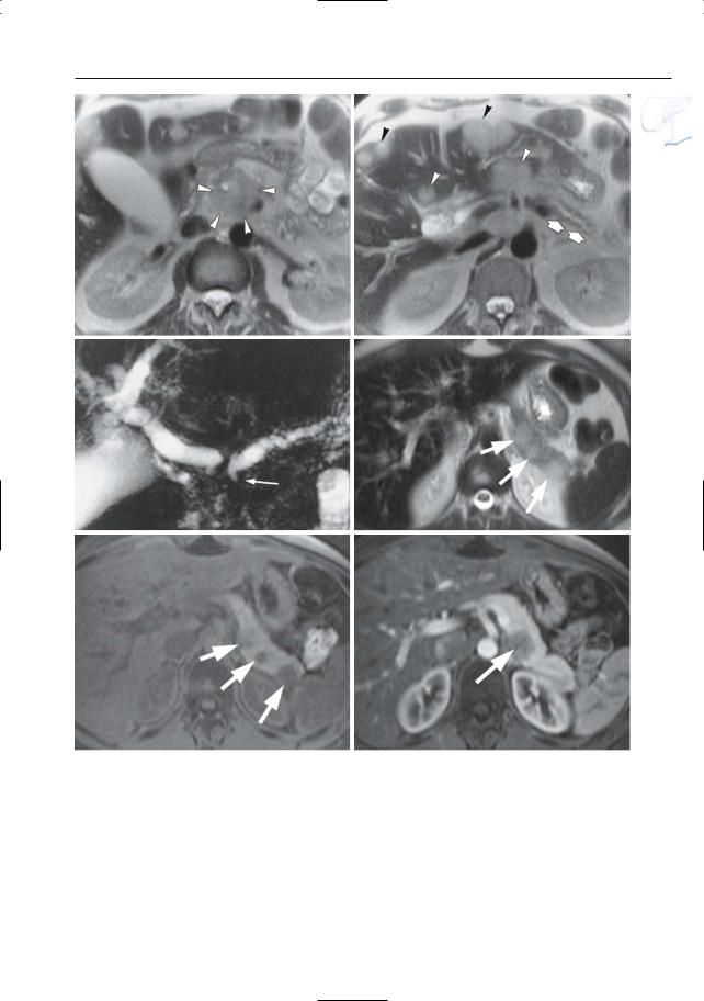
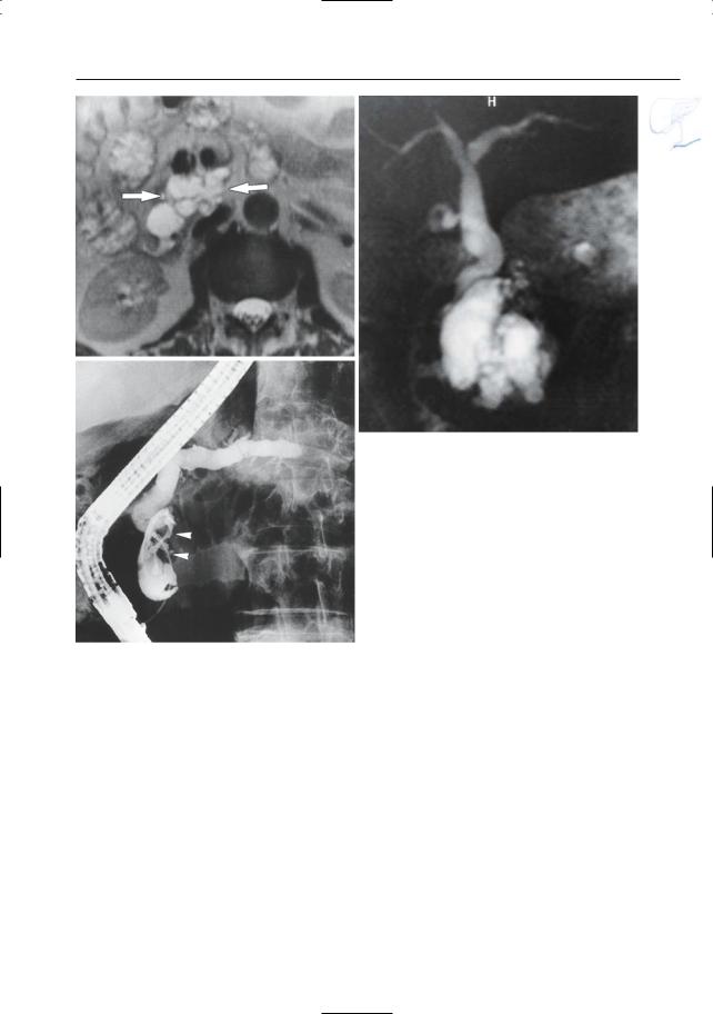
Fig. 187 a – j. (continued) e Projective image showing obliteration of the pancreatic duct associated with a short stricture of the common bile duct (double duct sign, arrows). f–h Axial T2-weighted HASTE image (f) and axial T1-weighted image (g) showing a small adenocarcinoma in the pancreatic head (arrow). As in most cases, the lesion is nearly iso-

intense to normal pancreatic parenchyma on the T2-weighted images and clearly T1-hypointense. h Projective image showing dilatation of the proximal main pancreatic and common bile duct with an abrupt obliteration of the ducts (arrow). The distal main pancreatic and common bile duct have a normal appearance (four segment sign)

6 Pancreatic Ducts 381

i

j

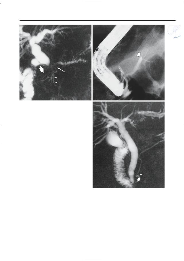
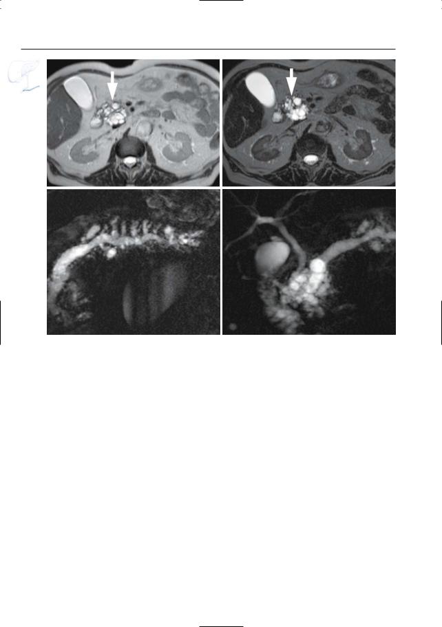
Fig. 187 a – j. (continued) i, j Axial T2-weighted HASTE image (i) showing a small adenocarcinoma in the pancreatic body (arrow) with narrowing of the main pancreatic duct and proximal dilatation

(arrow–head). j Projective image showing dilatation of the proximal main pancreatic duct (arrowheads) with irregular narrowing in the pancreatic body

(arrow)

382 6.4 Malignant Tumors and Tumors with Malignant Potential

#188 Variant Morphology

of Adenocarcinoma (1):

Degree of Obstruction

KEY FACTS: MRI

Since most pancreatic adenocarcinomas originate in the main pancreatic duct, ductal obstruction occurs early in the course of disease. MRI correlates:

Dilation of the proximal main pancreatic duct

More or less uniform dilation of the proximal side branches

Variable degree of parenchymal atrophy

Small intrapancreatic pseudocysts in 10%

!However, the absence of proximal dilation does not rule out pancreatic carcinoma (Fig. 188c):

Tumor may develop in a side branch and invade the main pancreatic duct only in a more advanced stage

In patients with “dual” drainage (“bifid” ductal configuration, see also

# 136), obstruction of the ventral duct does not necessarily implicate dilation of the dorsal ducts

Pancreas divisum (see # 190)

References

Fukumoto K, Nakajima M, Murakami K, Kawai K (1974) Diagnosis of pancreatic cancer by endoscopic cholangiopancreatography (ERCP). Am J Gastroenterol 60 : 210–213

Kim JH, Kim MJ, Chung JJ et al. (2002) Differential diagnosis of periampullary carcinomas at MR imaging. Radiographics 22 : 1335–1352

Muranaka T (1990) Morphologic changes in the body of the pancreas secondary to a mass in the pancreatic head. Analysis by CT. Acta Radiol 31 : 483–488

6 Pancreatic Ducts 383

a

Fig. 188 a, b. Patient with adenocarcinoma. a Projective MR image showing apparent occlusion of the pancreatic duct and bile duct (arrows).Also note the slightly dilated side branch in the pancreatic head (arrowheads). b ERCP showing occlusion of the main pancreatic duct (arrow). c Patient with a small adenocarcinoma in the pancreatic head. Projective MR image showing narrowing of the distal part of the pancreatic duct (arrow), without prestenotic dilation. Also note tumor necrosis (arrowhead). Bile duct dilation was caused by involvement of the papilla by the tumor

b

c

384 6.4 Malignant Tumors and Tumors with Malignant Potential

#189 Variant Morphology

of Adenocarcinoma (2):

Necrosis/Cystic Degeneration

KEY FACTS: MRI

Adenocarcinomas commonly contain T2-hyperintense areas

These may correspond to:

Necrosis

Cystic components

Occasionally, cystic/necrotic areas may be quite large (cystic adenocarcinoma; Fig. 189)

Differential diagnosis:

Pseudocysts in chronic pancreatitis

Other cystic tumors (e.g., mucinous neoplasms)

Dilated side branches (see # 187)

ERCP correlate of necrosis: extraductal extravasation of contrast material (38%) (Bilabao and Katon 1977)

References

Bilabao MK, Katon RM (1977) Neoplasms of the pancreas. In: Stewart E, Vennes J, Geenen J (eds) Atlas of endoscopic retrograde cholangiopancreatography. Mosby, St Louis, pp 181–192

Sahani D, Prasad S, Saini S, Mueller P (2002) Cystic pancreatic neoplasms: evaluation by CT and magnetic resonance cholangiopancreatography. Gastrointest Endoscopy Clin N Am 12 : 657–672

6 Pancreatic Ducts 385

a

Fig. 189 a–c. Projective image (a), axial (b) and coronal (c) T2-weighted HASTE images showing a large cystic lesion in the pancreatic tail (arrow) representing adenocarcinoma with marked cystic/ necrotic degeneration

b

c

386 6.4 Malignant Tumors and Tumors with Malignant Potential

#190 Adenocarcinoma

in Pancreas Divisum

Related topic: # 140 (pancreas divisum)

KEY FACTS: MRI

!The “double duct sign” usually refers to concomitant stenosis or occlusion of the “main” pancreatic duct and common bile duct. It may, however, also refer to:

Stenosis of the common bile duct and a side branch of the main pancreatic duct

Stenosis of the common bile duct and the ventral duct (in pancreas divisum)

Note: In complete pancreas divisum, obstruction of the ventral pancreatic

duct is typically not associated with ductal dilation in the body and tail, except in cases where the tumor extends to the minor papilla or accessory duct (Fig. 190)

References

Kelekis NL, Semelka RC (1995) Carcinoma of the pancreatic head area. Diagnostic imaging: magnetic resonance imaging. Rays 20 : 289–303

6 Pancreatic Ducts 387

a

b

Fig. 190 a, b. Projective image (a) showing a normal pancreatic duct in the body/tail with a patent duct of Santorini (small arrows). Note the dilation of the common bile duct with abrupt narrowing (arrow). Also note the marked obstructive dilation

of a side branch in the pancreatic head/neck (asterisk). b Axial T1-weighted image showing a small tumor in the posterior part of the pancreatic head (arrows). (Figure courtesy of C. Feys)

388 6.4 Malignant Tumors and Tumors with Malignant Potential

#191 Spread of Adenocarci-

noma (1): Vascular Invasion

KEY FACTS: DISEASE

Invasion of peripancreatic vessels usually indicates unresectability

Incidence: ± 33%

Arterial encasement involves the superior mesenteric, splenic, celiac, hepatic, gastroduodenal, and left renal arteries, in descending order of frequency

The veins most commonly affected are the splenic vein and portosplenic confluence

KEY FACTS: MRI

Criteria for vascular invasion:

Narrowing/occlusion

Vessel partially or completely encircled by tumor (Loyer et al. 1996; Mitchell et al. 1987)

!In patients with pancreatic head cancer invading or compressing the superior mesenteric vein, visualization of dilated peripancreatic veins (that act as collaterals) may be another useful sign of unresectability (Fig. 191; Hommeyer et al. 1995):

Gastrocolic trunk

Anterior superior pancreaticoduodenal vein

Posterior superior pancreaticoduodenal vein

Comments:

In many cases, diagnosis of vascular invasion is evident on non-contrast- enhanced scans (Fig. 191)

Dual-phase MRA can be used in conjunction with cross-sectional MRI and MRCP to provide a “complete” preoperative assessment of pancreatic neoplasms (Gaa et al. 1997)

References

Gaa J, Wendl K, Trede M, Georgi M (1997) New concepts in MR imaging of pancreatic carcinoma: the all-in-one approach. In: Oudkerk M, Edelman R (eds) High-power gradient MR-imaging. Blackwell Science, Berlin, pp 425–430

Hommeyer S, Freeny PC, Crabo LG (1995) Carcinoma of the head of the pancreas: evaluation of the pancreaticoduodenal veins with dynamic CT

– potential for improved accuracy in staging. Radiology 196 : 233–238

Loyer EM,David CL,Dubrow RA,Evans DB,Charnsangavej C (1996) Vascular involvement in pancreatic adenocarcinoma: reassessment by thin-section CT. Abdom Imaging 21 : 202–206

Mitchell DG,Hill MC,Cooper R et al.(1987) The superior mesenteric artery fat plane: is obliteration pathognomonic of pancreatic carcinoma? J Comp Assist Tomogr 11 : 247–253

6 Pancreatic Ducts 389

a

b

c

d

Fig. 191 a–d. Axial T1-weighted image (a) showing a large hypointense adenocarcinoma with clear extrapancreatic extension (arrow). b–d Axial contrastenhanced T1-weighted VIBE images obtained in the venous phase show dilatation of the gastrocolic venous trunc with mesenteric venous collaterals

[arrowheads in (b)].Also seen is displacement of the celiac trunc [large arrow in (c)], and invasion of the venous confluence and splenic vein [small arrows in (d)]. Note the hypovascular adenocarcinoma [open arrow in (d)]

390 6.4 Malignant Tumors and Tumors with Malignant Potential

#192 Spread of Adenocarci-

noma (2): Lymph Nodes

KEY FACTS: DISEASE

Lymph node metastases are found in

± 50% of patients (Megibow et al. 1995)

Lymph nodes most commonly involved:

– Cancer of the pancreatic head: retroperitoneal and superior pancreatic head groups

– Cancer of the pancreatic body and tail: superior body and head groups

KEY FACTS: MRI

Signal intensity of (enlarged) lymph nodes relative to surrounding fat:

T2 without fat suppression: variable, often more or less isointense

T2 with fat suppression: variable, commonly hyperintense

T1 without fat suppression: hypointense

T1 with fat suppression: commonly isointense

Contrast-enhanced images with fat suppression: hyperintense

Limitations:


!

Detection

of slightly

enlarged peri-


pancreatic nodes may be difficult


because of limitations of spatial reso-


lution (particularly in the third


dimension; volume averaging), multi-


plicity of small structures present in


the peripancreatic space (e.g., arter-


ies, veins), and subtle differences in


signal intensity from surrounding tis-


sues


Detection of micrometastases in nor-


mal-sized nodes is impossible

Importance:

detection

of enlarged

lymph nodes by imaging studies has little clinical relevance because, in the absence of other signs of non-resectabil- ity, it does not represent a contra-indica- tion for surgery

References

Kelekis NL, Semelka RC (1997) MRI of pancreatic tumors. Eur Radiol 7 : 875–886

Megibow AJ, Zhou XH, Rotterdam H et al. (1995) Pancreatic adenocarcinoma: CT versus MR imaging in the evaluation of resectability – report of the Radiology Diagnostic Oncology Group. Radiology 195 : 327–332

6 Pancreatic Ducts 391

a

b

Fig. 192 a, b. Patient with pancreatic carcinoma. Images obtained using the “black blood” technique showing several enlarged a superior body and

b portocaval nodes (arrows). Note that these nodes were invisible on the classical HASTE sequence

392 6.4 Malignant Tumors and Tumors with Malignant Potential

#193 Spread of Adenocarci-

noma (3): Distant Metastases

KEY FACTS: DISEASE

Incidence of hematogenous metastases in autopsy series: 85%–95%

Location (in descending order of frequency): liver, lungs, adrenals, kidneys, bones

KEY FACTS: MRI

T2: (minimally) hyperintense with or without central necrosis

Hypointense on contrast-enhanced scans, sometimes with peripheral rim enhancement

Peritoneal metastases are usually not directly visualized; presence of ascites in patients with a pancreatic neoplasm and without signs of cirrhosis or portal vein thrombosis may be an indirect sign (Fig. 1)

Hepatic metastases of pancreatic adenocarcinoma have a nonspecific appearance (see also #60):

– T1: hypointense

References

Baert AL, Rigauts H, Marchal G (1994) Ductal adenocarcinoma. In: Baert AL (ed) Radiology of the pancreas. Springer, Berlin Heidelberg New York, pp 129–172

Kelekis NL, Semelka RC (1997) MRI of pancreatic tumors. Eur Radiol 7 : 875–886

6 Pancreatic Ducts 393

a

b

c

d

e

f

Fig. 193 a, b. a T1and b T2-weighted images showing hepatic metastases as hypoand slightly hyperintense, respectively (arrowheads). Note that the T1weighted image shows more lesions. c, d Axial contrast-enhanced T1-weighted VIBE images obtained in the portovenous phase showing a hypoenhancing adenocarcinoma in the pancreatic head [large arrow in (c)], with retro-obstructive dilatation of the main pancreatic duct [arrowheads in (c)], and two rim-enhancing liver metastases in the right lobe [small arrows in (d)]. Because of the subtle enhance-

ment, these lesions could be mistaken for cysts. e, f Coronal T2-weighted HASTE image (e) showing a hyperintense adenocarcinoma in the pancreatic head (large arrow) with obliteration and proximal dilatation of the common bile duct (small arrows), and multiple slightly hyperintense hepatic metastases (arrowheads). f Axial contrast-enhanced T1-weighted VIBE image obtained in the venous phase confirming the presence of multiple metastatic lesions in the liver

(arrows)

394 6.4 Malignant Tumors and Tumors with Malignant Potential

#194 Adenocarcinoma

with Retro-obstructive

Pancreatitis

Related topic: # 168 (chronic obstructive pancreatitis)

KEY FACTS: DISEASE

Ductal obstruction by adenocarcinoma may cause:

Chronic obstructive pancreatitis (see

# 168)

Acute pancreatitis

Note: Pancreatic cancer may also develop in patients with preexisting chronic pancreatitis (occurring in 2–4% of patients)

KEY FACTS: MRI

!Pancreatitis associated with adenocarcinoma may cause diagnostic problems for several reasons:

Ductal changes in chronic pancreatitis may be so dominant that the associated carcinoma is overlooked

Peripancreatic inflammatory changes in acute pancreatitis may jeopardize the diagnostic quality of projective MR images

Tumor detection may be difficult due to loss of the normal hyperintensity of surrounding pancreatic tissue on T1weighted images

Signs of pancreatitis indicate the need for obtaining immediate postcontrast gadolinium-enhanced scans for tumor detection (Fig. 194)

Signs suggestive of carcinoma (see also

# 167):

Focal irregular ductal stenosis in a duct with an otherwise normal appearance (not absolute, see # 1)

Length of stenosis: > 1–2 cm (idem)

Abrupt termination of stenosis (idem)

Double duct sign (not absolute, see

# 164)

Focal markedly T1-hypointense mass, with parenchyma with an otherwise normal appearance

Markedly hypovascular solid focal mass causing ductal obstruction

References

Delmaschio A, Vanzulli A, Sironi S et al. (1991) Pancreatic cancer versus chronic pancreatitis: diagnosis with CA 19-9 assessment, US, CT, and CTguided fine-needle biopsy. Radiology 178 : 95–99 Kelekis NL, Semelka RC (1997) MRI of pancreatic

tumors. Eur Radiol 7 : 875–886

Shemesh E, Czerniak A, Nass S, Klein E (1990) Role of endoscopic retrograde cholangiopancreatography in differentiating pancreatic cancer coexisting with chronic pancreatitis. Cancer 65 : 893–896

!

!

6 Pancreatic Ducts 395

a

b

c

d

Fig. 194 a–d. Patient with adenocarcinoma and retro-obstructive pancreatitis. a Projective image showing a stenosis of the pancreatic duct (arrow) with proximal dilatation (arrowheads). b CT image showing a small pancreatic tumor (large arrow), prestenotic duct dilatation (small arrows),and subtle peripancreatic infiltration (arrowheads). c Axial T2weighted HASTE image (TE 360) showing slightly

increased signal intensity of the fat around pancreatic tail. d Axial contrast-enhanced T1-weighted VIBE image obtained in the pancreatic phase showing decreased enhancement of the pancreatic tail (small arrows) when compared to the head (large arrow). The findings in (c) and (d) point to the presence of retro-obstructive pancreatitis. Final diagnosis: adenocarcinoma

396 6.4 Malignant Tumors and Tumors with Malignant Potential

#195 Recurrent Adenocarcinoma

KEY FACTS: GENERAL

Incidence: not rare:

Only 14% of patients with adenocarcinoma have tumor confined to the pancreas

Approximately 35% of metastatic lymph nodes cannot be removed during surgery; similarly, tumors encasing retroperitoneal vessels can only partially be removed

Location: most commonly in close proximity to the superior mesenteric artery or celiac trunk

KEY FACTS: MRI

Typical feature: infiltrating mass surrounding and encasing the superior mesenteric artery and/or celiac trunk

References

Coombs RJ, Zeiss J, Howard JM, Thomford NR, Merrick HW (1990) CT of the abdomen after the Whipple procedure: value of depicting postoperative anatomy, surgical complications and tumor recurrence. AJR Am J Roentgenol 154 : 1011–1014

6 Pancreatic Ducts 397

a

b

Fig. 195 a, b. Patient who underwent a Whipple operation for carcinoma in the pancreatic head. a T2-weighted image showing soft tissue mass (arrows) between the superior mesenteric artery

and the aorta. b CT image obtained at a more caudal level also showing soft tissue cuff around the superior mesenteric artery (arrowheads)

398 6.4 Malignant Tumors and Tumors with Malignant Potential

#196 Secondary Pancreatic Tumors

Related topics: #60 (intrahepatic bile ducts: metastases), #91 (extrahepatic bile duct: involvement by other extrabiliary neoplasms)

KEY FACTS: DISEASE

Most common secondary pancreatic tumors:

Lymphoma

Metastases from breast cancer, lung cancer (24% of patients with small cell lung cancer), cancer of the colon, renal cell carcinoma, and malignant melanoma

Usually end-stage disease

KEY FACTS: MRI

Pancreatic duct:

Typically no involvement or displacement only

Narrowing may occur but is less frequent

Parenchyma: usually multiple locations

Specific features:

Melanoma metastases may be T1hyperintense

Renal cell carcinoma metastases are typically hypervascular (differential diagnosis with endocrine tumors)

Key elements: clinical history/locations in other organs

References

Ehrlich A, Stalder G, Geller W, Sherlock P (1968) Gastrointestinal manifestations of malignant lymphoma. Gastroenterology 54 : 1115–1121

Roland CF, Van Heerden JA (1989) Nonpancreatic primary tumors with metastases to the pancreas. Surg Gynecol Obstet 168 : 345–347

6 Pancreatic Ducts 399

a

b

c

d

e

f

Fig.196 a – c. Patient with lung cancer. a, b T2weighted images showing a large mass in the pancreatic head (arrowheads), atrophy of the pancreatic tail (arrows), and multiple hepatic metastases (arrowheads). c Projective MR image showing double duct sign (arrow), which is an unusual finding in patients with pancreatic metastases. d–f Patient with esophageal myosarcoma and pancreatic metastases.

Axial T2-weighted HASTE image (d) and axial T1 weighted image (e) showing multiple focal lesions in the pancreatic tail (arrows). Note the peripheral location of these masses and the absence of dilatation of the pancreatic duct. f Axial contrast-enhan- ced T1-weighted VIBE image (different position) obtained in the pancreatic phase showing poor vascularization of the metastatic lesion (arrow)

400 6.4 Malignant Tumors and Tumors with Malignant Potential

#197 Malignant Neuroendocrine

Tumors

Related topic: # 177 (benign neuroendocrine tumors)

KEY FACTS: DISEASE

Incidence of malignancy with respect to subtype: see Table 197

Other features:

Gastrinomas found in patients with type-1 multiple endocrine neoplasia syndrome are less likely to be malignant than isolated gastrinomas

Larger tumors (> 2 cm) are more commonly malignant

KEY FACTS: MRI

Secreting tumors: see # 177 (no morphologic features to distinguish between benign and malignant tumors)

Nonsecreting tumors are usually large and centrally necrotic

Note: Hepatic metastases of gastrinoma


!

typically have a very high signal inten-

sity on T2-weighted images and, there-


fore, may mimic hemangiomas. Solu-


tion: delayed contrast-enhanced MRI


(Berger et al. 1996):


Gastrinoma metastases: hypoor isointense

Hemangiomas: hyperintense

References

Berger JF, Laissy JP, Limot O et al. (1996) Differentiation between multiple liver hemangiomas and liver metastases of gastrinomas: value of enhanced MRI. J Comp Assist Tomogr 20 : 349–355 Marcos HB, Libutti SK, Alexander HR, et al. (2002) Neuroendocrine tumors of the pancreas in von Hippel-Lindau disease: spectrum of appearances at CT and MR imaging with histopathologic

comparison. Radiology 225 : 751–758

Van Hoe L, Vanbeckevoort D, Baert AL (1997) Endocrine tumors of the pancreas: computed tomography and magnetic resonance imaging. Chir Gastroenterol 13 : 301–306

6 Pancreatic Ducts 401

Table 197. Incidence of malignancy in neuroendocrine tumors

Tumor

Relative frequency (%)

Malignant (%)


Insulinoma

56

5–10

Gastrinoma

21

60

Glucagonoma

2.3

80

Vipoma

1.8

60

Nonfunctioning tumors

15

80–100


402 6.4 Malignant Tumors and Tumors with Malignant Potential

a

b


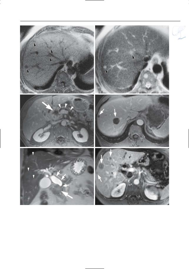
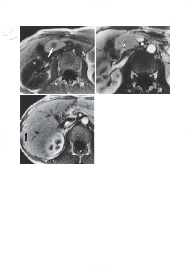
Fig. 197 a – c. Patient with metastatic gastrinoma.


a T1-weighted image showing a hypointense lesion


in the pancreatic head, corresponding to a gastri-


noma (arrow). b Contrast-enhanced fat-suppressed


T1-weighted image only showing the nonenhancing


central part of the tumor (arrowhead). c Contrast-


enhanced image obtained at a more cranial level

c

showing liver metastasis with typical rim enhance-

ment (arrow)


6 Pancreatic Ducts 403

d

e

f

g

Fig. 197 d– g. Patient with a malignant cystic neuroendocrine tumor with hypervascular liver metastases. d, e Axial and coronal T2-weighted HASTE images showing a hyperintense cystic mass in the pancreatic tail with a thick irregular wall (large arrow), and multiple T2-hyperintense liver lesions

(small arrows). f, g Axial contrast-enhanced T1weighted VIBE images obtained in the arterial phase showing strong uptake of contrast medium in these lesions (small arrows). Note the nonenhancing cystic neuroendocrine tumor in the pancreatic tail [large arrow in (g)]

404 6.4 Malignant Tumors and Tumors with Malignant Potential

#198 Mucinous Pancreatic

Neoplasm (Peripheral Type)

Related topics: # 175, 176 (serous cystadenoma), # 199 (intraductal neoplasm)

KEY FACTS: DISEASE

Synonyms:

Macrocystic pancreatic neoplasm

Mucinous cystadenoma/cystadenocarcinoma

Cystic lesions lined by columnar, mucinproducing cells

Macroscopic appearance:

Unilocular or multilocular

Cystic components usually greater than 2 cm in diameter

Papillary protrusions may be seen

Sometimes peripheral calcifications

To be distinguished from intraductal mucinous neoplasms (see # 199)

Mean age: 50 years

a

Ratio of women to men: 6 : 1–9 : 1

Location: 85% in the pancreatic tail

Usually large

All tumors are malignant or have malignant potential

Local recurrence is common

KEY FACTS: MRI (FIG. 198)

Unilocular or multilocular cystic lesion

Cysts greater than 2 cm in diameter

Signal intensity of contents of cystic spaces: reflects varying concentrations of mucin

Usually like fluid

Sometimes T1-hyperintense (high concentration of mucin)

Sometimes lower signal intensity in dependent portions on T2 (“layering”)

The intensities may vary in different loculi

No enhancing components

Malignant tumors have a propensity for invasion of local organs and vessels

b

Fig. 198 a, b. a Axial T2-weighted image showing a cystic lesion in the pancreatic body/tail, containing a septum (arrows). b Projective image showing a

septum and small mural nodule (arrowhead), which would be unusual for a (pseudo-)cyst.

6 Pancreatic Ducts 405

References

Compagno J, Oertel JE (1978) Mucinous cystic neoplasms of the pancreas with overt and latent malignancy (cystadenocarcinoma and cystadenoma): a clinicopathologic study of 41 cases. Am J Clin Pathol 69 : 573–580

Kim YH, Saini S, Sahani D, Hahn PF, Mueller P, Auh Y (2005) Imaging diagnosis of cystic pancreatic lesions: pseudocyst versus nonpseudocyst. RadioGraphics 25 : 671–685

c

e

Fig. 198 c–g. c Axial T2-weighted HASTE image (TE 60) showing a large cystic lesion in the pancreatic body/tail with two hyperintense components (arrows) and debris (arrowheads). d–f Axial T2weighted HASTE image (TE 360), axial T1-weighted image and projective image clearly show the different signal intensities in the different components of the lesion (arrows). g Axial contrast-enhanced T1weighted VIBE image obtained in the venous phase showing no internal enhancing components

Minami M,Itai Y,Ohtono K,Yoshida H,Yoshikawa K, Lio M (1989) Cystic neoplasms of the pancreas: comparison of MR imaging with CT. Radiology 171 : 53–56

Sahani D, Prasad S, Saini S, Mueller P (2002) Cystic pancreatic neoplasms: evaluation by CT and magnetic resonance cholangiopancreatography. Gastrointest Endoscopy Clin N Am 12 : 657–672

d

f

g

406 6.4 Malignant Tumors and Tumors with Malignant Potential

#199 Intraductal Papillary

Mucinous Tumor (IPMT)

Related topic: # 198 (mucinous pancreatic neoplasm, peripheral type)

KEY FACTS: DISEASE

Mucin-producing tumor of intraductal origin

No preponderance for men or women

Belongs to the group of mucinous pancreatic tumors. Classification (Itai and Ohtomo 1996):

Branch duct type: mucin-secreting intraductal tumor usually located in the uncinate process and causing localized dilation of a side branch

Main duct type: mucin-secreting intraductal tumor characterized by dilation of the main pancreatic duct

Like tumors of the peripheral type, these tumors have malignant potential

The prognosis depends on the presence or absence of local invasion

KEY FACTS: MRI

General remarks:

The mucin-secreting tumor is often too small to be visualized; the hallmark of the disease is marked focal or diffuse ductal dilation

Mucin has a very high signal intensity on T2-weighted images and cannot usually be differentiated from fluid

!Typical appearance of the branch duct type (Fig. 199):

“Polycystic” mass in the uncinate process, actually representing focally dilated side branches filled with mucin

Differential diagnosis: pseudocyst(s)

Typical appearance of the main duct ! type:

Usually marked global dilation of the main pancreatic duct without a demonstrable cause of obstruction

Clue to diagnosis on ERCP: endoscopic visualization of a protruding gelatinous substance (mucin)

Differential diagnosis: other diseases causing ductal dilation (ductal or orificial stenosis, lithiasis)

Signs of malignancy (Irie et al. 2000)

Branch duct type: filling defects visible at MRI; main pancreatic duct dilation

Main duct type: filling defects visible at MRI; diffuse main pancreatic duct dilatation greater than 15 mm

References

Irie H, Honda H, Aibe H et al. (2000) MR cholangiopancreatographic differentiation of benign and malignant intraductal mucin-producing tumors of the pancreas. AJR Am J Roentgenol 174 : 1403–1408

Irie H, Yoshimitsu K, Aibe H et al. (2004) Natural history of pancreatic intraductal papillary mucinous tumor of branch duct type: follow-up study by magnetic resonance cholangiopancreatography. J Comput Assist Tomogr 28 : 117–122

Itai Y, Ohtomo K (1996) Cystic tumors of the pancreas. Eur Radiol 6 : 844–850

Itai Y, Ohhashi K, Nagai H et al. (1986) ‘Ductectatic’ mucinous cystadenoma and cystadenocarcinoma of the pancreas. Radiology 161 : 697–700

Lim JH (2003) Cholangiocarcinoma: morphologic classification according to growth pattern and imaging findings. AJR Am J Roentgenol 181 : 819–827

Procacci C, Graziani R, Bicego E et al. (1996) Intraductal mucin-producing tumors of the pancreas: imaging findings. Radiology 198 : 249–257

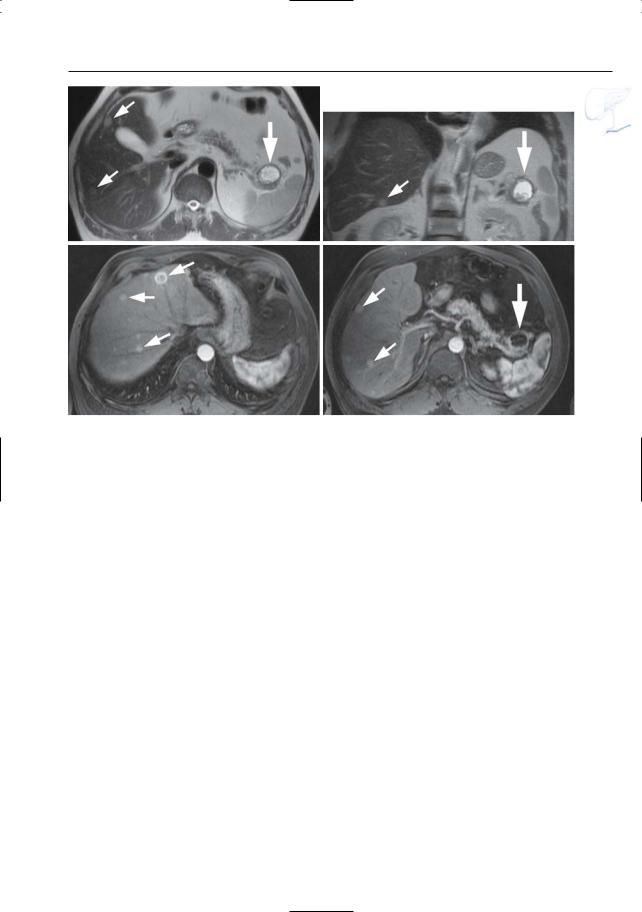
6 Pancreatic Ducts 407

a

b

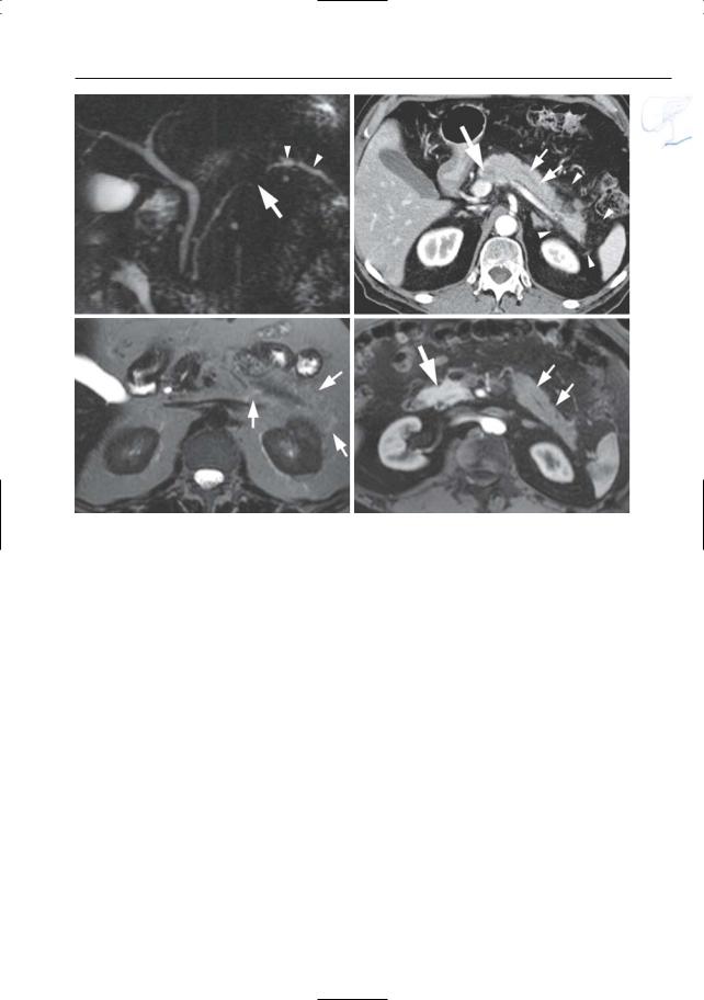
Fig. 199 a–c. a Moderately T2-weighted image showing “polycystic” mass in the uncinate process (arrows), representing a conglomerate of dilated side branches. b Projective MR image showing compression of the common bile duct. c ERCP image showing a dilated side branch containing a large filling defect (mucin plug; arrowheads). Note that the mucin plug is invisible on MRI (same intensity as fluid). Surgery was not performed in this patient because of medical contraindications.

c One year later, he presented with a large inoperable malignant mass in the pancreatic head

408 6.4 Malignant Tumors and Tumors with Malignant Potential

d

e

f

g

Fig. 199 d–g. IPMT branch duct type with associated dilatation of main duct. (d, e) Axial T2-weighted HASTE image (TE 60 and 360) showing a “polycystic” mass in the pancreatic head (arrow), represen-

ting a conglomerate of dilated side branches. f, g Projective images showing marked global dilatation of the main pancreatic duct and all of the side branches without a demonstrable cause of obstruction

6 Pancreatic Ducts 409

h

i

j

k

Fig. 199 h–l. Patient with a malignant IPMT of the main duct type.h Coronal T2-weighted HASTE image showing marked ductal dilatation (arrow). Axial (i, j) and coronal (k) reconstructed contrast-enhanced T1weighted VIBE images showing multiple contrast enhancing papillary protrusions in one of the larger “cystic” components due to malignant degeneration [arrowheads in (i)]. Note the presence of mucus in the main and left portal vein secondary to vascular invasion [arrows in (j, k)].l Projective image showing marked cystic pancreatic duct dilatation in the pancreatic

l head and secondary intraand extrahepatic biliary duct dilatation due to mass-effect