


272 5.4 Malignant Tumors
5.4 Malignant Tumors
#132 (Peri-)Ampullary Carcinoma
KEY FACTS: DISEASE
●Carcinomas of the papilla, ampulla, and adjacent structures are collectively referred to as periampullary carcinomas
●Usually adenocarcinomata; tend to be smaller than more proximal biliary neoplasms
●In many cases, it is impossible to determine the exact origin (e.g., duodenum, papilla proper, ampulla)
●Age peak: 60 years
●Symptoms: icterus, duodenal obstruction, weight loss
●Resectable in 80%–95%
●Relatively good prognosis (5-year survival rate, 35%–60%)
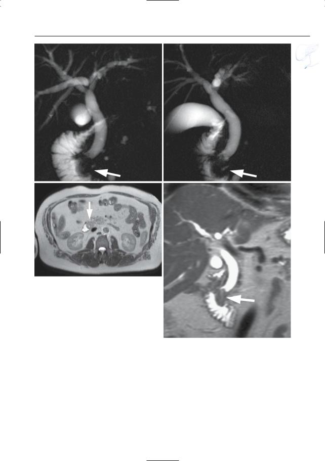
KEY FACTS: MRI (FIG. 132)
●Typical features on projective images:
–Dilation of the common bile duct and pancreatic duct
–Abrupt irregular narrowing or occlusion of the distalmost portions of both ducts (“double duct” sign)
–Signs of fixation (no contractile activity)
–Enlarged “bulging” papilla with or without soft tissue mass (Fig. 132)
●Cross-sectional images:
–Sometimes small mass lesion in the lateral part of the pancreatic head
–Signal intensity and contrast uptake pattern: similar to pancreatic carcinoma
●Differential diagnosis:
–Adenocarcinoma of pancreatic head invading the vaterian sphincter complex (see # 133)
–Distal cholangiocarcinoma (diagnosis more likely if the pancreatic duct diameter is normal)
–Other tumors (e.g., gastrinoma, carcinoid, adenoma, metastasis)
–Non-neoplastic causes of papillary obstruction (e.g., impacted stone with edema)
References
Geier A, Nguyen HN, Gartung C et al. (2000) MRCP and ERCP to detect small ampullary carcinoma. Lancet 356 : 1607–1608
Irie H, Honda H, Shinozaki K et al. (2002) MR imaging of ampullary carcinomas. J Comput Assist Tomogr 26 : 711–717
Kim JH, Kim MJ, Chung JJ et al. (2002) Differential diagnosis of periampullary carcinomas at MR imaging. Radiographics 22 : 1335–1352
Semelka RC, Kelekis N, John G et al. (1997) Ampullary carcinoma: demonstration by current MR techniques. J Magn Reson Imaging 7 : 153–156
5 Vaterian Sphincter Complex 273
a
c
Fig. 132 a–d. Projective images (a, b) showing dilatation of the common bile duct with an abrupt stenosis in the distal most part of the duct. Note the enlarged papilla bulging into the duodenal lumen (arrows). c, d Axial and coronal T2-weighted HASTE images confirm the presence of a mass lesion located in the area of the sphincter and bulging into the duodenal lumen (arrows). Final diagnosis: adenocarcinoma
b
d
274 5.4 Malignant Tumors
#133 Carcinoma of the Pancreatic
Head Invading the Vaterian
Sphincter Complex
Related topics: #90 (extrahepatic duct: involvement by pancreatic carcinoma),
# 185–195 (pancreatic adenocarcinoma)
KEY FACTS: DISEASE
●Differential diagnosis with primary ampullary carcinoma (see # 132): usually difficult or even impossible
●Pancreatic carcinomas with a more proximal location may present with the “four-segment sign” or involve the pancreatic duct only,thus making ampullary carcinoma unlikely (Kim et al. 2002)
● See # 185
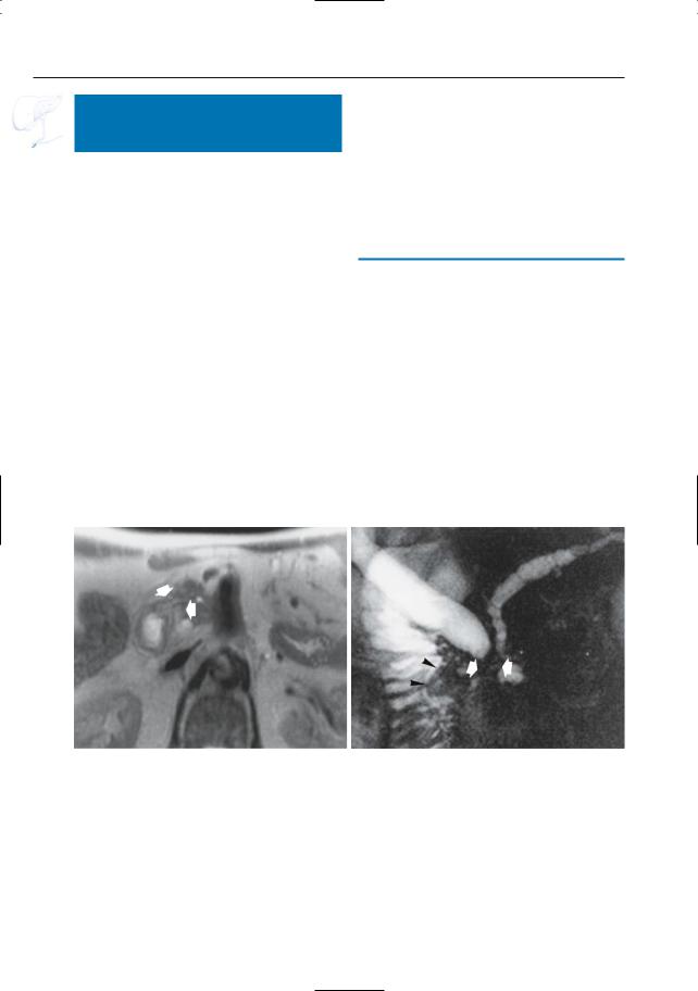
KEY FACTS: MRI
●See also #90
●Typical features:
–Stenosis of the distal common bile duct and pancreatic duct, commonly manifested as “invisible ducts”
–Unchanged aspect during dynamic evaluation
–Mass lesion in the pancreatic head
a
References
Kelekis NL, Semelka RC (1995) Carcinoma of the pancreatic head area. Diagnostic imaging: magnetic resonance imaging. Rays 20 : 289–303
Kim JH, Kim MJ, Chung JJ et al. (2002) Differential diagnosis of periampullary carcinomas at MR imaging. Radiographics 22 : 1335–1352
b
Fig. 133 a, b. Axial T2-weighted image (a) showing slightly hyperintense area in the lateral part of the pancreatic head, corresponding to pancreatic adenocarcinoma. b Projective image obtained in the
same patient showing a dilated common bile duct and pancreatic duct with abrupt narrowing in the pancreatic head (double duct sign; arrows). Also note prominent papilla (arrowheads)