


Pancreatic Ducts

276
1 Pancreatic Ducts
6.1 Normal Anatomy and Variants
#134 Normal Pancreas: Location
and Signal Intensity
KEY FACTS
●The pancreas is a retroperitoneal organ crossing the lumbar spine at level L2
●The head of the pancreas lies within the duodenal sweep, while the tail is usually directed towards the splenic hilum
●After splenectomy, the pancreatic tail migrates cranially and dorsally (Fig. 134)
●Length of duct: ranging from 9.5 to over 25 cm (average, 16 cm)
●Normal maximal anteroposterior diameters of the head and body: 3.5 and 2.2 cm, respectively
●Signal intensity of normal pancreatic parenchyma:
–T1: high intensity (equal to or higher than signal intensity of liver, due to aqueous protein in the acini; see also Fig. 10)
–T2: relatively hypointense (lower than signal intensity of spleen)
●T1-weighted images with fat suppression provide the best delineation of the pancreatic borders
References
Mitchell DG, Winston CB, Outwater EK, Ehrlich SM (1995) Delineation of the pancreas with MR imaging: multiobserver comparison of five pulse sequences. J Magn Reson Imaging 5 : 193–199
Semelka RC, Ascher SM (1993) MR imaging of the pancreas. Radiology 188 : 593–602
Winston CB, Mitchell DG, Outwater EK, Ehrlich SM (1995) Pancreatic signal intensity on T1weighted fat saturation MR images: clinical correlation. J Magn Reson Imaging 5 : 267–271
6 Pancreatic Ducts 277
a
c
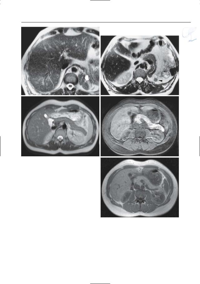
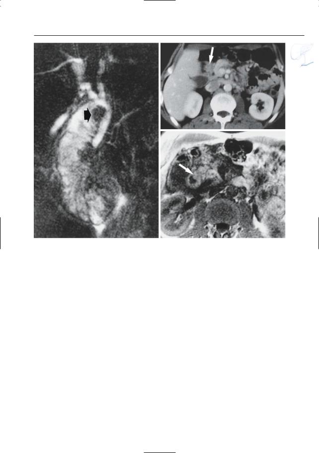
Fig. 134 a, b. Patient who underwent splenectomy and left nephrectomy. Axial T2-weighted images show aberrant position of the pancreatic tail (arrow in a). Also note the small accessory spleen in b (arrowhead). c–e. Signal characteristics of normal pancreatic parenchyma. c On T2-weighted images pancreatic tissue is hypointense when compared to the spleen. d, e On T1-weighted images with and without fat saturation pancreatic tissue is hyperand isointense to the liver, respectively
b
d
e

278 6.1 Normal Anatomy and Variants
#135 Classical Ductal Anatomy
KEY FACTS: ANATOMY
Features of the normal main pancreatic duct include:
●Size
–Reported maximum diameter: < 5 mm (increases with age)
–The caliber is greatest in the head and tapers smoothly to the tail
●Common types of course:
–“Sigmoid” configuration (ascending- horizontal-ascending; 34.5%)
–“Pistol” configuration (ascending- horizontal-horizontal; 27.6%)
Features of normal side branches include:
●Number: 15–30
●Taper gently as they course into the parenchyma
●In the body and tail, branch points appear at regular intervals
●All the body and tail side branches should be uniform (the presence of one or two mildly ectatic branches is still considered normal)
●The side branches in the pancreatic head that course inferiorly to serve the uncinate process are variable in number, length, and caliber
KEY FACTS: MRI
●The normal pancreatic duct is usually visible on MRCP, both on cross-sectional and projective images; the latter type of image is best suited to visualize the entire pancreatic duct
●Normal side branches are usually not ! seen on MRCP
●Occasionally,large normal side branches may be seen in the pancreatic head
References
Laubenberger J, Büchert M, Schneider B, Blum B, Hennig J, Langer M (1995) Breath-hold projection magnetic resonance cholangio-pancre- aticography (MRCP): a new method for the examination of the bile and pancreatic ducts. Magn Reson Med 33 : 18–23
Reinhold C, Bret P (1996) MR cholangiopancreatography. Abdom Imaging 21 : 105–116
6 Pancreatic Ducts 279
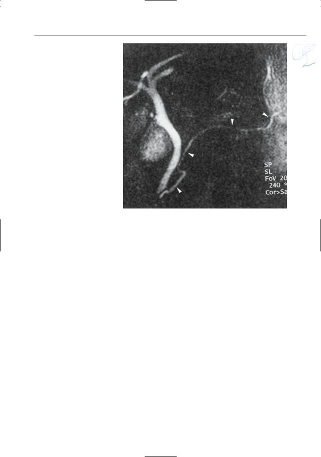
Fig. 135. Projective image shows sigmoid configuration of the pancreatic duct
(arrowheads)

280 6.1 Normal Anatomy and Variants
#136 Classical Ductal Anatomy
in Pancreatic Head
KEY FACTS: ANATOMY
●Normal development of the dorsal anlage (see Fig. 140a):
–Develops into the tail, body, and cranial portion of the head
–Drains into the minor papilla through the accessory duct of Santorini
●Normal development of the ventral anlage:
–Forms the caudal portion of the pancreatic head
–Drains into the major papilla through the distal portion of the duct of Wirsung
●Normal ductal fusion and variants (see also # 137):
–In 60% of patients, the duct of the ventral pancreas (Wirsung) fuses with the dorsal duct to become the primary continuation of the main pancreatic duct; the duct of Santorini remains patent but has a small diameter (usually 1 mm less than the diameter of the duct of Wirsung)
–In 30%, the duct of Santorini regresses further and loses its direct connection with the duodenum at the minor papilla
–In 3%–7%,there is pancreas divisum: no fusion (see # 140)
● Comment on anatomy: |
|
|
! |
||
– The ventral pancreas corresponds to |
||
the posterior part of the pancreatic |
|
|
head |
|
|
– The dorsal pancreas corresponds to |
|
|
the anterior part of pancreatic head |
|
|
and the body and tail |
|
KEY FACTS: MRI
●The duct of Wirsung is clearly visualized in the large majority of patients
●Visualization of the duct of Santorini is variable; in most patients, it is barely visible or not seen at all
References
Millbourn E (1960) Calibre and appearance of the pancreatic ducts and relevant clinical problems: a roentgenographic and anatomic study. Acta Chir Scand 118 : 286–303
Reinhold C, Bret P (1996) MR cholangiopancreatography. Abdom Imaging 21 : 105–116
Taylor AJ, Bohorfoush AG (1997) Interpretation of ERCP. Lippincott-Raven, Philadelphia, p 210
6 Pancreatic Ducts 281
a
Fig. 136. a Variation of accessory duct appearance: 1, bifid configuration (see # 137); 2, 3, incomplete duct of Santorini; 4, ansa pancreatica (see # 138). (From Taylor and Bohorfoush 1997, with permis-
b
sion). b Projective image showing normal aspect of the main pancreatic duct draining into the papilla major (arrows). The duct of Santorini is hardly visible (arrowheads)

282 6.1 Normal Anatomy and Variants
#137 Variant Anatomy (1):
“Bifid” Configuration
with a Prominent Duct of Santorini
Related topic: # 140 (pancreas divisum)
KEY FACTS: MRI
●In some patients, the duct of Santorini is clearly seen
●Types:
–“Bifid” ductal configuration (common): both ducts have approximately the same size and are connected (Fig. 137)
–“Dominant duct of Santorini” (± 3%): variant of the bifid configuration in which the duct of Santorini has the largest diameter
●Differential diagnosis with pancreas divisum (# 140):
–The ventral and dorsal systems are connected
–The ventral duct (Wirsung) is always clearly visible and does not arborize
References
Leyendecker JR, Elsayes KM, Gratz BI et al. (2002) MR cholangiopancreatography: spectrum of pancreatic duct abnormalities. AJR Am J Roentgenol 179 : 1465–1471
Sigfusson BF, Wehlin L, Lindström CG (1983) Variants of pancreatic duct system of importance in endoscopic retrograde cholangiopancreatography. Acta Radiol Diagn 24 : 113–128
6 Pancreatic Ducts 283
a
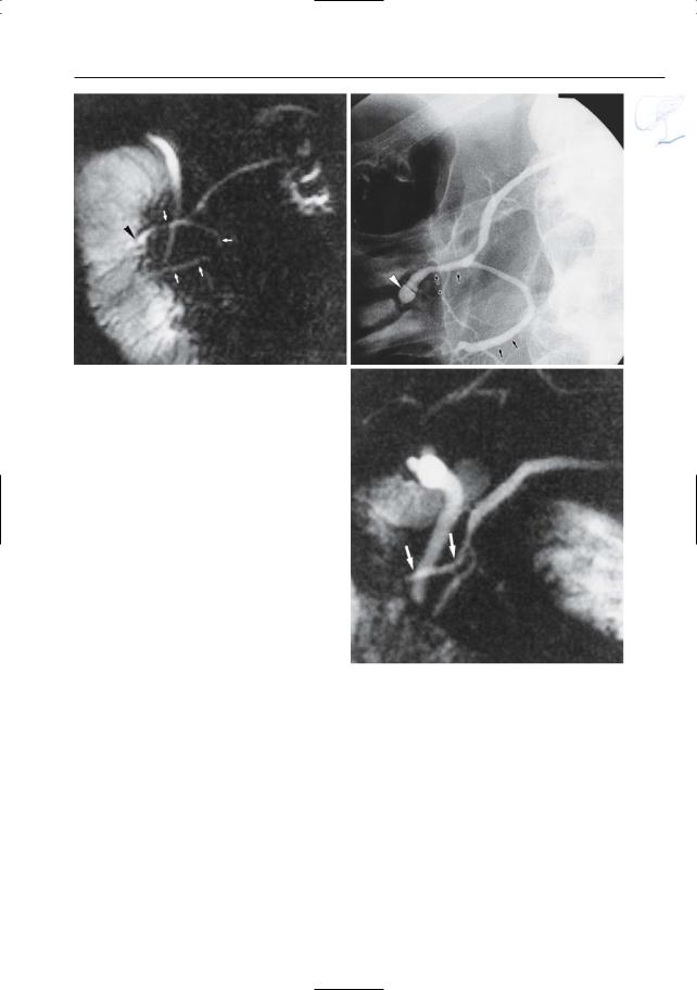
Fig. 137. a Projective MR image and b ERCP showing “bifid” ductal configuration with prominent duct of Santorini (arrows). Also note santorinicele (arrowheads; see # 150). c Another example of a prominent duct of Santorini (arrows)
b
c
284 6.1 Normal Anatomy and Variants
#138 Variant Anatomy (2):
Ansa Pancreatica
and Ductal Loops
KEY FACTS
●Loops may be seen in the head,neck,and body (Fig. 138)
●Ansa pancreatica may be associated with chronic pancreatitis (Tanaka et al. 1992)
●The accessory duct of Santorini usually has a fairly direct course from the pancreatic neck to the duodenum
●Occasionally, this duct has a reversed S-shaped appearance and is connected with an uncinate process side branch: ansa pancreatica (see Fig. 136a)
References
Dawson W, Langman J (1961) An anatomical-radio- logical study on the pancreatic duct pattern in man. Anat Rec 139 : 51–56
Tanaka T, Ichiba Y, Miura Y, Itoh H, Dohi K (1992) Variations of the pancreatic ducts as a cause of chronic alcoholic pancreatitis; ansa pancreatica. Am J Gastroenterol 87 : 806
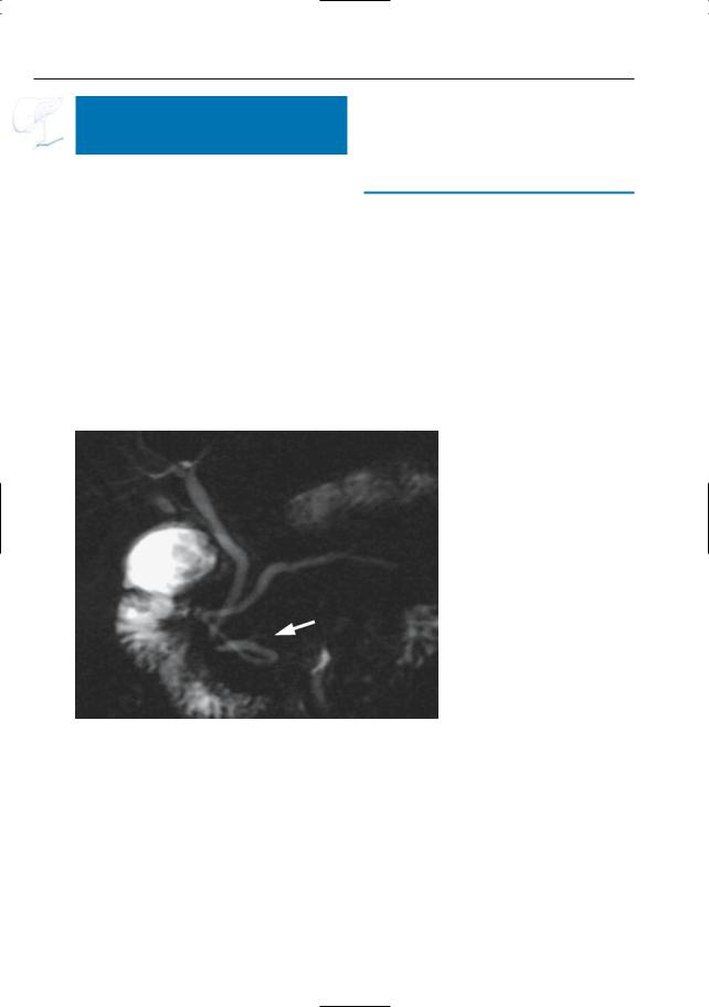
Fig. 138. Projective image obtained in a asymptomatic volunteer showing a prominent ductal loop (arrow)
6 Pancreatic Ducts 285
#139 Variant Anatomy (3):
Focal Narrowing
at the Junction
KEY FACTS
●Narrowing at the site of fusion of the dorsal and ventral ducts (“fusion narrowing”)
●3% in autopsy series
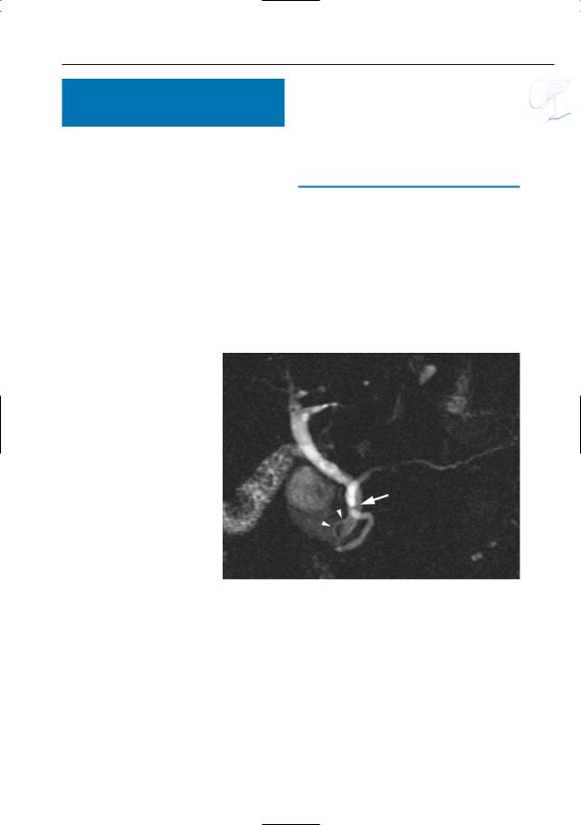
●Typical features (Fig. 139):
–Length: usually a few millimeters
–Characteristic location: takeoff of the accessory duct, the original drainage conduit for the dorsal pancreas
Fig. 139. Projective image showing typical fusion narrowing (arrow) located at the takeoff of the accessory duct (arrowheads)
●Differential diagnosis with pathologic stenosis:
–Typical location
–Subtle narrowing, short length
–Lack of proximal dilation, abnormal side branches,or other ductal changes
References
Belber JP, Bill K (1977) Fusion anomalies of the pancreatic ductal system: differentiation from pathologic states. Radiology 122 : 637–642
Sivak MV,Sullivan BH (1976) Endoscopic retrograde pancreatography: analysis of the normal pancreatogram. Am J Dig Dis 21 : 263–269

286 6.1 Normal Anatomy and Variants
#140 Variant Anatomy (4):
Pancreas Divisum
Related topics: # 137 (bifid configuration with a prominent duct of Santorini), # 141 (uneven lipomatosis), # 150 (santorinicele)
KEY FACTS: GENERAL
●The dorsal and ventral pancreatic anlage fail to fuse in the eighth week of fetal life (Fig. 140a)
●Incidence:
–3%–7% of the normal population
–12%–26% of patients with idiopathic recurrent pancreatitis
●Ductal anatomy:
–Duct of Wirsung (ventral duct) originates from the major papilla and terminates within 1–7 cm
–The duct of Wirsung typically has a small diameter and an arborizing pattern
–The dorsal duct originates from the papilla minor and drains the entire dorsal pancreas
●Types:
–Complete pancreas divisum (most common)
–Incomplete pancreas divisum (much less common): the ventral and dorsal systems remain connected through small-caliber branch ducts
●Clinical significance: increased incidence of acute and chronic pancreatitis (the orifice of the minor papilla is too small to adequately drain the volume of secretions produced by the pancreatic body and tail; Delhaye et al. 1988)
KEY FACTS: MRI (FIG. 140)
●Pancreas divisum can accurately be demonstrated by MRI
●Key cholangiographic findings:
–Visualization of the dorsal duct draining into the duodenum via the papilla minor, separated from the distal common bile duct (see also Figs. 63, 67)
–No connection between the dorsal and ventral ductal system
–The ventral duct may be small (29%) or invisible (71%)
●Signs on axial cross-sectional images:
–Visualization of the duct of Santorini ventrally in the pancreatic head clearly separated from the common bile duct. The duct of Santorini is usually clearly seen because of its more or less horizontal course
–Enlargement of the pancreatic head (not always present)
–Flat cleft separating the dorsal and ventral pancreatic moieties (not always present; see also # 141)
●Note: MRCP is probably less accurate than ERCP in the differentiation of complete and incomplete pancreas divisum
!
!
6 Pancreatic Ducts 287
References
Bret P, Reinhold C, Taourel P, Guibaud L, Atri M, Barkun AN (1996) Pancreas divisum: evaluation with MRCP. Radiology 199 : 99–103
Delhaye M, Engelholm L, Cremer M (1988) Pancreas divisum: controversial clinical significance. Dig Dis 6 : 30–39
Gregg JA (1977) Pancreas divisum: its association with pancreatitis. Am J Surg 134 : 539–543
Mortelé KJ, Segatto E, Ros PR (2004) The infected liver: radiologic-pathologic correlation. RadioGraphics 24 : 937–955
Taylor AJ, Bohorfoush AG (1997) Interpretation of ERCP. Lippincott-Raven, Philadelphia, p 210
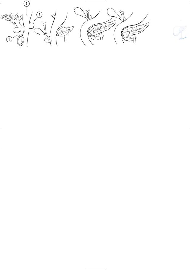
Fig. 140. a Embryologic development of the pancreas (from left to right). Both the ventral pancreas (1) and dorsal pancreas (2) are buds off the foregut (3). The ventral pancreas develops as a separate anlage and rotates posteriorly as maturation occurs. In pancreas divisum, the ventral and dorsal pancreas are adjacent to each other; however, the two ductal systems remain separated. (From Stewart et al. 1997, with permission).
288 6.1 Normal Anatomy and Variants
b |
c |
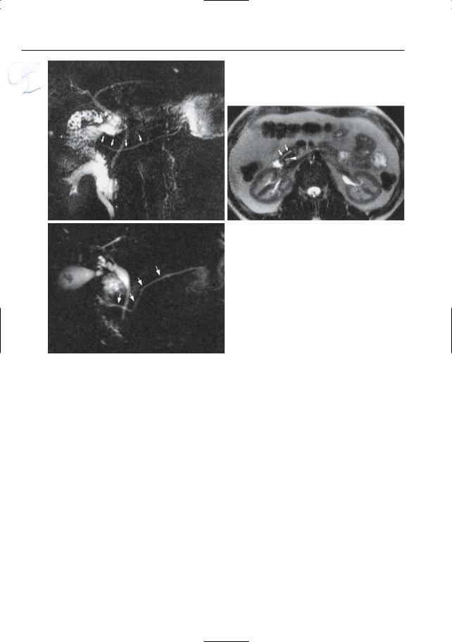
Fig. 140 b–c. b Projective MR image showing classical ductal anatomy in pancreas divisum. The duct of Wirsung is not visible. c Heavily T2-weighted axial image showing duct of Santorini located ventrally in the pancreatic head (arrows) and clearly separated from the common bile duct (arrowhead). d Projective image showing the classical ductal anatomy in pancreas divisum, with a clearly visible dor-
d sal ductal system (duct of Santorini) and an invisible ventral ductal system (duct of Wirsung).
6 Pancreatic Ducts 289
e
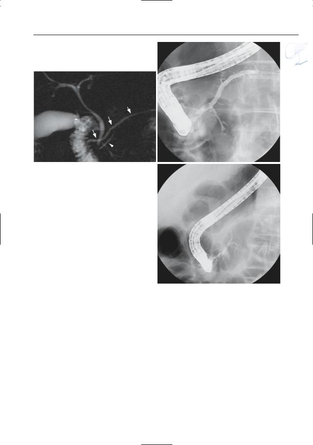
Fig. 140. (continued) e–g Projective image (e) showing a separate dorsal (arrows) and ventral (arrowhead) pancreatic ductal system. Note that the ventral ductal system is small but still visible. ERCP images with contrast injection in the papilla minor (f) and major (g) showing the separate dorsal and ventral ductal system.
f
g
|
|
|
|
|
|
KEY FACTS: MRI (FIG. 141) |
|
|||
|
|
|
#141 Variant Anatomy (5): |
|
|
|||||
|
|
|
|
|
|
|
|
|
||
|
|
|
|
Uneven Lipomatosis |
|
● |
Signal intensity (reflecting the degree of |
|
||
|
|
|
|
|
|
|
||||
|
|
KEY FACTS: GENERAL |
|
lipomatosis): |
|
|
||||
|
|
|
|
|
||||||
|
|
|
– |
T1-weighted images: anterior part of |
|
|||||
|
|
|
|
|
|
|
|
|||
|
|
● |
Marked difference in fat content be- |
|
|
the pancreatic |
head more hyperin- |
|
||
|
|
|
|
tense than the posterior part |
|
|||||
|
|
|
tween the dorsal and ventral pancreas |
|
|
|
||||
|
|
|
|
– |
T1-weighted images with fat suppres- |
|
||||
|
|
|
(the dorsal moiety contains more fat) |
|
|
|||||
|
|
|
|
|
sion: anterior part less hyperintense |
|
||||
|
|
● |
Incidence: ± 3% |
|
|
|
||||
|
|
|
– |
T2-weighted images: aspect depends |
|
|||||
|
|
● |
May be associated with pancreas divi- |
|
|
|||||
|
|
|
|
on the technique used |
|
|||||
|
|
|
sum |
|
|
|
||||
|
|
|
● |
Key finding: straight demarcation be- |
|
|||||
|
|
● |
Etiology: |
! |
||||||
|
|
|
tween the ventral and dorsal parts of the |
|||||||
|
|
|
– |
Related to separate embryologic de- |
|
|||||
|
|
|
|
pancreatic head. This line of transition |
||||||
|
|
|
|
velopment of both moieties |
|
|
||||
|
|
|
|
|
corresponds to |
the level of fusion |
|
|||
|
|
|
– |
Precise mechanism not known |
|
|
||||
! |
|
|
|
between the two embryonal portions of |
|
|||||
|
● |
Importance: may mimic focal pancreatic |
|
|
||||||
290 |
|
6.1 Normal Anatomy and Variants |
|
|
|
|
|
|||
|
mass on ultrasonography, CT, and MRI |
the organ |
|
|
|
||
|
(Marchal et al. 1989; Matsumoto et al. |
|
|
1995) |
|
||
References |
|||
|
|
||
|
|
Marchal G, Verbeken E, Van Steenbergen W, Baert |
|
|
|
AL, Lauweryns J (1989) Uneven lipomatosis: a |
|
|
|
pitfall in pancreatic sonography. Gastrointest |
|
|
|
Radiol 14 : 233–237 |
|
|
|
Matsumoto S, Mori H, Miyake H et al. (1995) Uneven |
|
|
|
fatty replacement of the pancreas: evaluation |
|
|
|
with CT. Radiology 194 : 453–458 |
|
a |
b |
Fig. 141 a–f. a T1-weighted image with fat suppres- |
b T2-weighed image showing the opposite rela– |
sion showing ventral part of the pancreatic head as |
tionship. |
hypointense compared to dorsal part (arrowheads). |
|
6 Pancreatic Ducts 291
d
c
e |
f |
Fig. 141. (continued) c Non-fat-suppressed T1weighted image showing no difference in signal intensity. d CT image showing ventral part as hypodense. Note the straight demarcation line (arrows). Same patient. All these “abnormalities” are consistent with increased fat content in the anterior part of the pancreatic head. e Same patient. Projective
image showing a clearly visible dorsal ductal system (duct of Santorini) (arrows), with Santorinicele, and an invisible ventral ductal system (duct of Wirsung). f ERCP image with contrast injection in the papilla major show opacification of the small ventral ductal system (arrow). In this patient, the uneven lipomatosis was associated with pancreas divisum
292 6.1 Normal Anatomy and Variants
#142 Variant Anatomy (6):
Lobulations
of the Pancreatic Head
KEY FACTS: GENERAL
●Variants in the lateral contour of the pancreatic head (and neck)
●Incidence: up to 35%
●Classification (taking into account the orientation of the lobule):
–Anterior (29%)
–Posterior (56%)
–Horizontal (15%) (Ross et al. 1996)
●Mechanism: probably a spectrum of fusion patterns of the dorsal and ventral embryonal parts of the pancreas
!● Importance: marked focal convexity can be mistaken for tumor
a
KEY FACTS: MRI
●Cross-sectional images show marked focal convexity of the lateral contour of the pancreatic head (Fig. 142a)
●Projective images may show a duct branch in an unusual lateral position (Fig. 142b)
●Key finding: isointensity with the rest of the pancreas on all pulse sequences
References
Ross B, Brooke Jeffrey R, Mindelzun R (1996) Normal variations in the lateral contour of the head and neck of the pancreas mimicking neoplasm: evaluation with dual-phase helical CT. AJR Am J Roentgenol 166 : 799–801
b
Fig. 142. a T1-weighted image showing lobulated lateral contour of the pancreatic head (arrow). b Different patient. Projective image showing duct of
Santorini projecting to the right of the common bile duct (arrowheads), which is an indirect sign of a far lateral position of (part of) the pancreatic head
6 Pancreatic Ducts 293
#143 Developmental
Abnormalities (1):
Agenesis of the
Dorsal Pancreas
KEY FACTS: DISEASE
●Types of agenesis:
–Agenesis of the ventral pancreas (extremely rare)
–Complete agenesis of the dorsal pancreas (rare)
–Incomplete development or hypoplasia of the dorsal pancreas: short pancreas and duct system (rare)
–Complete pancreatic agenesis: (extremely rare; early neonatal death)
●Complications:
–Endocrine insufficiency (diabetes)
–Exocrine insufficiency (steatorrhea)
KEY FACTS: MRI (FIG. 143)
●Cross-sectional images show absence or hypoplasia of the dorsal pancreas
●Projective images show a short pancreatic duct
●Note: On ERCP, agenesis may be misinterpreted as a pathologic obstruction
References
Macari M, Giovanniello G, Blair L, Krinsky G (1998) Diagnosis of agenesis of the dorsal pancreas with MR pancreatography. AJR Am J Roentgenol 170 : 144–146
Wang JT, Lin JT, Chuang CN et al. (1990) Complete agenesis of the dorsal pancreas: a case report and review of the literature. Pancreas 5 : 493–497
a |
|
b |
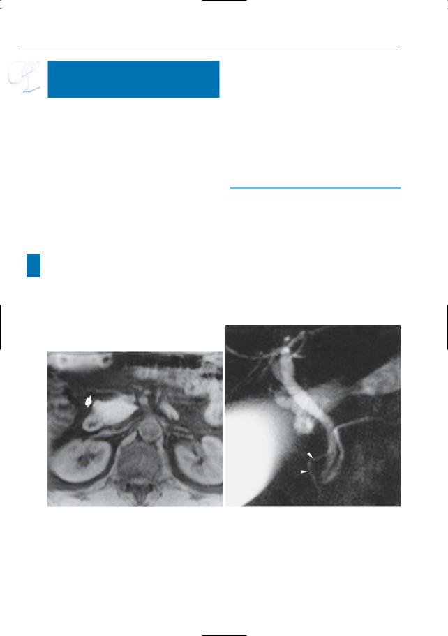
Fig. 143. a Projective |
image showing short pan- |
obtained in coronal plane showing normal pancrea- |
creatic duct with abrupt termination (arrow). Note |
tic head (arrows) and absence of pancreatic |
|
the incidental finding of a small duodenal diverticu- |
body/tail (asterisk). (Figure courtesy of L. Storms |
|
lum (arrowheads in |
a). b Cross-sectional image |
and J. Agneessens) |

294 6.1 Normal Anatomy and Variants
#144 Developmental
Abnormalities (2):
Annular Pancreas
KEY FACTS: DISEASE
●Ring of normal pancreatic tissue encircles the duodenum
●Incidence: 0.05%
●Age at presentation (bimodal peak):
–Childhood (usually neonatal period): 50%
–Adulthood: 50%
●Pathogenesis: abnormal migration of the ventral pancreas
●Location:
–Second portion of the duodenum: 85%
–First portion: 15%
●Associated with other congenital anomalies in 75%
–Duodenal atresia, stenosis or diaphragm
–Down syndrome
–Situs inversus (Fig. 144d, e)
–Pancreas divisum (36%) (the annular duct drains into the ventral duct system)
●Symptoms:
–Persistent vomiting (neonate)
–Frequently no symptoms
●Complications (40%–50%)
–Ulcer disease
–Pancreatitis
KEY FACTS: MRI (FIG. 144)
●Key features:
–The pancreatic duct surrounds the duodenum
–Apparent wall thickening of the duodenum
References
Choi JY, Kim MJ, Kim JH et al. (2004) Annular pancreas: emphasis on magnetic resonance cholangiopancreatography findings. J Comput Assist Tomogr 28 : 528–532
Glazer GM, Margulis AR (1979) Annular pancreas: etiology and diagnosis using ERCP. Radiology 133 : 303–306
6 Pancreatic Ducts 295
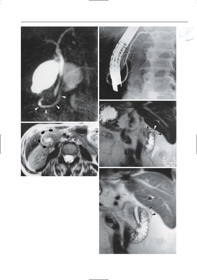
a
c
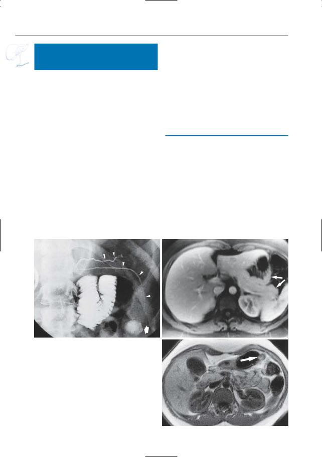
Fig. 144 a–c. a Projective MR image and b ERCP showing loop configuration of the distal pancreatic duct (arrowheads). In b, the duct encircles the endoscope. c Axial T2-weighted image showing distal pancreatic duct located in the wall of the duodenum and encircling the duodenal lumen (arrows). d, e Sixty-year-old woman with suspected esophageal tumor. Coronal T2-weighted images show (complete) situs inversus. The distal pancreatic duct encircles the duodenum (arrowheads): annular pancreas. Other images showed associated pancreas divisum. The patient had no complaints related to these abnormalities
b
d
e

296 6.1 Normal Anatomy and Variants
#145 Developmental
Abnormalities (3):
Ectopic Pancreas
KEY FACTS: DISEASE
●Pancreatic tissue that is present outside the normal confines of the pancreas
●Incidence: 1%–2% of autopsies
●Location:
–Wall of stomach (26%–38%)
–Duodenum (28%–36%)
–Jejunum (16%)
–Many other sites
●Size: usually less than 2 cm
●Usually asymptomatic
●Complications:
–Acute or chronic pancreatitis
–Ulceration with bleeding
–Neoplastic transformation
–Cystic dystrophy (secondary to obstruction)
KEY FACTS: MRI
●Abnormal mass in the gastric or duodenal wall, with characteristically high signal intensity on T1-weighted images and relatively low signal intensity on T2weighted images (Fig. 145)
●Cystic dystrophy is characterized by the presence of cystic formations surrounded by inflammation (Fékété et al. 1996)
●Note: In view of its small size, it is likely that ectopic pancreatic tissue is commonly overlooked on MRI
References
Fékété F, Noun R, Sauvanet A, Réjou JF, Bernades P, Belghiti J (1996) Pseudotumor developing in heterotopic pancreas. World J Surg 20 : 295–298
Lai EC, Tompkins RK. Heterotopic pancreas: review of a 26-year experience. Am J Surg 151 : 697–700
6 Pancreatic Ducts 297
b
a |
c |
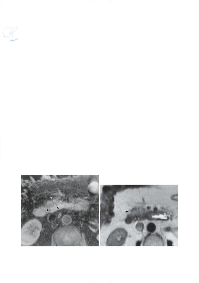
Fig. 145 a–c. a Projective MR image showing filling defect at medial border of the duodenum (arrow). Note increased signal intensity of background tissue, related to pancreatitis. b CT image showing a
soft tissue mass at this location (arrow), initially interpreted as a duodenal tumor. c T1-weighted MR image showing this“mass”to have high signal intensity (arrow), similar to normal pancreatic tissue
298 6.1 Normal Anatomy and Variants
#146 Developmental
Abnormalities (4):
Partial Duplication
KEY FACTS: DISEASE
●Incidence depends on type:
–Duplication of the main duct in the body and tail: 10%
–Duplication of both duct and parenchyma: very rare
●Association: duplication of stomach or intestine communicating with the main pancreatic duct through an anomalous duct and pancreatic lobe (Hoffman et al. 1987)
KEY FACTS: MRI
●Ductal duplication: projective images show two parallel ducts in the pancreatic body/tail (see Fig. 168)
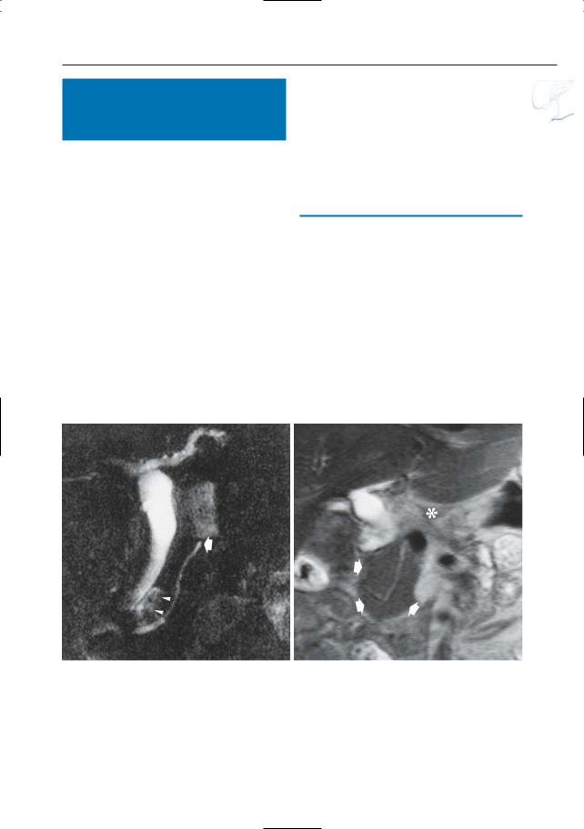
a
Fig. 146 a–c. a ERCP showing “normal” upper pancreatic duct (arrowheads) and second lower pancreatic duct coursing in a caudal direction (arrowheads) and connected with a cystic lesion (either pseudocyst or gastric duplication cyst; arrow). b Axial T1-weighted contrast-enhanced MR image showing bifurcation of the pancreatic tail (arrows). The ventral part of the pancreatic tail is situated in close proximity to the wall of the stomach. c Non-contrast-enhanced T1-weighted image obtained at a more caudal level showing the caudal end of the aberrant pancreatic tail, still in close proximity to the wall of the stomach (arrow)
●Parenchymal duplication:
–Projective images: visualization of a second duct with variable course
–Cross-sectional images: visualization of an accessory pancreatic lobe, most commonly in close connection with the stomach
–The aberrant duct may terminate in a cystic structure representing a gastric duplication cyst (Hoffman et al. 1987)
References
Agha FP (1987) Duplex ventral pancreas. Gastrointest Radiol 12 : 23–25
Hoffman M, Sugerman HJ, Heuman D, Turner MA, Kisloff B (1987) Gastric duplication cyst communicating with aberrant pancreatic duct:a rare cause of recurrent acute pancreatitis. Surgery 101 : 369–372 Kikuchi K, Nomiyama T, Miwa M, Harasawa S, Miwa T (1983) Bifid tail of the pancreas: a case presenting as a gastric submucosal tumor. Am J Gastro-
enterol 78 : 23–27
b
c
6 Pancreatic Ducts 299
#147 The Elderly Pancreas
KEY FACTS: GENERAL
●Common changes in elderly patients: fatty infiltration, periductal and lobular fibrosis, hyperplasia of the ductular epithelium
KEY FACTS: MRI
●The following changes may be seen:
–Atrophy of pancreatic parenchyma
–Decreased signal intensity on T1weighted images (fibrosis)
–Changes in the main pancreatic duct: discrete caliber irregularities, diffuse ectasia,strikingly straight appearance (Fig. 147)
–Dilation of side branches
●Significance: differentiation with chronic pancreatitis may be difficult
References
Jones SN, McNeil NI, Lees WR (1989) The interpretation of retrograde pancreatography in the elderly. Clin Radiol 40 : 393–396
Schmitz-Moormann P, Himmelmann GW, Brandes JW et al. (1985) Comparative radiological and morphological study of the human pancreas. Pancreatitis-like changes in postmortem ductograms. Possible implications for ERCP. Gut 26 : 406–414
Winston CB, Mitchell DG, Outwater EK, Ehrlich SM (1995) Pancreatic signal intensity on T1weighted fat saturation MR images: clinical correlation. J Magn Reson Imaging 5 : 267–271
Fig. 147. Seventy-six-year old patient. Projective image showing markedly straight appearance of the pancreatic duct (arrows). This patient had no clinical signs that suggested the presence of pancreatic disease

300 6.1 Normal Anatomy and Variants
|
|
|
|
|
|
KEY FACTS: MRI |
|
||
|
|
#148 Postoperative Anatomy (1): |
|
|
|||||
|
|
|
|
|
|
|
|||
|
|
|
After Partial Resection |
|
|
The exact appearance depends on the |
|
||
|
|
|
|
|
|
● |
! |
||
|
|
|
|
|
|
||||
|
|
Related topics: #66 (extrahepatic bile duct: |
|
type of surgery performed; close corre- |
|||||
|
|
|
|||||||
|
|
|
lation with the surgical report is impera- |
||||||
|
|
anatomy after the |
Whipple procedure), |
|
|
||||
|
|
|
tive when defining the normal postoper- |
|
|||||
|
|
# 184 (complications of partial pancreatic |
|
|
|||||
|
|
|
ative anatomy |
|
|||||
|
|
resection) |
|
|
|
|
|||
|
|
|
|
● |
Technical requirements for a successful |
|
|||
|
|
|
|
|
|
|
|||
|
|
KEY FACTS: TECHNIQUE |
|
|
|
MRCP study: adequate distention of the |
|
||
|
|
|
|
|
anastomosed jejunal loop (recognition |
|
|||
|
|
|
|
|
|
|
|
||
|
|
● Indications: usually tumor or conditions |
|
of valvulae conniventes) |
|
||||
|
|
● |
Differential diagnosis: collapsed bowel |
|
|||||
|
|
requiring ductal |
decompression (e.g., |
|
|||||
|
|
|
loops may mimic abscesses and fluid |
|
|||||
|
|
chronic pancreatitis with stricture for- |
|
|
|||||
|
|
|
collections |
|
|||||
|
|
mation and proximal ductal dilation) |
|
|
|||||
|
|
|
|
|
|
||||
|
|
● Surgical techniques: |
|
|
|
|
|||
|
|
– |
Whipple procedure (see #66) |
|
|
|
|
||
|
|
References |
|
||||||
|
|
– |
“Simple” resection of the pancreatic |
|
|||||
|
|
|
|
|
|
||||
|
|
|
tail |
|
|
Freed KS, Paulson EK, Frederick MG, Keogan MT, |
|
||
|
|
– Duval procedure: resection of the |
|
Pappas TN (1997) Abdomen after a Puestow pro- |
|
||||
|
|
|
pancreatic tail with end-to-end pan- |
|
cedure: postoperative CT appearance, complica- |
|
|||
|
|
|
creatojejunostomy |
|
tions, and potential pitfalls. Radiology 203 : 790– |
|
|||
|
|
|
|
794 |
|
|
|||
|
|
– |
Puestow procedure: side-to-side pan- |
|
|
|
|||
|
|
Rossi RL, Heiss FW, Braasch JW (1985) Surgical |
|
||||||
|
|
|
creaticojejunostomy |
|
management of chronic pancreatitis. Surg Clin |
|
|||
North Am 65 : 79–101
Taylor AJ, Bohorfoush AG (1997) Pancreatic duct – miscellaneous. In: Taylor AJ, Bohorfoush AG (eds) Interpretation of ERCP. Lippincott-Raven, Philadelphia, pp 291–303
6 Pancreatic Ducts 301
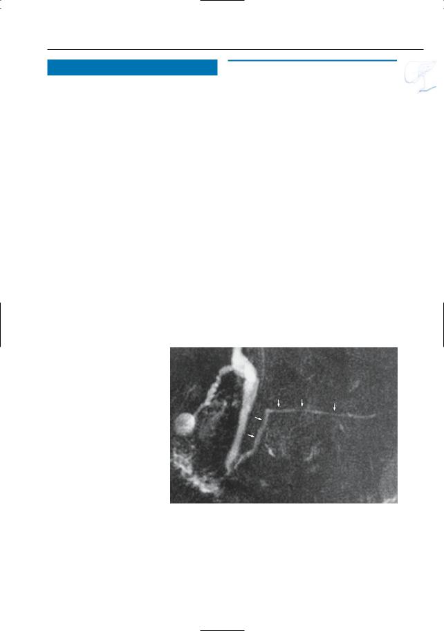
Fig. 148. Projective image obtained in a patient who underwent a partial pancreatectomy showing absence of the panc– reatic duct in the body of the pancreas (arrowheads). Note
the presence of the pancreatic duct in the pancreatic head and tail (arrows). The pancreatoje– junal anastomosis is also visu– alized (small arrows), although the jejunal loop is suboptimally filled with fluid. Note the slightly dilated aspect of the pancreatic duct in the tail of the pancreas

302 6.1 Normal Anatomy and Variants
#149 Postoperative Anatomy (2):
After Pancreatic
Transplantation
Related topics: # 182, 183 (complications after pancreatic transplantation)
KEY FACTS: TECHNIQUE
●Indication: restoration of endogenous secretion of insulin in patients with severe diabetes
●Commonly performed together with renal transplantation (diabetic nephropathy)
●Procedure:
–The graft is placed within the peritoneal cavity in the pelvis
–Exocrine pancreatic secretions are managed by bladder drainage via an interposed duodenal segment
–The celiac trunk and superior mesenteric artery of the donor are usually implanted with a common aortic patch on the common iliac artery
KEY FACTS: MRI
●The graft is usually seen in the right iliac fossa extending cephalad, parallel to the ascending colon
●The diameter of the graft depends on surgical factors (grafts that are stretched within the flank appear thinner) and may be temporarily increased in the immediate postoperative period (edema)
●Signal intensity is similar to pancreatic tissue in situ:
–T1: approximately as intense as the renal cortex
–T2: intermediate
●The pancreatic duct should show regular contours and gradual tapering towards the tail
References
Gore RM, Levine MS, Laufer I (eds) (1994) Textbook of gastrointestinal radiology. Saunders, Philadelphia, p 2204
Kelcz F, Sollinger HW, Pirsch JD (1991) MRI of the pancreas transplant: lack of correlation between imaging and clinical status. Magn Reson Med 21 : 30–38
Pozniak MA, Propeck PA, Kelcz F, Sollinger H (1995) Imaging of pancreas transplants. Rad Clin North Am 33 : 581–594
6 Pancreatic Ducts 303
a |
b |
|
c |
d |
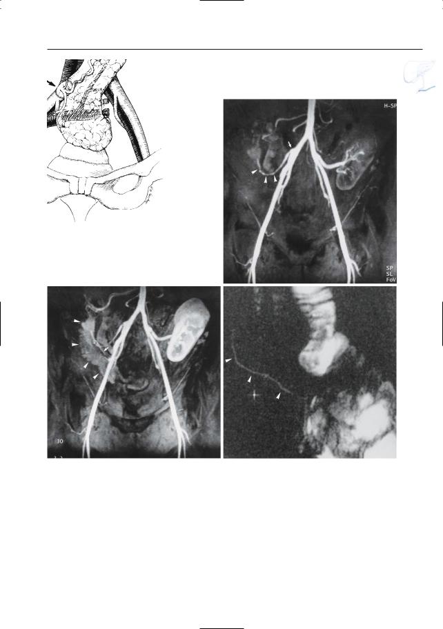
Fig. 149. a Pancreatic transplant anatomy.A duodenocystostomy is used for exocrine drainage.An aortic patch and the portal vein are used for anastomoses with the iliac vessels (arrows). (Reprinted with permission from Gore et al. 1994). b–d Normal vascular and ductal anatomy. In this patient, the transplant pancreas is situated in the right iliac fossa. b Contrast-enhanced MR angiogram obtained in the “early” arterial phase showing aorta, iliac arte-
ries, aortic patch (arrow), and splenic artery (arrowheads). Note initial cortical enhancement of the renal transplant in the left fossa. The pancreas is invisible. c Contrast-enhanced MR angiogram obtained in the “late” arterial phase showing homogeneous enhancement of the pancreatic parenchyma (arrowheads) and discrete opacification of the splenic vein (arrow). d Projective image showing pancreatic duct of normal size (arrowheads)