


258 5.2 Benign Nontraumatic Abnormalities
5.2Benign Nontraumatic Abnormalities
#125 Impacted Stone
Related topics: # 73 (stones in common bile duct: pitfalls in diagnosis), # 122 (pseudocalculus sign)
KEY FACTS: DISEASE
●Stone located in the intramural portion of the common bile duct, leading to either transient or persistent blockage and edema of the ampulla of Vater
●Clinical signs/complications:
–Acute painful jaundice
–Acute pancreatitis
–Fistula formation
●Note: In the majority of cases, gallstones pass into the duodenum after a few days (Kelly 1980)
KEY FACTS: MRI
●Stones that are not surrounded by fluid are invisible on MRCP
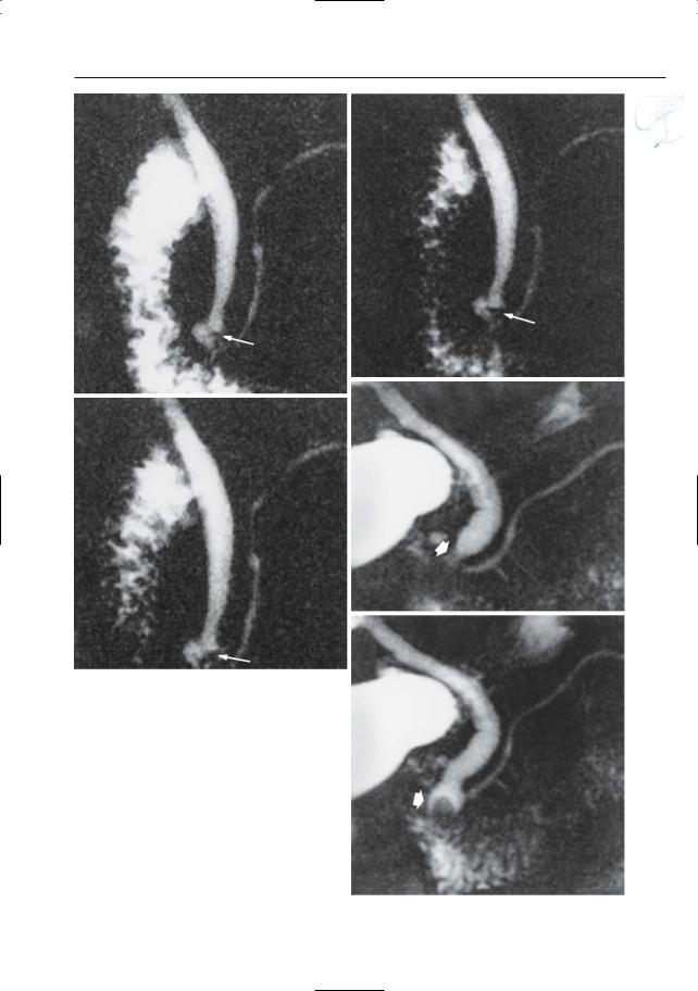
●Dynamic (repetitive) scanning is mandatory to diagnose small stones impacted in the distalmost portion of the common bile duct and to differentiate true lithiasis from the “pseudocalculus” effect (true lithiasis has an unchanged shape throughout the study) (Fig. 125a–c; see also # 122)
●Dynamic scanning also helps to avoid false-negative diagnoses: stones are best seen on images obtained during sphincter relaxation (Fig. 125d, e)
References
Chan Y-L, Chan ACW, Lam WWM et al. (1996) Choledocholithiasis: comparison of MR cholangiography and endoscopic retrograde cholangiography. Radiology 200 : 85–89
Guibaud L, Bret PM, Reinhold C, Atri M, Barkun AN (1995) Bile duct obstruction and choledocholithiasis: diagnosis with MR cholangiography. Radiology 197 : 109–115
Kelly TR (1980) Gallstone pancreatitis: the timing of surgery. Surgery 88 : 345–350
Van Hoe L, Mermuys K, Vanhoenacker P (2004) MRCP pitfalls. Abdom Imaging 29 : 360–387
!
!
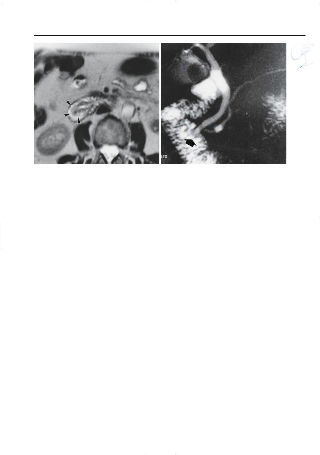
5 Vaterian Sphincter Complex 259
a
c
Fig. 125. a–c Three projective images out of a series of 20 showing a small hypointense “lesion” in the distal common bile duct (arrows). The unchanged shape of the abnormality during dynamic scanning virtually rules out a pseudocalculus phenomenon. In this case, the lesion represented a small stone. d, e Two consecutively obtained projective images in a different patient.d Image obtained during contraction of the sphincter showing no abnormalities (arrow). Note absence of fluid in the adjacent duodenum. e Relatively large stone impacted in the distalmost portion of the extrahepatic bile duct (arrow). This stone is invisible in a because it is not surrounded by fluid.Note that
the pseudocalculus sign appears during contraction, and not during relaxation (see # 122)
b
d
e

260 5.2 Benign Nontraumatic Abnormalities
#126 Choledochocele
Related topics: #30 (Caroli’s disease), #68 (choledochal cysts)
KEY FACTS: DISEASE
●Cystic dilation of the distal/intramural portion of the common bile duct with herniation of the common bile duct into the duodenum (similar to ureterocele)
●Represents type III bile duct cyst (see #30)
●Often manifests in adulthood
●Etiology:
–Congenital: diverticulum of the distal common bile duct; stenosis of the ductal orifice
–Acquired: stone passage Æ inflammation Æ stenosis of the orifice
●Symptoms: nausea, vomiting, episodic abdominal pain
●Stones/sludge often present
KEY FACTS: MRI (FIG. 126)
●Typical appearance:
–Rather than being narrowed, the intramural portion of the common bile duct is broadened and clubbed
–The papilla bulges into the duodenum
References
De Backer AI, Van den Abbeele K, De Schepper AM et al. (2000) Choledochocele: diagnosis by magnetic resonance imaging. Abdom Imaging 25 : 508–510
Venu RP, Geenen JE, Hogan WJ (1984) Role of ERCP in the diagnosis and treatment of choledochocele. Gastroenterology 87 : 1144–1149
5 Vaterian Sphincter Complex 261
Fig. 126. Axial T2-weighted image showing broadened aspect of the distal (intramural) part of the common bile duct
(arrow)

262 5.1 Normal Anatomy and Variants
#127 Sphincter Dysfunction,
General
KEY FACTS: DISEASE
●Clinical entity resulting from the impedance of biliary and/or pancreatic flow
●Most common in middle-aged women
●Underlying disorder:
–Structural narrowing (fibrosis): papillary stenosis
–Motor abnormality: dyskinesia
●Predisposing factors: prior cholecystectomy; stone passage
●Symptoms: biliary colic
●Objective diagnostic criteria (Hogan et al. 1987):
–Elevation of transaminases and alkaline phosphatase (twice the normal value), documented on two different occasions
–Dilation of extrahepatic bile duct (>12 mm)
–Delayed emptying on ERCP (> 45 min)
●Classification:
–Type I: all three criteria fulfilled
–Type II: one or two criteria fulfilled
–Type III: no criteria fulfilled
●Gold standard for diagnosis: manometry. If dyskinesia is present, abnormalities include (Toonly et al. 1997):
–Most common: elevation of basal pressure (> 40 mmHg)
–Increased frequency of phasic wave contractions (more than eight per min)
● Other diagnostic techniques that have been proposed:
– 99mTc-hepato-iminodiacetic acid (HIDA) scan (delayed transit from the common bile duct to the bowel)
–Secretin test with measurement of pancreatic duct diameter (see also # 19)
–Fatty meal test with measurement of bile duct diameter (see also # 19)
●Treatment:
–Type I: sphincterotomy
–Type II: sphincterotomy if manometry is positive
–Type III: medication
●Complications:
–Chronic obstructive pancreatitis (see
# 168)
–Secondary sclerosing cholangitis (see #45; Tarnasky et al. 1997)
References
Hogan WJ, Geenen JE, Dodds WJ (1987) Dysmotility disturbances of the biliary tract: classification, diagnosis, and treatment. Semin Liver Dis 7 : 302–311
Tarnasky PR, Hoffman B, Aabakken L et al. (1997) Sphincter of Oddi dysfunction is associated with chronic pancreatitis. Am J Gastroenterol 92 : 1125–1129
Toonly J, Roberts-Thomson IC, Dent J, Lee J (1985) Manometric studies in patients with suspected sphincter of Oddi dysfunction. Gastroenterology 88 : 1243–1250

5 Vaterian Sphincter Complex 263
#128 Sphincter Dysfunction,
Features on Dynamic MRCP
Related topic: # 120 (vaterian sphincter complex: normal contractile activity)
KEY FACTS
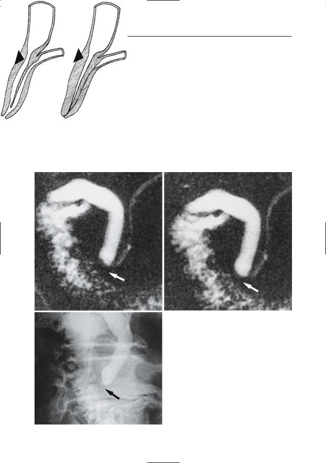
!● The following morphologic patterns may be seen on dynamic MRCP:
–“Type A”: nonvisualization of sphincter segment (Fig. 128a, c–e)
–“Type B”: absence of variability in shape on serial breathhold images (Fig. 128b, f–j)
●These morphologic abnormalities of the vaterian sphincter complex may be associated with dilation of the common bile duct and pancreatic duct (not always present)
●Note: Detection of these abnormalities requires use of a dedicated MR technique and specific attention to the morphology of the vaterian sphincter complex on consecutive images
References
Van Hoe L, Vanbeckevoort D, Gryspeerdt S et al. (1997) MR cholangiography of periampullary lesions: potential pitfalls in diagnosis. Radiology 205 (P) : 709
264 5.2 Benign Nontraumatic Abnormalities
a |
b |
c |
d |
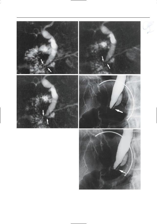
Fig. 128. a Sphincter dysfunction: nonvisualization of sphincter segment (type A). The drawings represent the vaterian sphincter complex imaged during contraction (left) and relaxation (right). In this type of dysfunction, elevation of basal pressure and/or fibrosis result in an unchanged“contracted”aspect of the sphincter. b Sphincter dysfunction: absence of variability in shape (type B). The drawings represent the vaterian sphincter complex during relaxation (left) and contraction (right). A portion of the sphincter (here the cranial portion) does not contract adequately and fluid remains within the lumen. Type A. c–e Projective MR images showing a dilated common bile duct with nonvisualization of the intramural portion of both ducts (arrows). The other images in
e the dynamic series showed similar features. e ERCP image confirming these findings (arrow). Type B
5 Vaterian Sphincter Complex 265
f |
g |
h |
i |
Fig. 128. (continued) f–j Projective MR images |
|
|
(f–h) showing the intramural portions of both ducts |
|
|
(arrows); however, there is no variability in shape. |
j |
|
i, j ERCP images confirming these findings (arrows) |
||
|

266 5.2 Benign Nontraumatic Abnormalities
#129 Sphincter Dysfunction, False-Negative Diagnosis on Dynamic MRCP
KEY FACTS: MRI
●While dynamic MRCP provides information on the presence of contractile activity, it does not actually show the passage of bile into the duodenum
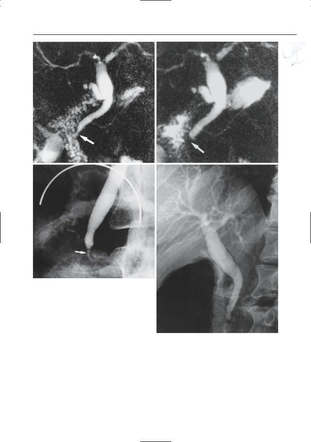
●In some patients with papillary dyskinesia, ERCP shows delayed emptying of the common bile duct despite the presence of contractile activity; this feature is not shown on MRCP (Fig. 129)
●Note: In order to overcome this limitation, the use of provocative tests has been proposed (Mariani et al. 2003).
Further studies are required to determine the role of MRCP in comparison with ERCP/manometry.
References
Mariani A,Curioni S,Zanello A,et al. (2003) Secretin MRCP and endoscopic pancreatic manometry in the evaluation of sphincter of Oddi function: a comparative pilot study in patients with idiopathic recurrent pancreatitis. Gastrointest Endosc 58 : 847–852
5 Vaterian Sphincter Complex 267
a |
b |
c
d
Fig. 129 a–d. Projective MR images showing apparently normal contractility of the sphincter: a sphincter relaxation with visualization of the intramural segment; b contraction (arrows). c ERCP image showing apparently normal distal (intramural) portion of bile duct (arrow). d However, 1-h delayed ERCP image shows bile ducts still filled with
contrast medium, thus suggesting sphincter dysfunction (see # 127,diagnostic criteria). Surprisingly, manometry showed normal contractility and normal baseline pressure. The final diagnosis was sphincter dysfunction, however, mainly because the symptoms disappeared after sphincterotomy

268 5.2 Benign Nontraumatic Abnormalities
#130 Benign Tumors
KEY FACTS: DISEASE
●Histology of ampullary tumors:
–Adenoma most common
–Other types: leiomyoma,neurofibroma, hemangioma, hamartoma
●Incidence: 0.04%–0.62%
●Age peak: 50–70 years
●Associations (adenoma):
–Familial polyposis coli
–Gardner syndrome
●Adenoma is considered a precancerous lesion (Yamaguchi and Enjoji 1991)
KEY FACTS: MRI
●Typical features (Fig. 130a):
–Dilation of pancreatic duct and bile ducts
–Bulging papilla (= generously sized papilla protruding into the lumen) with or without soft tissue mass
●Differential diagnosis (usually impossible by MRI):
–Non-neoplastic causes of bulging papilla: impacted gallstone; choledochocele; papillitis (Fig. 130b)
–Malignant tumors (see # 132, 133)
References
Seifert E, Schulte F, Stolte M (1992) Adenoma and carcinoma of the duodenum and papilla of Vater: a clinicopathologic study. Am J Gastroenterol 87 : 37–42
Yamaguchi K, Enjoji M (1991) Adenoma of the ampulla of Vater: putative precancerous lesion. Gut 32 : 1558–1561
5 Vaterian Sphincter Complex 269
a |
b |
Fig. 130. a Axial T2-weighted image showing enlarged papilla bulging into the duodenal lumen (arrowheads). The final diagnosis was tubulovillous adenoma. b Different patient. Projective image sho-
wing prominent papilla (“bulging papilla”; arrow). In this patient, the final diagnosis was papillary edema, most likely secondary to stone passage

270 5.3 Traumatic, Postoperative, and Iatrogenic Abnormalities
5.3Traumatic, Postoperative, and Iatrogenic Abnormalities
#131 Sequelae/Complications
of ERCP
KEY FACTS: DISEASE
●Complication rate: between 4.6% and 12%, depending on the type of procedure (diagnostic, manometry, sphincterotomy) (Cotton and Chong 1995)
●Complications of diagnostic ERCP:
–Pancreatitis (most common)
–Infection
–Sepsis (0.8%)
–Aspiration pneumonia (0.1%)
●Complications of sphincterotomy (Catalano et al. 1997):
–Overall complication rate: 10%
–Most common: hemorrhage, perforation (0.4%–2.1%), pancreatitis
–Mortality rate: 0.3%
KEY FACTS: MRI
●Pancreatitis (see # 151)
●Perforation:
–Paraduodenal collection containing fluid and/or air
–May spread in retroperitoneum
–A defect may be demonstrated in the wall of the distal common bile duct
●Bleeding:
–Collection with typical signal inten-
sity of blood (see #51)
● Note: After sphincterotomy, the distal- ! most portions of the bile duct and pancreatic duct may have an unusual appearance (Fig. 131):
–Abnormally “broad” aspect
–Absence of normal contractions
–Irregular contours
–Intraluminal air
References
Catalano O, Lapiccirella G, Rotondo A (1997) Papillary injuries and duodenal perforation during endoscopic retrograde sphincterotomy: radiological findings. Clin Rad 52 : 688–691
Cotton P, Chong W (1995) Complications of ERCP and therapy. In: Silvis S, Rohrmann C, Ansel H (eds) Endoscopic retrograde cholangiopancreatography. Igaku-Shoin, New York, pp 446–469
5 Vaterian Sphincter Complex 271
Fig. 131. Projective image obtained in a patient who underwent papillotomy showing abnormally broad aspect of the distal common bile duct (arrow). Note decreased signal intensity of the distalmost portion of the common bile duct, most likely caused by presence of air