

6
1 Technique
1.1 Magnetic Resonance Sequences
#1 Overview of Imaging Protocol
For practical purposes, our standard protocol for pancreaticobiliary MRI and MRCP using the Siemens platform (1.5-T Symphony or Sonata) is given below.
Phased array body coil and rectangular FOV are used. No specific preparation is required. No drugs or oral contrast media are given for routine studies.
1.Coronal free breathing T2-weighted HASTE (TR infinite, TE 60 ms, 5-mm slices, 1-mm gap, matrix 256×230, acquisition time ±400 ms/slice, no fat saturation).
Purpose: characterisation of focal parenchymal lesions, visualisation of fluidcontaining structures, general anatomical overview.
2.Same sequence as in (1) but with TE = 360 ms. Alternatively, if the double-echo HASTE sequence is available, both sets of images may be obtained using one single acquisition.
!Purpose: characterisation of focal parenchymal lesions, detection of small amounts of fluid, evaluation of biliary and pancreatic ducts.
3.Same sequence as in (1) but images obtained in the axial plane.
4.Same sequence as in (2) but images obtained in the axial plane.
5.Axial free-breathing magnetisation prepared gradient echo (turboFLASH) (TR 7 ms, TE 4.3 ms, 5-mm slices, 1-mm gap, matrix 192×256, acquisition time ±700 ms/slice). Alternatively, a spoiled gradient echo sequence may be used during breath-holding. No fat saturation. Care should be taken to optimise T1 contrast.
Purpose: detection of hepatic and pancreatic tumors, evaluation of extrapancreatic spread of pancreatic neoplasms, lesion characterisation (together with T2-weighted images).
6.Breath-hold single-slice MR cholangiography using the RARE sequence (TR 2800, TE 1100, acquisition time ±3 s; matrix 256×256,3-cm slice). Imaging should be repeated during consecutive breathholding episodes using different slice position and/or orientation in order to obtain visualisation of the entire biliary and pancreatic ductal system. Also, in the case of suspected biliary obstruction, kinematic imaging can be performed by repeating the acquisition without changing the parameters for slice position and slice orientation (see Fig. 3).
Later in the book, images obtained with this sequence are referred to as projective MR images (see # 5).
Purpose: overview images of biliary and pancreatic ducts.
Optional (according to clinical information and/or findings at non-contrast imaging):
7.Coronal breath-hold multislice 3D VIBE T1-weighted gradient echo before IV contrast administration, and in the pancreatic and portal venous phases (±30 and 70 s after initiation of contrast injection, respectively) (TR 4.8, TE 1.6, acquisition time ±20 s, 3.5-mm partitions, matrix 384×512). Fat saturation is used to optimise contrast.
Purpose: differentiation of pancreatic neoplasm from surrounding pancreatitis, characterisation of focal lesions, detection of hypervascular lesions, assessment of vascular anatomy.
1 MRCP Technique |
7 |
These sequences provide complementary information. Together, and with experience, they allow accurate diagnosis of most conditions encountered in the clinical setting. The complementarity of the different
types of images is illustrated in Fig. 1 a–d (diagnosis and staging of pancreatic cancer) and Fig. 1e–k (characterisation of pancreatic mass).
a |
b |
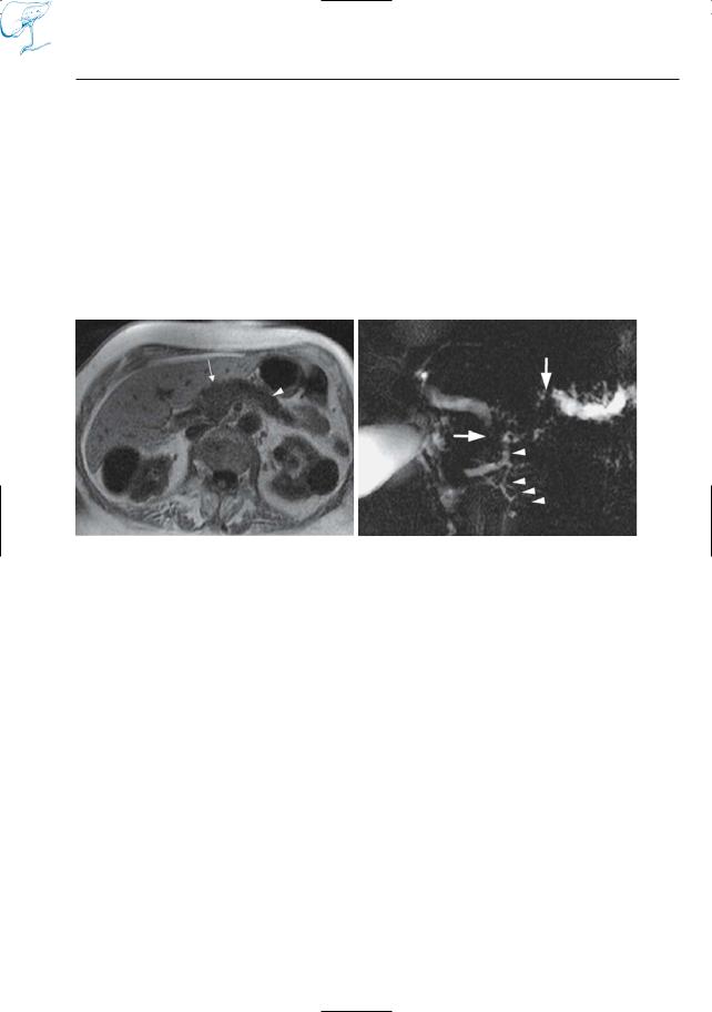
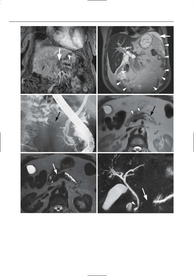
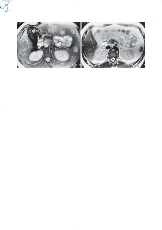
Fig. 1 a–c. Patient with obstructive jaundice. a T1weighted, non-fat-suppressed snapshot image. Although the image is noisy, the contrast is excellent, and the image has a high diagnostic value. The pancreas has a diffusely low signal intensity (arrow), which is aspecific. Atrophy of pancreatic tail is also observed (arrowhead). Projective cholangiographic (RARE) image (b) shows dilatation of biliary duct and proximal pancreatic duct with severe stenosis of distal biliary and pancreatic ducts (arrows). Note dilated side branch in pancreatic head (arrowheads). Contrast-enhanced VIBE image (c) shows hypointense lesion in pancreatic neck (arrow) with dilatation of proximal pancreatic duct. Note enhancement of pancreatic parenchyma in the body/tail (arrowhead). Based on these findings, the diagnosis of adenocarcinoma with retro-obstruc- tive pancreatitis and biliary invasion can be made. In another patient with pancreatic carcinoma, coronal HASTE image (d) shows intraperitoneal free fluid (arrow) and omental cake (arrowheads): peritoneal/omental metastatic disease. e–k Patient presenting with weight loss and dorsal back pain. e ERCP image shows long and severe stenosis of pancreatic duct (arrow). Because of these findings, pancreatic cancer is the first diagnosis. Cross-sec- tional T2-weighted image (f) shows marked atrophy
of pancreatic parenchyma in the tail (arrow), associated ductal dilatation,and a mass-like lesion in the pancreatic neck (arrowheads) (all supportive of the presumed diagnosis of malignancy). Heavily T2weighted image and projective MRCP (g, h) show absence of penetrating main pancreatic duct, again a suspect finding. There is possibly some focal narrowing of the distal common bile duct (arrowhead in h), a finding that can also be observed at ERCP. Snapshot T1-weighted image (i) surprisingly reveals high signal intensity of the process causing the ductal narrowing (arrow). Such a finding is surprising in this context because it virtually excludes adenocarcinoma. Note that this crucial finding is not well seen on the fat-suppressed T1-weighted VIBE image, where the lesion is grey (arrow), just like the remaining parenchyma in the tail (j). Dynamic con- trast-enhanced imaging reveals contrast uptake of the lesion, which is another argument against carcinoma (carcinomas present as “black holes” on dynamic contrast-enhanced VIBE images). Based on integration of all these pieces of information and on experience with the relative value of the different findings, chronic pancreatitis with “pseudotumor” was proposed as first diagnosis. Surgery was not performed. The diagnosis was confirmed by followup (unchanged appearance at 2-year interval)
81.1 MR Sequences
c |
d |
e |
f |
g |
h |
Fig. 1 a–k. (continued)
1 MRCP Technique |
9 |
i
Fig. 1 a–k. (continued)
References
Fayad LM, Kowalski T, Mitchell DG (2003) MR cholangiopancreatography: evaluation of common pancreatic diseases. Radiol Clin North Am 41: 97–114
Fulcher AS, Turner MA (2002) MR cholangiopancreatography. Radiol Clin North Am 40: 1363– 1376
Hundt W, Petsch R, Scheidler J et al. (2002) Clinical evaluation of further-developed MRCP sequences in comparison with standard MRCP sequences. Eur Radiol 12: 1768–1777
j
k
Kim MJ, Mitchell DG, Ito K et al. (2000) Biliary dilatation: differentiation of benign from malignant causes – value of adding conventional MR imaging to MR cholangiopancreatography. Radiology 214: 173–181
Lopez Hanninen E, Amthauer H, Hosten N et al. (2002) Prospective evaluation of pancreatic tumors: accuracy of MR imaging with MR cholangiopancreatography and MR angiography. Radiology 224: 34–41
Ly JN,Miller FH (2002) MR imaging of the pancreas: a practical approach. Radiol Clin North Am 40: 1289–1306
Van Hoe L, Mermuys K, Vanhoenacker P (2004) MRCP pitfalls. Abdom Imaging 29: 360–387

10 1.1 MR Sequences
!
!
#2 Snapshot T2-Weighted MRI
KEY FACTS
●“Snapshot” MRI: acquisition of individual slices in less than 1 s
●Advantages:
–Short examination times
–Image quality independent of patient cooperation
●Commonly used sequences:
–Echo planar imaging (EPI) (see # 8)
–Half-Fourier acquisition single-shot turbo spin echo (HASTE) = halfFourier rapid acquisition with relaxation enhancement (RARE; acquisition time per slice, ±400 ms) (Kiefer et al. 1994; Van Hoe et al. 1996)
●Technical realization of HASTE (Fig. 2a; Kiefer et al. 1994; Van Hoe et al. 1996):
–Generation of multiple echoes after a single 90° excitation pulse
–Each echo fills a horizontal line in k-space
–Approximately 56 % of k-space is filled, starting from a few lines above the center of k-space to the bottom
–Half-Fourier reconstruction is used
●Advantages of HASTE:
–General advantages of snapshot imaging (see above)
–Images nearly unaffected by specific artifacts (magnetic susceptibility, chemical shift)
●Disadvantages of HASTE:
–Intrinsic limitations in signal-to- noise ratio and spatial resolution
–Inconsistent signal intensity of blood vessels: a typical “flow void” is seen only in case of high blood velocity
●Clinical applications:
–Excellent technique for cross-section- al T2-weighted MRI
–Can also be used to obtain cholangiographic images (Morrin et al. 2000)
References
Morrin MM, Farrell RJ, McEntee G et al. (2000) MR cholangiopancreatography of pancreaticobiliary diseases: comparison of single-shot RARE and multislice HASTE sequences. Clin Radiol 55 : 866–873
Kiefer B,Grässner J,Hausman R (1994) Image acquisition in a second with half-Fourier acquisition single-shot turbo spin echo. J Magn Reson Imaging (P) Suppl: 86–87
Tang Y,Yamashita Y,Arakawa A et al. (2000) Pancreaticobiliary ductal system: value of halfFourier rapid acquisition with relaxation enhancement MR cholangiopancreatography for postoperative evaluation. Radiology 215 : 81–88
Van Epps K, Regan F (1999) MR cholangiopancreatography using HASTE sequences. Clin Radiol 54 : 588–594
Van Hoe L, Bosmans H, Aerts P, Baert AL, Fevery J, Kiefer B,Marchal G (1996) Focal liver lesions: fast T2-weighted MR imaging with half-Fourier rapid acquisition with relaxation enhancement. Radiology 201: 817–823
Vitellas KM, El-Dieb A, Vaswani KK, et al. (2002) Using contrast-enhanced MR cholangiography with IV mangafodipir trisodium (Teslascan) to evaluate bile duct leaks after cholecystectomy: a prospective study of 11 patients. AJR Am J Roentgenol 179 : 409–416
1 MRCP Technique |
11 |
a
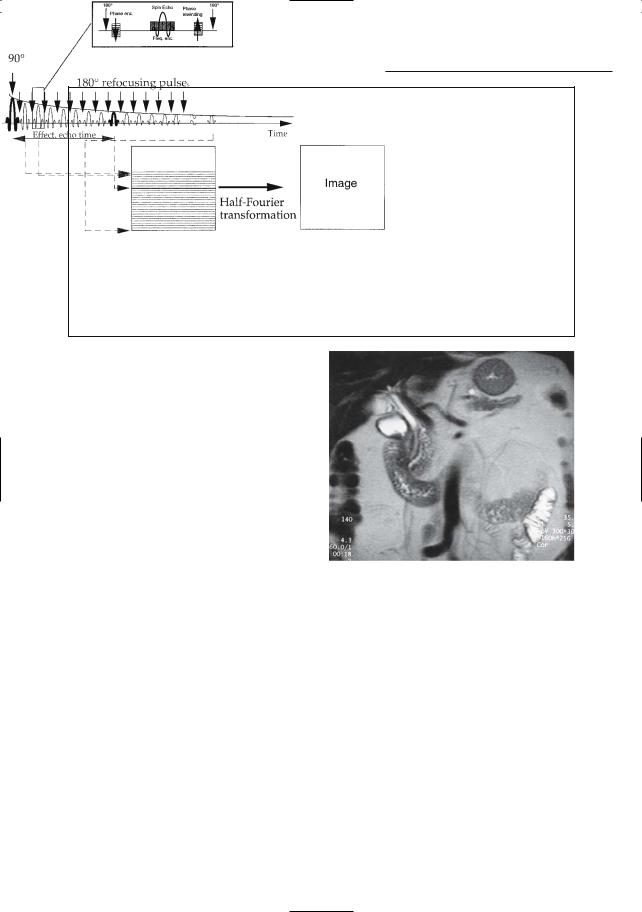
Fig. 2. a The HASTE pulse sequence. The 90° radio frequency excitation pulse is followed by a long b series of spin echoes. The interval between succes-
sive echoes is as short as possible. Phase encoding, 180° refocusing pulse, frequency encoding, and phase rewinding are performed within a window of about 4 ms. The k-space is filled from eight lines above the center to the bottom. The effective TE of the image is defined as the time between the radio frequency excitation pulse and the acquisition of the line of the center of k-space. b Coronal T2weighted image obtained with HASTE. The bile duct,pancreatic duct,and the fluid within the lumen of the duodenum and small bowel are markedly hyperintense. Solid organs and bowel walls have relatively low signal intensity. Fat has intermediate to high signal intensity. Flowing blood and air have very low signal intensity. Note the absence of artifacts related to motion and susceptibility and the good delineation of the contours of the stomach, pancreas, duodenum, and colon

12 1.1 MR Sequences
#3 Snapshot T 2-Weighted MRI:
Double-Echo Technique
KEY FACTS
●Modification of the HASTE technique (see # 2)
●Two images are calculated per anatomical slice position
●Image characteristics:
–First image: moderately T2-weighted
–Second image: heavily T2-weighted
●Acquisition time per slice: ± 700 ms
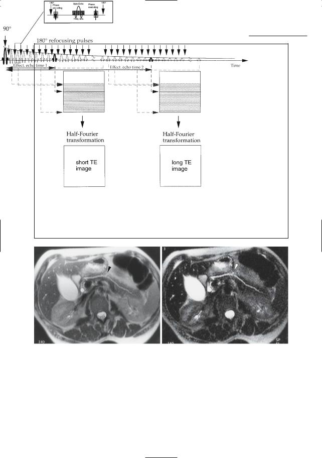
●Technical realization (see Fig. 3a; Bosmans et al. 1997a, b):
–Each excitation pulse is followed by a long echo train
–The first part of the echo train is used to construct an image with short TE (e.g., 60 ms)
–The second part of the echo train is used to construct an image with long TE (e.g., 350 ms)
●Theoretical advantages:
–Excellent visualization of fluid-con- taining structures and lesions on images with long TE
●Clinical applications:
–Characterization of focal liver lesions (Bosmans et al. 1997a; Table 3.1)
–Detection of acute pancreatitis, acute
Table 3.1. Characterization of focal liver lesions
cholecystitis, biliary fistulas, subtle ! anomalies of bile ducts, and pancreatic ducts (Fig. 3b, c)
–Differentiation between acute and chronic cholecystitis (see # 104)
–Part of routine imaging protocol at our institution
●Note: Unless specifically indicated, all T2-weighted MR images shown in the book are obtained using this sequence
●Note: as an alternative, two separate acquisitions with short and long TE can be obtained (see # 1)
References
Bosmans H, Gryspeerdt S,Van Hoe L,Van Oostende S, De Jaegere T, Kiefer B, Baert AL, Marchal G (1997a) Preliminary experience with a new double echo half-Fourier single-shot turbo spin echo acquisition in the characterisation of liver lesions. MAGMA 5 : 79–84
Bosmans H, Van Hoe L, Gryspeerdt S, Kiefer B, Van Steenbergen W, Baert AL, Marchal G (1997b) Technical note. Single-shot T2-weighted MR imaging of the upper abdomen: preliminary experience with the double echo HASTE technique. AJR Am J Roentgenol 169 :1291–1293
Pawluk RS, Borrello JA, Brown JJ, McFarland EG, Mirowitz SA, Tsao LY (2000) A prospective assessment of breath-hold fast spin echo and inversion recovery fast spin echo techniques for detection and characterization of focal hepatic lesions. Magn Reson Imaging 18 : 543–551
|
Signal intensity (relative to normal liver) |
|
|
First echo (TE 60 ms) |
Second echo (TE 250–350 ms) |
|
|
|
Cyst |
Hyperintense ++/+++ |
Hyperintense ++++ |
Hemangioma |
Hyperintense ++ |
Hyperintense + |
Solid lesion |
Hyperintense +/++ |
Isointensea |
! |
a Lesions containing cystic components or necrosis may be partially hyperintense. |
|
1 MRCP Technique |
13 |
a
b |
c |
Fig. 3. a The double-echo HASTE pulse sequence.A 90° radio frequency excitation pulse is followed by a very long echo train. Two k-spaces are filled successively and transformed into an image with a halfFourier reconstruction technique. The effective TE of the resulting images is the time between the radio frequency pulse and the acquisition of the echo attributed to the center of the first and second
k-space, respectively. b, c Corresponding “doubleecho” HASTE images with b a TE of 60 ms and c a TE of 439 ms showing a slightly dilated pancreatic duct with a small pseudocyst (or dilated side branch) in the pancreatic body (arrowheads). The pseudocyst is better seen in c. Note that in b the signal intensity of fat and fluid is comparable. In c, fluid really stands out

14 1.1 MR Sequences
#4 Snapshot T2-Weighted MRI:
Is Fat Suppression Required?
KEY FACTS
●Use of fat suppression improves the quality of spin echo MR images of the upper abdomen (Lu et al. 1994)
●Advantages of fat suppression in spin echo MRI (Lu et al. 1994):
–Reduction of image noise, mainly because fat signal does not contribute to motion-related phase-encoding artifacts
–Improved conspicuity of focal liver lesions, particularly in the case of fatty liver
!● In snapshot T2-weighted MRI, the advantages of fat suppression are questionable (Bosmans et al. 1997):
–Motion-related artifacts are virtually absent (see # 2)
–Use of fat suppression significantly decreases the conspicuity of the contours of several extrahepatic structures (e.g., bile duct, gastric wall, pancreas, retroperitoneal vessels; Fig. 4)
–The double-echo technique offers effective fat suppression while highlighting fluid (long TE)
References
Bosmans H, Van Hoe L, Gryspeerdt S et al. (1997) Technical note. Single-shot T2-weighted MR imaging of the upper abdomen: preliminary experience with the double echo HASTE technique. AJR Am J Roentgenol 169 :1291–1293
Lu DS, Saini S, Hahn PF et al. (1994) T2-weighted MR imaging of the upper part of the abdomen: should fat suppression be used routinely? AJR Am J Roentgenol 162 :1095–1100
1 MRCP Technique |
15 |
a |
b |
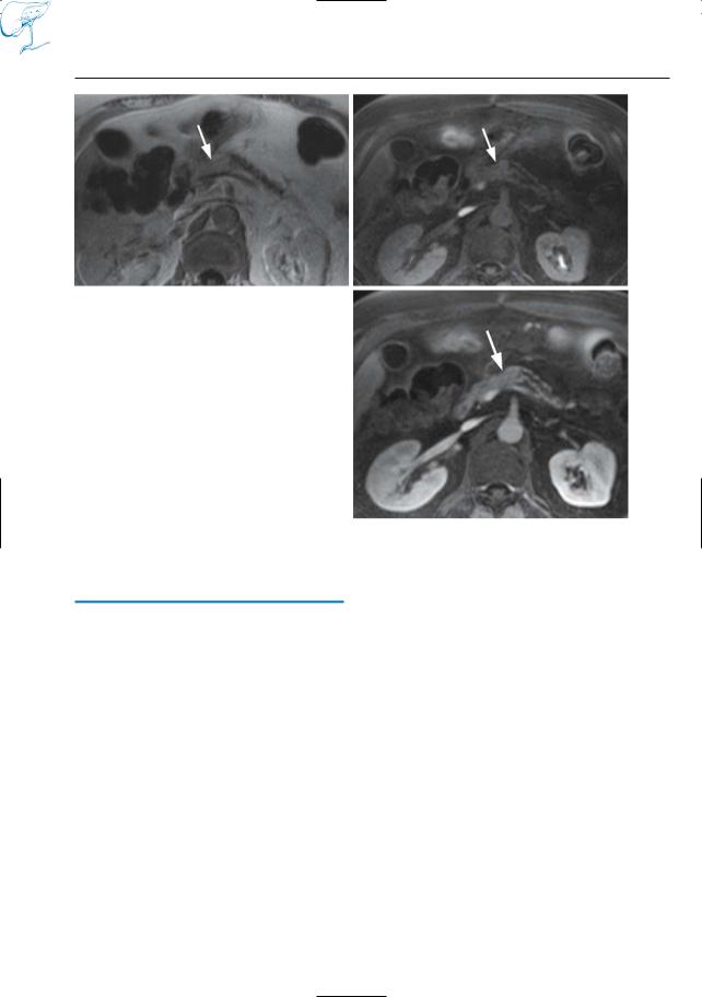
Fig. 4 a, b. Corresponding images a with and b without fat suppression. Note better visualization of the contours of the pancreas, colon, and adrenals in b
16 1.1 MR Sequences
#5 “Projective” Cholangiography
KEY FACTS
●Principle:
–A projection image is obtained through the structure of interest with a relatively large slice thickness (2–10 cm)
–One voxel dimension is significantly larger than the other two dimensions
–In order to obtain sufficiently high contrast, the signal obtained from the structure of interest (e.g., bile ducts) should be much higher than the signal of background tissue
●Sequence commonly used: RARE (Laubenberger et al. 1995; Matos et al. 1997; Van Hoe et al. 2004)
!● Image characteristics of RARE:
–Only structures/lesions containing pure water are displayed
●Technical realization of RARE (Fig. 5a):
–All echoes are obtained after a single excitation pulse
–A very long echo train is used (usually
± 250 echoes)
–The resulting image has an extremely long effective TE (e.g., 1100 ms)
●Acquisition time: ± 3–5 s (breathholding required)
●Advantages:
– Excellent display of bile duct and/or
pancreatic ducts on one image (Fig. 5b)
– Relatively high in-plane resolution
●Note: Unless specifically indicated, all projective cholangiographic MR images shown in the book are obtained using this technique
References
Laubenberger J, Büchert M, Schneider B, Blum B, Hennig J, Langer M (1995) Breath-hold projection magnetic resonance cholangio-pancre- aticography (MRCP): a new method for the examination of the bile and pancreatic ducts. Magn Reson Med 33 : 18–23
Matos C, Metens T, Devière J et al. (1997) Pancreatic duct: morphologic and functional evaluation with dynamic MR pancreatography after secretin stimulation. Radiology 203 : 435–441
Keogan MT, Edelman RR (2001) Technologic advances in abdominal MR imaging. Radiology 220 : 310–320
Van Hoe L, Mermuys K, Vanhoenacker P (2004) MRCP pitfalls. Abdom Imaging 29 : 360–387
1 MRCP Technique |
17 |
a
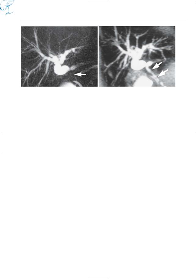
Fig. 5. a RARE acquisition is a single-shot technique in which a 90° radio frequency excitation pulse is followed by an extremely long echo train. The k-space is completely filled, with the center of k-space acquired half way along the acquisition. The effective TE can be as long as 1100 ms. b RARE image obtained in a patient with papillary stenosis after cholecystectomy, showing entire common bile duct, pancreatic duct, and cystic duct remnant. A 3-cm slice thickness was used
b

18 1.1 MR Sequences
#6 “Projective” Cholangiography:
Limitations
KEY FACTS
●Image quality dependent on patient cooperation (breathholding required) (Fig. 6a, b)
●Suboptimal visualization of bile duct and/or pancreatic ducts in the case of elevation of signal intensity of background tissue (presence of free water or edema), e.g.:
–Acute pancreatitis with peripancreatic exudate
–Liver transplant with hilar biloma/ hematoma
–Ascites (Fig. 6c, d)
!● Suboptimal visualization of intraluminal structures/lesions that are not surrounded by fluid, e.g.:
–Stone impacted in the distal common bile duct or cystic duct
–Blood clots, sludge, or necrotic debris filling a large part of the bile duct (see also #23)
●Medical interaction required during image acquisition
–Selection of slice position depending on the clinical question and findings on cross-sectional images (see # 11)
References
Laubenberger J, Büchert M, Schneider B, Blum B, Hennig J, Langer M (1995) Breath-hold projection magnetic resonance cholangio-pancre- aticography (MRCP): a new method for the examination of the bile and pancreatic ducts. Magn Reson Med 33 : 18–23
Matos C, Metens T, Devière J et al. (1997) Pancreatic duct: morphologic and functional evaluation with dynamic MR pancreatography after secretin stimulation. Radiology 203 : 435–41
Van Hoe L, Mermuys K, Vanhoenacker P (2004) MRCP pitfalls. Abdom Imaging 29 : 360–387
1 MRCP Technique |
19 |
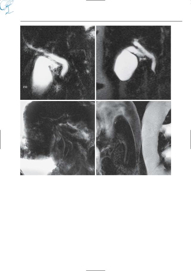
a |
b |
c |
d |
Fig. 6 a,b. Elderly patient. a The quality of the projective (RARE) image is severely degraded by respiratory artifact. Choledocholithiasis cannot be ruled out. b Heavily T2-weighted snapshot (HASTE) image adequately showing the anatomy of the distal
common bile duct; no stone. c Projective image severely degraded by the presence of ascites. Adequate interpretation is not possible. d Projective image severely degraded by the presence of ascites and wrap-around artifact

20 1.1 MR Sequences
#7 An Alternative Approach:
Calculation of Maximum-
Intensity Projection Images
KEY FACTS
●Maximum-intensity projection (MIP): postprocessing technique that calculates projective images from a stack of crosssectional images
●Principle (Laub 1990):
–Mathematical rays are projected in the desired viewing direction through a stack of reconstructed sections.
–The density of each pixel in the resulting image is the maximum intensity encountered along the ray as it traverses the volume
●Advantages over RARE:
–If used in combination with snapshot techniques for T2-weighted MRI, image quality is less dependent on patient cooperation
–If used in combination with a threedimensional acquisition technique, high-resolution projective images can be calculated a posteriori in any desired viewing direction
●Disadvantages compared with RARE:
–Time required for postprocessing
–Elevation of background intensity caused by the algorithm, leading to lower levels of contrast and a less sharp delineation of small biliary and pancreatic ducts (Fig. 7)
–If used in combination with a twodimensional sequence, high-resolu- tion images can be calculated in one viewing direction only
●Applications:
–Tends to be replaced by direct RARE projective cholangiography (Bearcroft and Lomas 1997)
–Maximum-intensity projection processing of heavily T2-weighted HASTE images can be used as an alternative to RARE projective cholangiography in patients unable to cooperate
References
Bearcroft P, Lomas D (1997) Magnetic resonance cholangiopancreatography. Gut 41 : 135–137
Laub G (1990) Displays for MR angiography. Magn Reson Med 14 : 222–229
1 MRCP Technique |
21 |
a |
b |
Fig. 7 a, b. Limitations of MIP images. Patient with jaundice after gallbladder surgery. Projective (RARE) image with 3-cm slice thickness (a) compared with MIP image obtained after postprocessing of a series of thin slices (b). Both images show
intrahepatic biliary dilatation. The single-slice MRCP image clearly shows occlusion of the common hepatic duct (arrow). This diagnosis is missed on the MIP image because an intrahepatic bile duct superimposes on the common hepatic duct (arrows)

22 1.1 MR Sequences
#8 Other Techniques
for Obtaining
T2-Weighted Images
KEY FACTS
Techniques that have been used to obtain T2-weighted MR images in pancreatobiliary MRI can be classified as follows:
Spin Echo Versus Gradient
Echo Techniques
Spin Echo Techniques
●Use a 180° radio frequency pulse for rephasing
●Provide images relatively unaffected by magnetic field inhomogenities and magnetic susceptibility
●Examples: spin echo, fast spin echo (FSE) or turbo spin echo (TSE), RARE (see #5), and HASTE (see # 2)
Gradient Echo Techniques
●Use gradients instead of 180° radio frequency pulses. Consequently, images are more susceptible to different types of artifacts (Fig. 8)
●Examples: steady state free precession (SSFP) gradient echo (e.g., FISP) (Morimoto et al. 1992), and echo planar imaging
Breathhold Versus Non-breathhold
Techniques Versus Snapshot
Techniques
Breathhold Techniques
●Advantages: reduction of artifacts caused by respiration
●Disadvantages: intrinsic limitations in sig- nal-to-noise ratio and spatial resolution
●Examples: breathhold fast spin echo, RARE
Non-breathhold Techniques
●Advantages: possible to obtain multiple averages with a high signal-to-noise ratio and spatial resolution
●Disadvantages: speed, respiratory artifacts
●Examples: spin echo, all three-dimen- sional techniques
Snapshot Techniques
● See # 2
TwoVersus Three-Dimensional
Techniques
Two-Dimensional Techniques
●Advantages: shorter imaging time
●Disadvantages: limited resolution in the third dimension (slice thickness)
●Examples: two-dimensional fast spin echo, RARE
Three-Dimensional Techniques
●Advantages: high resolution in all three dimensions
●Disadvantages: long acquisition times
●Examples: three-dimensional steady state free precession (Morimoto et al. 1992), three-dimensional fast spin echo (Barish et al. 1995)
References
Barish MA,Yucel EK, Soto JA, Chuttani R, Ferruci JT (1995) MR cholangiopancreatography: efficacity of a three-dimensional turbo spin-echo technique. AJR Am J Roentgenol 165 : 295–300
Morimoto K, Shimoi M, Shirakawa T et al. (1992) Biliary obstruction: evaluation with three-di- mensional MR cholangiography. Radiology 183 : 578–80
Reinhold C, Bret P (1996) MR cholangiopancreatography. Abdom Imaging 21:105–116
1 MRCP Technique |
23 |
|
a |
b |
Fig. 8 a, b. T2-weighted images obtained with a HASTE and b fast imaging with steadystate precession. Note black borders of all abdominal organs in b, related to intravoxel dephasing. The
hepatic abnormalities (peribiliary fibrosis in patient with primary sclerosing cholangitis) are more clearly seen in a

24 1.1 MR Sequences
#9 T1-Weighted MRI:
Non-fat-suppressed
Magnetizationprepared
Snapshot Gradient Echo
KEY FACTS
●Abbreviation: MP snapshot GE T1
●Synonyms: snapshot fast low-angle shot (snapshot FLASH), turbo FLASH
●Acquisition time per slice: ± 700 ms
●Principle and technical realization (Fig. 9a; Haase 1990; Holsinger-Bamp- ton et al. 1991):
–Application of multiple small flip angle excitation pulses
–Generation of one echo after each excitation pulse
–Short TR (e.g., 7 ms)
–Use of an inversion recovery pulse for contrast preparation
●Theoretical advantages:
!– Good T1 contrast (excellent detection of hepatic and pancreatic neoplasms)
–Image quality independent of patient motion
●Limitations:
–Not applicable for fat-suppressed T1weighted imaging
–Sensitive to susceptibility artifact (no 180° refocusing pulses)
●Clinical use: part of our standard protocol for routine upper abdominal imaging together with the double-echo HASTE sequence
●Alternatively, a breath-hold T1-weighted sequence can be used
References
Haase A (1990) Snapshot FLASH MRI. Applications to T1,T2 and chemical shift imaging. Magn Reson Med 13 : 77–89
Holsinger-Bampton AE, Riederer SJ, Campeau NG, Ehman RL, Johnson CD (1991) T1-weighted snapshot gradient-echo MR imaging of the abdomen. Radiology 181 : 25–32
Keogan MT, Edelman RR (2001) Technologic advances in abdominal MR imaging. Radiology 220 : 310–320
1 MRCP Technique |
25 |
|
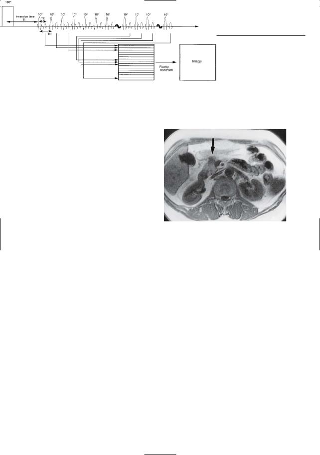
a
Fig. 9. a Magnetization-prepared snapshot gradient echo. This technique is basically an ultrafast gradient echo acquisition that uses a small flip angle and short TR. An 180° inversion recovery pulse prepares the magnetization of the spins prior to the acquisition. The inversion time is chosen so that
adequate T1 contrast is obtained. b Magnetization- |
|
prepared snapshot gradient echo T1-weighted |
|
image showing liver and pancreatic head as relati- |
|
vely hyperintense. A small tumor is shown as a |
|
hypointense lesion in the pancreatic neck (arrow). |
|
Although this sequence does not offer spectacular |
|
contrast, it is very sensitive in the detection of hepa- |
|
tic and pancreatic tumors |
b |
26 1.2 Practical Setup of an MRCP Study
#10 T1-Weighted MRI:
Use of Fat Suppression
KEY FACTS
●For unenhanced T1-weighted imaging use of fat suppression is not recommended (decreased liver-to-spleen contrast)
●For dynamic contrast-enhanced imaging, fat-suppressed images are routinely obtained
●Out-of-phase images can be useful for confirmation of fatty infiltration
a
c
References
Keogan MT, Edelman RR (2001) Technologic advances in abdominal MR imaging. Radiology 220 : 310–320
Semelka SR, Asher SM (1993) MR imaging of the pancreas. Radiology 188 : 593–602
Rofsky, NM, Lee VS, Laub G, et al. (1999) Abdominal MR imaging with a volumetric interpolated breath-hold examination. Radiology 212 : 876– 884
b
d
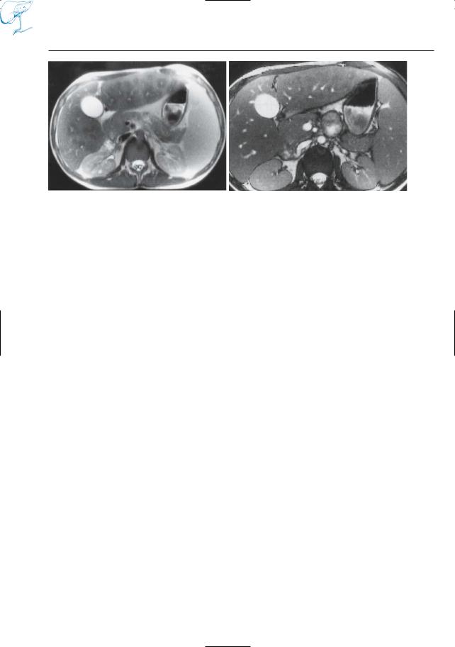
Fig. 10 a, b. T1-weighted MR images a with and b without fat suppression. Both images adequately show the normal pancreas and the large liver tumor. Note inversion of intensity of normal pancreas versus surrounding fat when b is compared to a.
c, d Different patient. Contrast-enhanced images obtained c with and d without fat suppression. Part of the pancreatic head and body are clearly seen in d (arrows). In d, the pancreatic parenchyma and the surrounding retroperitoneal fat are nearly isointense