


304 6.2 Benign Nontraumatic Abnormalities
6.2Benign Nontraumatic Abnormalities
#150 Santorinicele
and Wirsungocele
Related topic: # 140 (pancreas divisum)
KEY FACTS: DISEASE
●Focal dilation at the termination of the dorsal or ventral duct
●Similar to choledochocele (see # 126) and ureterocele
●Most common in elderly patients
●Etiology: probably a combination of outflow obstruction (small accessory orifice) and weakness of the distal duct wall, either congenital or acquired
●Commonly, but not necessarily, associated with pancreas divisum (see also
# 140 and Fig. 137)
●May be the underlying disorder in patients with recurrent attacks of acute pancreatitis
●Symptoms may improve after accessory sphincterotomy
KEY FACTS: MRI
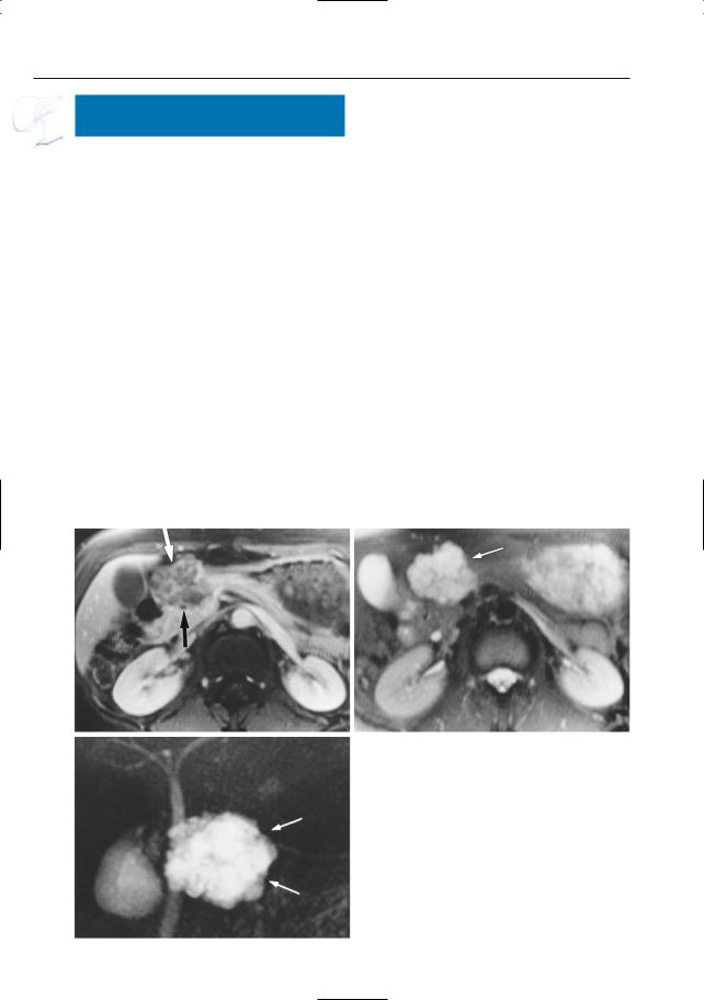
●Santorinicele (Fig. 150a,b):
–Pancreas divisum (see # 140)
–Focal dilation at the termination of the accessory duct
●Wirsungocele (Fig. 150c,d):
–Focal dilation of the termination of the duct of Wirsung
References
Manfredi R, Costamagna G, Brizi MG et al. (2000) Pancreas divisum and “santorinicele”: diagnosis with dynamic MR cholangiopancreatography with secretin stimulation. Radiology 217 : 403– 408
Eisen G, Schutz S, Metzler D, Baillie J, Cotton B (1994) Santorinicele: new evidence for obstruction in pancreas divisum. Gastrointest Endoscop 40 : 73–76
6 Pancreatic Ducts 305
a |
b |
d
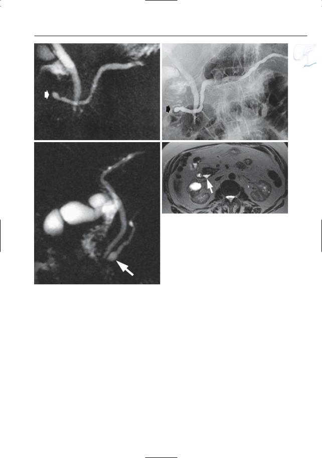
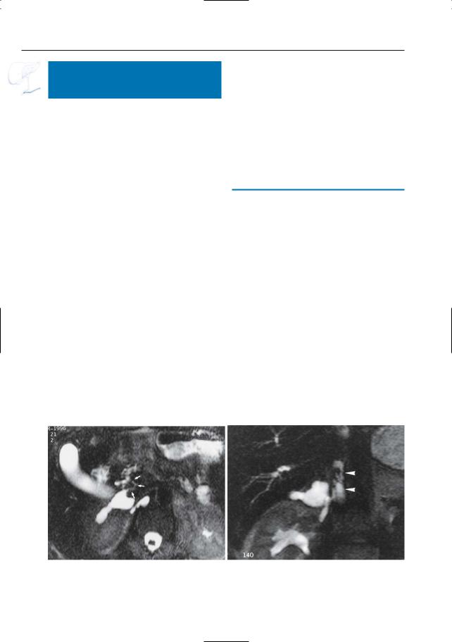
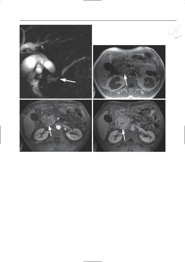
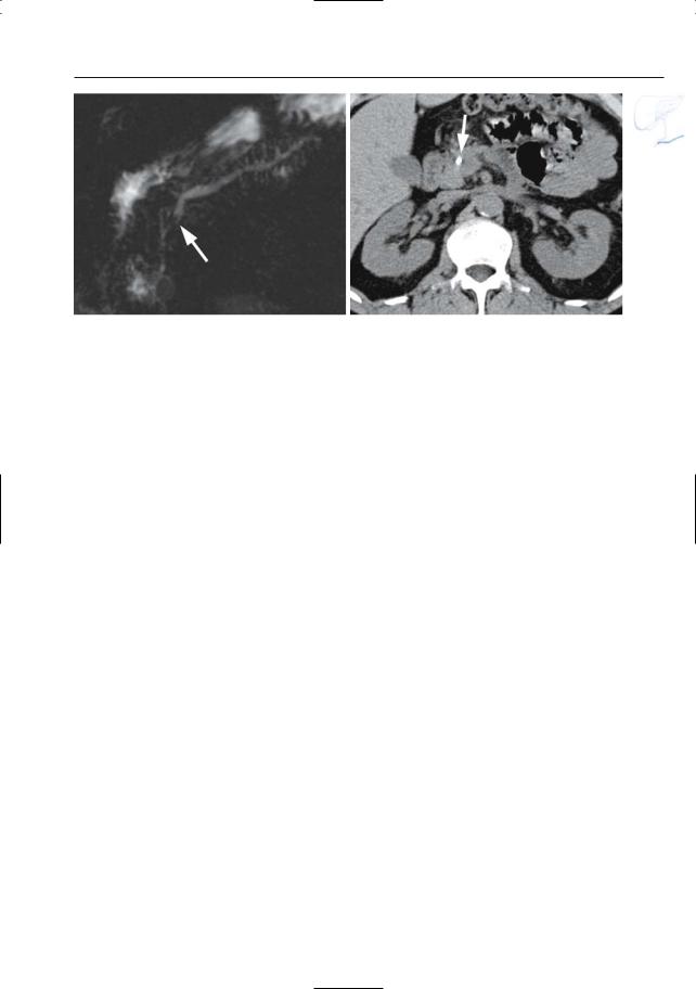
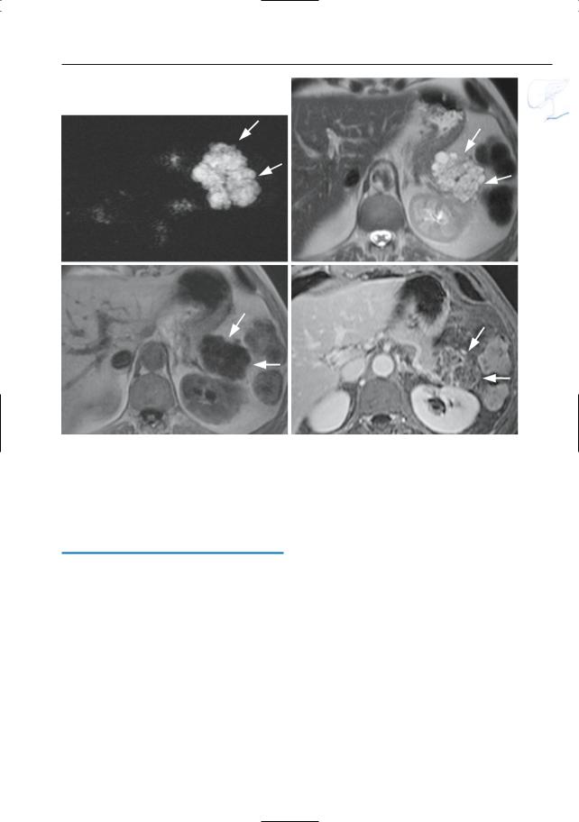
Fig. 150. a, b Projective MR image and b ERCP image showing pancreas divisum with focal dilation at the termination of the accessory duct (arrows). Wirsungocele. c, d Projective image (c) and axial T2weighted HASTE image (d) showing a focal dilata-
c tion of the distal main pancreatic duct (arrow): Wirsungocele

306 6.2 Benign Nontraumatic Abnormalities
#151 Acute Pancreatitis, General
KEY FACTS: DISEASE
●Acute inflammatory disease of the pancreas producing temporary changes with restoration of normal anatomy and function
●Causes:
–Biliary disease (± 60%)
–Alcoholism (± 30%)
–Other causes (e.g., hypercalcemia, hypertriglyceridemia, drugs)
●Clinical prognostic scores: Ranson score, Imrie score, Acute Physiology and Chronic Health Evaluation (APACHE) score
●Imaging classification (Balthazar 1994; Balthazar et al. 1985):
–Grades A, B: edematous pancreatitis (see # 152)
–Grade C: acute pancreatitis with associated peripancreatic inflammation (see # 153)
–Grades D, E: acute pancreatitis with ill-defined fluid collection/phlegmon (see # 154)
●This grading system was developed for CT (Balthazar 1994) and constitutes the basis for calculation of the severity index (see # 155)
●Associations/predisposing factors:
–Small gallstones (common)
–Wide cystic duct
–Common channel between the common bile duct and pancreatic duct
–Cholesterolosis of gallbladder
●Symptoms: upper abdominal pain, nausea, vomiting (often non-specific)
●Typically elevated amylase and lipase levels (sensitivity, 80%–90%)
●Complications (mainly occur in patients with stage D or E disease):
–Necrosis (see # 155)
–Pseudocyst (10%) (with or without infection; see # 156)
–Hemorrhage (3%)
–Venous thrombosis (see # 157)
–Pseudoaneurysm (see # 157)
–Biliary obstruction (see # 77)
–Abscess (2%–20%)
–Ascites
●Note: It has been stated that ERCP with sphincterotomy may be indicated in all patients with severe (biliary) pancreatitis; it is unclear whether MRCP might be useful in selecting candidates for sphincterotomy
References
Balthazar EJ (1994) Pancreatitis. In: Gore RM, Levine MS, Laufer I (eds) Textbook of gastrointestinal radiology. Saunders, Philadelphia, pp 2132–2160
Balthazar EJ, Ranson JHC, Naidich JP, Megibow AJ, Caccavale R, Cooper MM (1985) Acute pancreatitis: prognostic value of CT. Radiology 156 : 767–772
Chalmers AG (1997) The role of imaging in acute pancreatitis. Eur J Gastroenterol Hepatol 9 : 106– 116
Curran FT, Neoptolemos JP (1995) Acute biliary pancreatitis. Ann Ital Chir 66 : 197–202
6 Pancreatic Ducts 307
#152 Edematous Pancreatitis
(Grades A and B)
KEY FACTS: DISEASE
●Classification:
–Grade A: no macroscopic changes
–Grade B: disease confined to the pancreas
●Prognosis: most patients have a mild and uncomplicated clinical course
KEY FACTS: MRI
●Cross-sectional images (Fig. 152):
–Grade A: pancreas appears normal
–Grade B: focal or diffuse enlargement; contour irregularities; increased signal intensity on T2-weighted images (focal or diffuse); normal aspect of
a
c
peripancreatic fat; small intrapancreatic fluid collections may be seen together with hypoenhancing areas
●Projective images:
–Usually normal
–Pancreatic duct may be diffusely involved with stretching
–Conversely, it may show mild ectasia
–Occasionally, subtle intraluminal filling defects are seen
References
Amano Y, Oishi T, Takahashi M et al. (2001) Nonenhanced magnetic resonance imaging of mild acute pancreatitis. Abdom Imaging 26 : 59–63
Gryspeerdt S, Van Hoe L, Baert AL (1998) MRI of pancreatitis. In: Heuck A, Reiser M (eds) Magnetic resonance imaging of the abdomen and pelvis. Springer, Berlin Heidelberg New York, pp 91–108
b
d
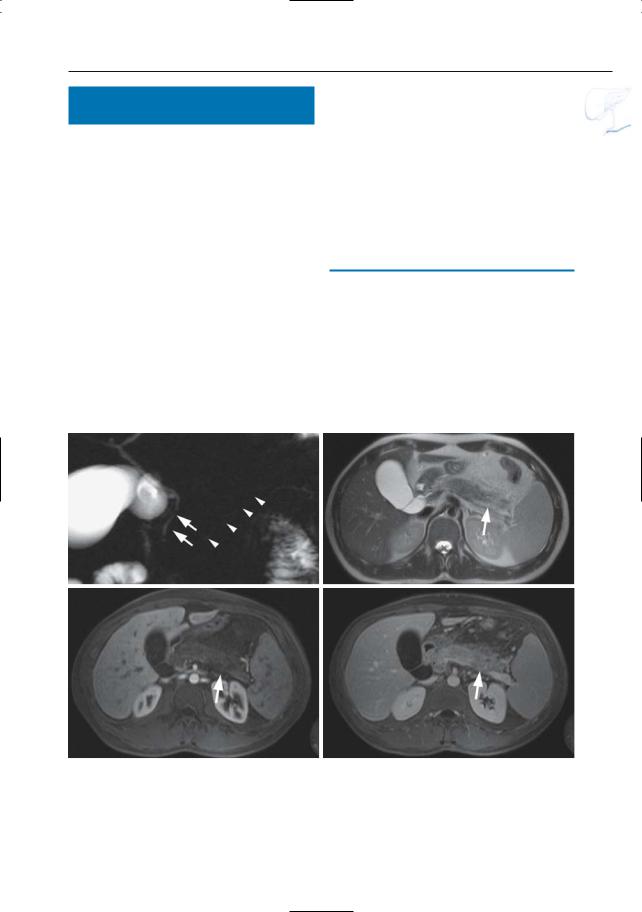
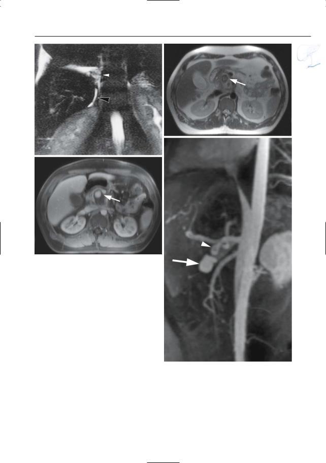
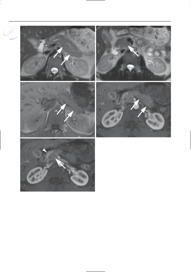
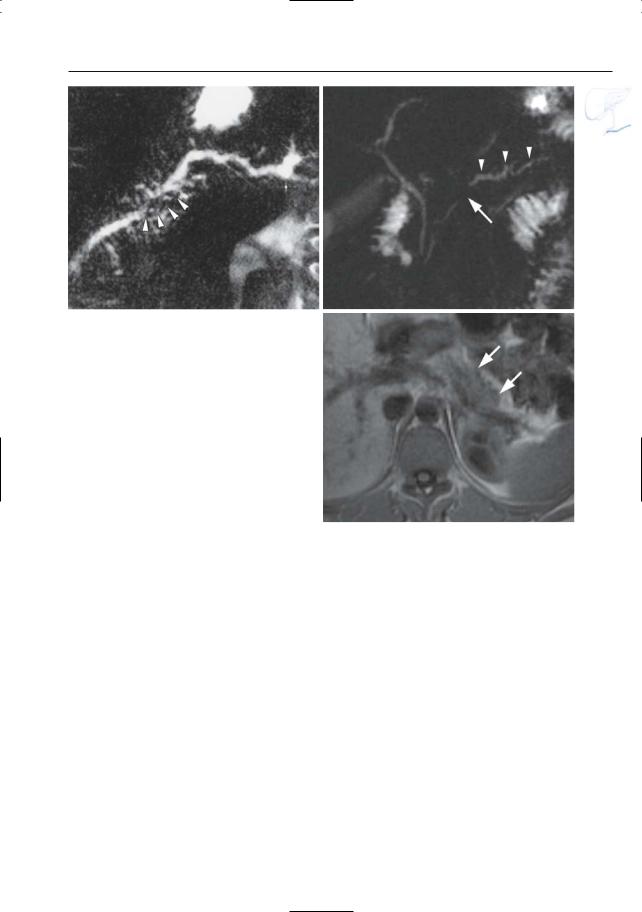
Fig. 152 a–d. Same patient as in Fig. 77 b,c with acute pancreatitis. a Projective image showing discrete narrowing of the distal common bile duct (arrow) and apparent partial absence of the main pancreatic duct due to pancreatic edema (arrowheads). b Axial T2-weighted HASTE image (TE 60) showing increased signal of the pancreatic tissue, particular-
ly in the tail (arrow). c, d Axial contrast-enhanced T1-weighted VIBE images obtained in the arterial (c) and venous (d) phase showing marked absence of pancreatic enhancement in the arterial phase, with delayed enhancement in the venous phase (arrows). This is secondary to acute pancreatitis and pancreatic edema

308 6.2 Benign Nontraumatic Abnormalities
#153 Acute Pancreatitis
with Peripancreatic
Inflammation (Grade C)
KEY FACTS: DISEASE
●Incidence: 40%–50% of patients with acute pancreatitis
●Mechanism: because the pancreas does not have a well-developed capsula,secretions are commonly discharged into the retroperitoneum
KEY FACTS: MRI
●Cross-sectional images: identical to grade B, plus peripancreatic fluid/edema (best appreciated on heavily T2-weighted MR images; Fig. 153)
●Projective images:
–Ductal changes: identical to grade B
–Not uncommonly, the pancreatic duct is obscured by overlying fluid
●Note: The presence of peripancreatic ! fluid may be the first and/or only sign of acute pancreatitis on MRI
References
Gryspeerdt S, Van Hoe L, Baert AL (1998) MRI of pancreatitis. In: Heuck A, Reiser M (eds) Magnetic resonance imaging of the abdomen and pelvis. Springer, Berlin Heidelberg New York, pp 91–108
Saïfuddin A, Ward J, Ridgway J, Chalmers AG (1993) Comparison of MR and CT scanning in severe acute pancreatitis: initial experience. Clin Radiol 48 : 111–116
6 Pancreatic Ducts 309
a |
b |
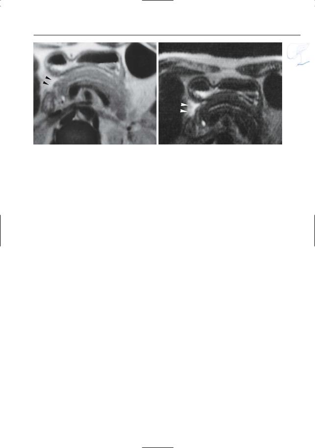
Fig. 153. a Moderately and b heavily T2-weighted MR images showing hyperintense aspect of fat adjacent to the pancreatic head (arrowheads): peripancreatic inflammation

310 6.2 Benign Nontraumatic Abnormalities
#154 Acute Pancreatitis
with Ill-Defined Fluid
Collection/Phlegmon
(Grades D and E)
KEY FACTS: DISEASE
●Definition of phlegmon: solid mass characterized by edema, infiltration of inflammatory cells, and necrosis
●Incidence: up to 50% of patients with acute pancreatitis
●Classification:
–Grade D: small and usually single, illdefined fluid collection or phlegmon
–Grade E: two or more large phlegmonous collections with or without the presence of gas
●Spread: primarily in anterior pararenal and interfascial spaces
●Evolution: commonly associated with abscess formation and necrosis; mortality rate between 4% and 40%
KEY FACTS: MRI
●Typical appearance of fluid collections (Fig. 154a–d):
–Pure water (remains visible on T2weighted images with very long TE)
–Typically located around the pancreas and in the anterior pararenal space. Other locations include the lesser sac, posterior pararenal space, mesocolon, mesenterium, psoas compartment, mediastinum, pleura, pericardium
●Typical appearance of phlegmon (Fig. 154b–d):
–Ill-defined structure with heterogeneous content
–Signal intensity on T2: mixed, usually with both hyperintense and hypointense portions (e.g.,necrotic debris,
blood)
● Note: MRI appears to be more accurate ! than CT for differentiating between pure fluid collections and phlegmons (these may exhibit fluid density on CT). Therefore, predrainage MRI should be performed in patients with subacute pancreatic collections to avoid attempts to drain “collections” actually representing necrotic debris (Morgan et al. 1997; Fig. 154)
References
Balthazar EJ (1994) Pancreatitis. In: Gore RM, Levine MS, Laufer I (eds) Textbook of gastrointestinal radiology. Saunders, Philadelphia, pp 2132–2160
Balthazar EJ, Ranson JHC, Naidich JP, Megibow AJ, Caccavale R, Cooper MM (1985) Acute pancreatitis: prognostic value of CT. Radiology 156 : 767–772
Morgan DE, Baron TH, Smith JK, Robbin ML, Kenney PJ (1997) Pancreatic fluid collections prior to intervention: evaluation with MR imaging compared with CT and US. Radiology 203 : 773–778
6 Pancreatic Ducts 311
a |
b |
c |
d |
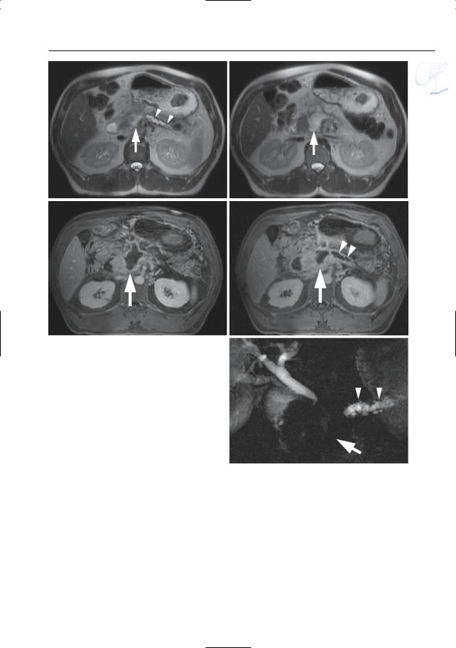
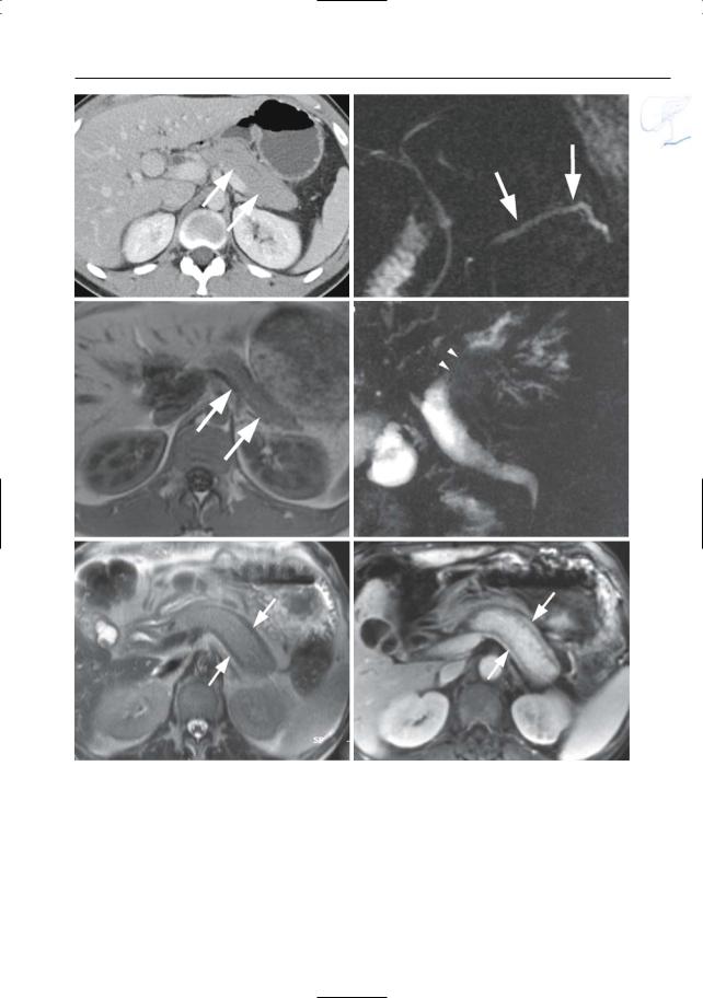
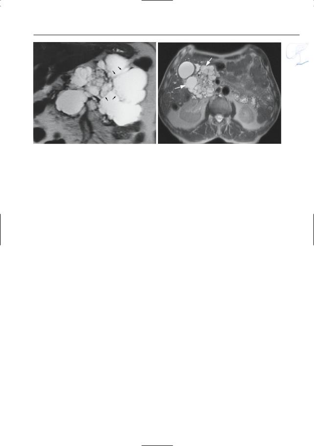
Fig. 154. a Heavily T2-weighted image showing classical spread of pancreatic fluid collections into the left and right interfascial space (arrows). b–d Axial T2-weighted HASTE images with short TE (TE 60) (b, c) and long TE (TE 360) (d) showing
an enlarged slightly hyperintense heterogeneous pancreas (small arrow), with peripancreatic exudate/phlegmon (arrowheads), and a fluid collection anterior to the pancreas (large arrow)

312 6.2 Benign Nontraumatic Abnormalities
|
|
|
|
KEY FACTS: MRI |
|
|
|
|
#155 Complications |
|
|
||
|
|
|
|
|
|
|
|
|
of Acute Pancreatitis (1): |
|
● Typical features: |
|
|
|
|
Necrosis |
|
! |
||
|
|
|
|
– Signal intensity: necrosis has no spe- |
||
|
|
|
|
|||
|
|
|
|
|||
|
|
KEY FACTS: DISEASE |
cific features on non-contrast-en- |
|||
|
|
hanced scans |
||||
|
|
|
|
|
||
|
|
● Classification: |
– Contrast-enhanced scans: focal or dif- |
|
||
|
|
fuse absence of enhancement (Fig.155) |
|
|||
|
|
– A: less than 30% of pancreatic par- |
|
|||
|
|
– Projective images: focal interruption |
|
|||
|
|
enchyma |
|
|||
|
|
of the pancreatic duct |
|
|||
|
|
– B: 30%–50% |
|
|||
|
|
● Note: Rapid injection of intravenous |
|
|||
|
|
– C: more than 50% |
|
|||
|
|
● Detection of necrosis has important |
contrast media and adequate bolus tim- |
|
||
|
|
ing are imperative for accurate detection |
|
|||
|
|
prognostic implications (Ranson et al. |
|
|||
|
|
of necrosis by MRI (Piironen et al. 1997) |
|
|||
|
|
1985): |
|
|
||
|
|
|
● Note: Necrosis may develop a few days |
|
||
|
|
– No necrosis: almost no mortality |
|
|||
|
|
– Necrosis: up to 23% mortality |
after the onset of acute pancreatitis and |
|
||
|
|
therefore may not be detected on the ini- |
|
|||
|
|
● The extent of necrosis also appears to be |
|
|||
|
|
tial contrast-enhanced study |
|
|||
|
|
important: |
|
|||
|
|
– Less than 30% of pancreatic par- |
|
|
|
|
|
|
enchyma: no mortality |
|
|
|
|
|
|
References |
|
|||
|
|
– More than 50%: 11%–25% mortality |
|
|||
|
|
|
|
|
||
|
|
● The severity index is calculated by |
Balthazar EJ, Robinson DL, Megibow AJ, Ranson JH |
|
||
|
|
adding together a morphologic score |
(1990) Acute pancreatitis: value of CT in estab- |
|
||
|
|
reflecting the grade of disease (Sg) (see |
lishing prognosis. Radiology 174 : 331–336 |
|
||
|
|
# 151) and a score reflecting the degree of |
Miller FH, Keppke AL, Dalal K et al. (2004) MRI of |
|
||
|
|
pancreatitis and its complications: part 1, acute |
|
|||
|
|
necrosis (Sn) (Balthazar et al. 1990; |
|
|||
|
|
pancreatitis. AJR Am J Roentgenol 183 : 1637– |
|
|||
|
|
Table 155) |
|
|||
|
|
1644 |
|
|
||
|
|
● Mortality as a function of the severity |
Piironen A, Kivisaari R, Pitkäranta P et al (1997) |
|
||
|
|
index: |
Contrast-enhanced MRI for the detection of |
|
||
|
|
– Severity index 0–3: 3% |
acute hemorrhagic necrotizing pancreatitis. Eur |
|
||
|
|
Radiol 7 : 17–20 |
|
|||
|
|
– Severity index 4–6: 6% |
|
|||
|
|
Ranson JHC, Balthazar E, Caccavale R, Cooper M |
|
|||
|
|
– Severity index 7–10: 17% |
(1985) Computed tomography and the predic- |
|
||
|
|
● If necrotic tissue becomes infected, the |
tion of abscess in acute pancreatitis. Ann Surg |
|
||
|
|
mortality rate rises to about 60% |
201 : 656–665 |
|
||
|
|
|
|
|
||
|
|
Table 155.1a. Grade of disease (Sg) |
Table 155.1b. Degree of necrosis (Sn) |
|
||
Grade |
Score (Sg) |
A |
0 |
B |
1 |
C |
2 |
D |
3 |
E |
4 |
Necrosis (%) |
Score (Sn) |
< 30 |
2 |
30–50 |
4 |
>50 |
6 |
6 Pancreatic Ducts 313
a
c
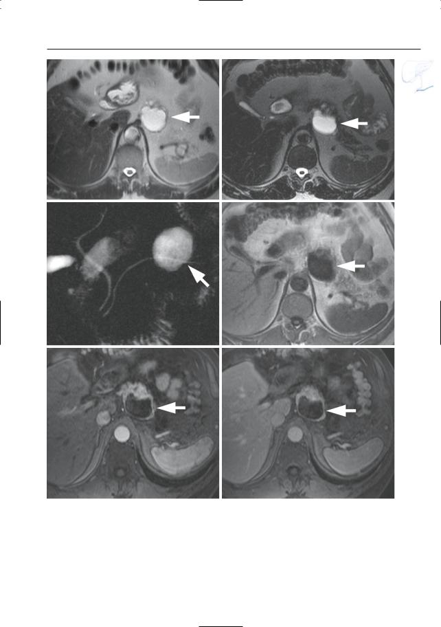
Fig. 155 a–e. Axial T2-weighted HASTE images (TE 60) (a, b) and axial contrast-enhanced T1weighted VIBE images (c, d) obtained in the venous phase showing a fluid collection in the pancreatic head/neck (arrow), with dilatation of the proximal pancreatic duct (arrowheads). e Projective image showing a dilated proximal pancreatic duct (arrowheads) and absence of a visible pancreatic duct in the pancreatic head/neck (arrow)
b
d
e

314 6.2 Benign Nontraumatic Abnormalities
#156 Complications
of Acute Pancreatitis (2):
Pseudocyst
Related topic: # 165 (pseudocyst in chronic pancreatitis)
KEY FACTS: DISEASE
●Collection of pancreatic fluid encapsulated by fibrous tissue (no true epithelial lining)
●The transition from fluid collection (without fibrous “wall”) to pseudocyst takes 4–6 weeks
●Incidence:
–90% of all cystic pancreatic masses
–2%–4% after initial attack of acute pancreatitis
–12% after several episodes of alcoholic pancreatitis
–10%–15% in chronic pancreatitis
●Other causes:
–Obstructive tumor
–Trauma
●Location:
–Most commonly in the anterior pararenal space and omental bursa
–Anywhere from the peripancreatic area to the pleura, groin, etc.
●Symptoms: persistent pain in a patient who is not seriously ill is typical
●Complications:
–Rupture/fistula formation (e.g., into abdominal cavity, stomach)
–Hemorrhage (with or without pseudoaneurysm)
–Infection
●Treatment:
–Surgery
–Percutaneous drainage
–Endoscopic drainage (cystogastrostomy or cystoduodenostomy)
KEY FACTS: MRI
●Typical features (Fig. 156):
–Unilocular in 94%
–Usually has the same intensity as water
–May contain internal material (e.g., blood clot, cellular debris)
–May have variable/mixed signal intensity if hemorrhagic
●Presence of air within a pseudocyst (hypointense on all sequences, sometimes with air–fluid level) suggests one of the following:
–Infection (usually anaerobe bacteria)
–Previous percutaneous, endoscopic, or surgical intervention
–Fistula with gastrointestinal tract
●Note: MRI is less accurate than ERCP in demonstrating duct-pseudocyst communication
References
Ahearne PM, Baillie JM, Cotton PB, Baker ME, Meyers WC, Pappas TN (1992) An endoscopic retrograde cholangiopancreatography (ERCP)-based algorithm for the management of pancreatic pseudocysts. Am J Surg 163 : 111–115
Miller FH, Keppke AL, Dalal K et al. (2004) MRI of pancreatitis and its complications: part 1, acute pancreatitis. AJR Am J Roentgenol 183 : 1637– 1644

6 Pancreatic Ducts 315
a
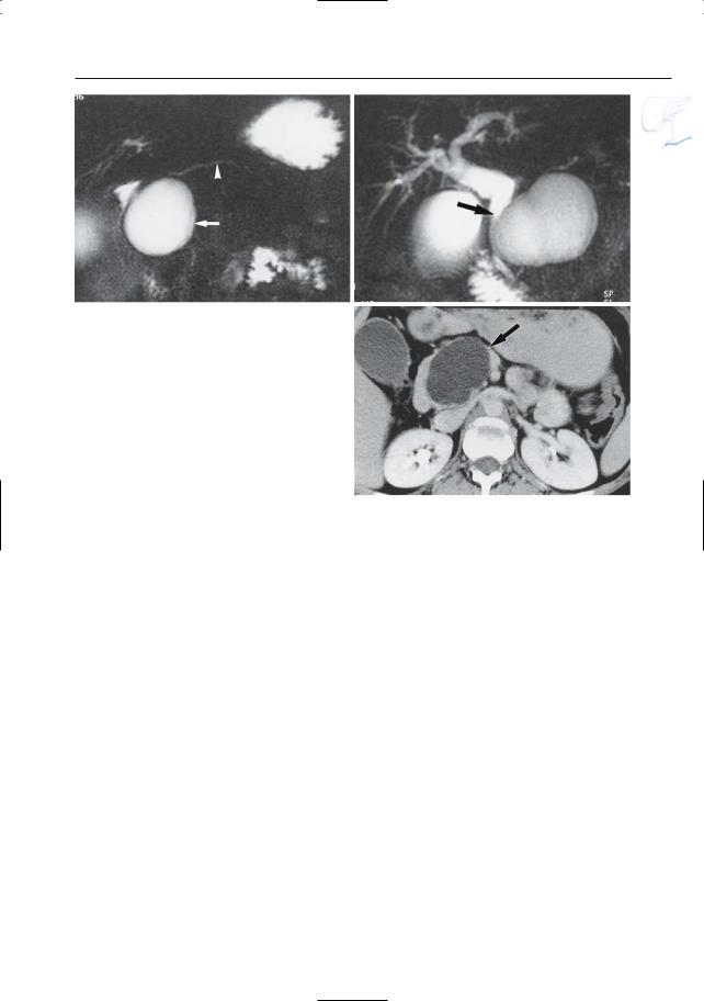
Fig. 156 a–c. Axial CT image (a) showing marked peripancreatic infiltration (arrows) due to acute biliary pancreatitis. b, c Same patient 1 month later. Axial T2-weighted HASTE image (b) and RARE image (c) showing a new large fluid collection in the pancreatic head/neck (arrow). Complicated acute pancreatitis with pseudocyst
b
c

316 6.2 Benign Nontraumatic Abnormalities
#157 Complications
of Acute Pancreatitis (3):
Vascular Complications
Related topic: # 166 (vascular complications of chronic pancreatitis)
KEY FACTS: DISEASE
Arterial Complications:
Pseudoaneurysm
●Up to 10% of severe cases of acute pancreatitis
●Mechanism: pancreatic or peripancreatic blood vessel erosion (autodigestion of arterial walls by enzymes liberated in pancreatitis)
●May occur within 2–3 weeks to several years after an acute episode of pancreatitis
●Location:
–Splenic and gastroduodenal arteries most commonly involved
–May develop within a preexisting pseudocyst
●Complications: rupture with free hemorrhage in the duodenum, bile duct, lesser sac, peritoneal cavity, pancreatic duct, portal vein, etc.
●Treatment: usually therapeutic angiographic embolization (Mandel et al. 1987)
Venous Complications:
Venous Thrombosis
●Incidence : up to 45% of patients with acute pancreatitis
●Commonly involves the mesenteric– splenic confluence and may extend into the main portal vein
●Complication: varices of the gastric fundus
●Treatment for bleeding gastric varices: splenectomy
KEY FACTS: MRI
Pseudoaneurysm (Fig. 157)
● Typical features:
– Signal intensity: variable, depends on |
! |
|
|
type of sequence and flow velocity |
|
(the typical flow void may be absent |
|
in pseudoaneurysms with a small |
|
connection to the feeding artery) |
|
–Key finding on contrast-enhanced MRA: enhancement in the arterial phase
●Note: Fast injection of contrast media and accurate bolus timing are mandatory (Gaa et al. 1997)
Venous Thrombosis
●Typical features:
–Absence of flow void
–No enhancement
–Presence of venous collaterals
References
Gaa J, Laub G, Georgi M (1997) Breath-hold threedimensional gadolinium-enhanced dual-phase MR angiography in the abdomen: first clinical results. In: Oudkerk M, Edelman R (eds) Highpower gradient MR-imaging. Blackwell Science, Berlin, pp 334–339
Mandel SR, Jaques PF, Sanofsky S, Mauro MA (1987) Non operative management of peripancreatic arterial aneurysms. A 10 years experience. Ann Surg 205 : 126–128
Vujic I (1989) Vascular complications of pancreatitis. Radiol Clin North Am 27 : 81–91
6 Pancreatic Ducts 317
b
a
c |
d |
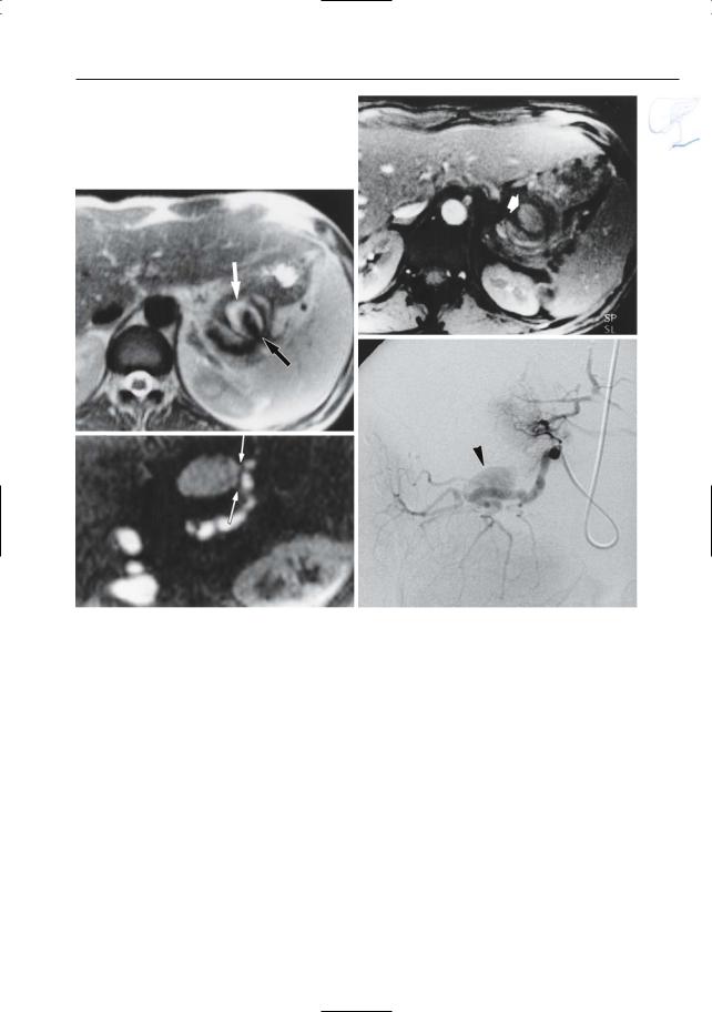
Fig. 157 a–d. Patient with chronic pancreatitis. a T2-weighted image showing pseudoaneurysm in close connection with the pancreatic tail (arrows). The hypointense signal in the center of the lesion is probably caused by flow-related signal void. b Time- of-flight (TOF) image confirming the presence of
flow in the center of the lesion (arrow). c Contrastenhanced image showing a connection with the splenic artery (arrows). d Catheter angiogram confirming the presence of a pseudoaneurysm
(arrowhead)

318 6.2 Benign Nontraumatic Abnormalities
#158 Recurrent (Relapsing)
Pancreatitis
KEY FACTS: DISEASE
●Two or more episodes of acute pancreatitis
●Causes/associated disease (found in up to 80% of patients):
–Segmental stenosis of the pancreatic duct
–Papillary obstruction/dysfunction (see # 127, 128)
–Common bile duct calculi (see # 72), biliary sludge
–Pancreas divisum (see # 140)
–Anomalous union of pancreatobiliary duct system (see # 123)
–Duodenal duplication cyst
KEY FACTS: MRI
●Signs of acute pancreatitis may be seen
●Signs of underlying disease (see respective entities)
●Note: The role of MRI is not to confirm the diagnosis of pancreatitis, but to detect (or rule out) underlying abnormalities
References
Asbun HJ, Rossi RL, Heiss FW, Shea JA (1993) Acute relapsing pancreatitis as a complication of papillary stenosis after sphincterotomy. Gastroenterology 104 : 1814–1817
Mori K, Nagakawa T, Ohta T et al. (1991) Acute pancreatitis associated with anomalous union of the pancreaticobiliary ductal system. J Clin Gastroenterol 13 : 673–677
Venu RP,Geenen JE,Hogan WJ,Stone J,Johnson GK, Soergel K (1989) Idiopathic recurrent pancreatitis. An approach to diagnosis and treatment. Dig Dis Sci 34 : 1943–1945

6 Pancreatic Ducts 319
a
Fig. 158 a–c. a Moderately and b heavily T2weighted MR images showing edematous pancreatitis (increased signal intensity) in the pancreatic tail, without exudate (arrows). The duct of Wirsung is focally dilated. c Same patient. Projective image showing narrowing of the pancreatic duct, not only in the pancreatic body/tail (arrows), but also in the pancreatic neck (arrowheads). These stenoses were considered to be the cause of the recurrent attacks of pancreatitis confined to the tail. Also note the presence of a small pseudocyst in c. The dilated portion of the duct of Wirsung is not visible on this image
b
c

320 6.2 Benign Nontraumatic Abnormalities
#159 Chronic Pancreatitis, General
KEY FACTS: DISEASE
●Continuing inflammatory disease characterized by irreversible damage to pancreatic anatomy and function
●Types:
–Chronic calcifying pancreatitis (most common)
–Chronic obstructive pancreatitis (see # 168)
–Nonalcoholic duct-destructive pancreatitis (see # 169)
–Hereditary pancreatitis (see # 170)
–Idiopathic
●Causes:
–Alcoholism (75%)
–Others: e.g., hyperlipidemia, hyperparathyroidism, trauma
●Pathophysiology (main mechanism): intraductal precipitation of (calcified) concretions, with secondary obstruction, scar formation, etc.
●Pathophysiology (possible underlying mechanisms):
–Alcohol-induced pancreatic secretion of protein
–Formation of a mucosubstance
●Symptoms: epigastric pain, weight loss, steatorhea (80%), diabetes (58%)
●Differential diagnosis with malignant tumor of the pancreas:
–Clinically, the differentiation between chronic pancreatitis and malignancy is often difficult
–The two diseases may coexist
●Complications:
–Pseudocyst (see # 165)
–Vascular (see # 166)
–Fistula (see # 166)
–Biliary (see # 78)
● Note: In accordance with most textbooks |
|
|
! |
||
and papers, the discussion on the pages |
||
that follow will refer to “chronic pancre- |
|
|
|
||
atitis” as a single disease. However, the |
|
|
reader should be aware of the fact that |
|
|
chronic obstructive pancreatitis and |
|
|
nonalcoholic duct-destructive pancre- |
|
|
atitis have quite characteristic imaging |
|
|
features that are clearly different from |
|
|
those seen in “classical” chronic pancre- |
|
|
atitis. For a discussion of these entities, |
|
|
the reader is referred to # 168 and # 169 |
|
6 Pancreatic Ducts 321
#160 Chronic Pancreatitis,
Signal Intensity Changes
and Enhancement Pattern
KEY FACTS: MRI
●Histology: normal pancreatic parenchyma is partially replaced by fibrotic tissue that contains no aqueous protein (no acini) and is less well vascularized
●Typical MR features (more commonly diffuse than focal; Fig. 160):
–Loss of the normal high signal intensity on T1-weighted images
–Decreased enhancement on dynamic contrast-enhanced sequences
●Differential diagnosis: the abnormalities described above are not specific and can be seen in:
–Pancreatic cancer (with or without retro-obstructive pancreatitis; clues for differentiation: see # 167)
–Acute pancreatitis
●Note: Punctate calcifications are the hallmark of chronic alcoholic pancreatitis. They are commonly invisible on MRI. Therefore, performing CT is always a good option when MRI findings suggest chronic pancreatitis,without being diagnostic
References
Semelka RC, Ascher SM (1993) MR imaging of the pancreas. Radiology 188 : 593–602
Sittek H, Heuck AF, Folsing C, Gieseke J, Reiser M (1995) Static and dynamic MR tomography of the pancreas: contrast media kinetics of the normal pancreatic parenchyma in pancreatic carcinoma and chronic pancreatitis. ROFO 162 : 396–403
Winston CB, Mitchell DG, Outwater EK, Ehrlich SM (1995) Pancreatic signal intensity on T1-weighted fat saturation MR images: clinical correlation. J Magn Reson Imaging 5 : 267–271
a |
b |
Fig. 160. a Non-contrast-enhanced T1-weighted MR image and b image obtained in the pancreatic phase of perfusion showing hypointense, hypoenhancing pancreas (arrows)

322 6.2 Benign Nontraumatic Abnormalities
#161 Chronic Pancreatitis,
Ductal Changes (1):
ERCP Classification
KEY FACTS
●Progressive changes in the main pancreatic duct: see Fig. 161a
●Progressive changes in side branches: see Fig. 161b
●Basic elements of chronic pancreatitis:
(a)changes in the main pancreatic duct,
(b)changes in side branches, and (c) intraductal concretions
●ERCP classification (Cambridge classification; Axon 1989):
– Mild CP: at least three abnormal side branches
– Moderate CP: addition of main pan-
creatic duct changes
– Marked CP: addition of one or more of the following: severe dilation (> 1 cm); marked mural irregularity; obstruction; opacification of large cavity (> 1 cm); intraductal filling defects
References
Axon AT (198) Endoscopic retrograde cholangiopancreatography in chronic pancreatitis. Cambridge classification. Radiol Clin North Am 27 : 39–50
Chari ST, Singer MV (1994) The problem of classification and staging of chronic pancreatitis. Proposals based on current knowledge of its natural history. Scand J Gastroenterol 29 : 949–960
Ponette E,Brys P,Van Steenbergen W (1994) Chronic pancreatitis: endoscopic retrograde cholangiopancreatography. In: Baert AL (ed) Radiology of the pancreas. Springer, Berlin Heidelberg New York, pp 106–115
Taylor AJ, Bohorfoush AG (1997) Pancreatic duct in inflammation of the pancreas. In: Taylor AJ, Bohorfoush AG (eds) Interpretation of ERCP. Lippincott-Raven, Philadelphia, pp 231–259

6 Pancreatic Ducts 323
Fig. 161. a Changes in the main pancreatic duct. 1, normal; 2, mild irregularity; 3, more pronounced irregularity; 4,“chain of lakes”.
b Changes in the side branches. 1, normal; 2, early changes: mild narrowing at the base; 3, marked cystic dilation of a side
branch; 4, cavitation and drainage from necrotic parenchyma;
5, intraductal concretions; 6, complete obstruction of side branch base. (From Taylor and Bohorfoush 1997, with permission)
a
b

324 6.2 Benign Nontraumatic Abnormalities
#162 Chronic Pancreatitis,
Ductal Changes (2):
Early Disease
KEY FACTS: MRI
●With the exception of unpaired branches in the pancreatic head, normal side branches are not visible on MRCP
●Signs of early chronic pancreatitis:
–Visualization of two or more side branches in the body and tail
–Discrete irregularities of the main pancreatic duct (Fig. 162; differential diagnosis: senescent changes – see
# 147)
●Note: Although subtle caliber changes of the main pancreatic duct are usually clearly seen on projective MR cholangiography, MRCP is certainly not as sensitive as ERCP in the detection of the earliest stages of the disease for the following reasons:
–Limitations in spatial resolution
–Absence of ductal distention during imaging (no intraductal injection of contrast medium). Drugs stimulating the secretion of pancreatic juice can be helpful in this respect (see # 19)
References
Fulcher AS, Capps GW, Turner MA (1999) Thoracopancreatic fistula: clinical and imaging findings. J Comput Assist Tomogr 23 : 181–187
Ponette E,Brys P,Van Steenbergen W (1994) Chronic pancreatitis: endoscopic retrograde cholangiopancreatography. In: Baert AL (ed) Radiology of the pancreas. Springer, Berlin Heidelberg New York, pp 106–115
Takehara Y, Ichijo K, Tooyama N et al. (1994) Breathhold MR cholangiopancreatography with a long- echo-train fast spin-echo sequence and a surface coil in chronic pancreatitis. Radiology 192 : 73–78
Taylor AJ, Bohorfoush AG (1997) Pancreatic duct in inflammation of the pancreas. In: Taylor AJ, Bohorfoush AG (eds) Interpretation of ERCP. Lippincott-Raven, Philadelphia, pp 231–259
6 Pancreatic Ducts 325
a
b
c |
d |
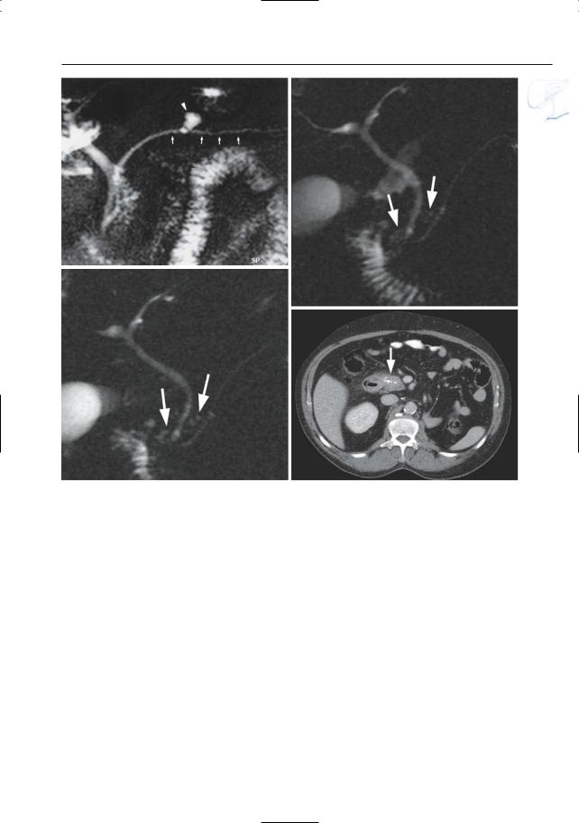
Fig. 162. a Projective MR image showing discrete ductal irregularities (arrows). Also note the presence of a small pseudocyst (arrowhead). b–d Projective images (b, c) showing multiple dilated side
branches in the pancreatic head (arrows). d Axial CT image showing multiple calcifications in the pancreatic head (arrow) confirming the diagnosis of chronic pancreatitis

326 6.2 Benign Nontraumatic Abnormalities
#163 Chronic Pancreatitis,
Ductal Changes (3):
Advanced Disease
Related topic: # 78 (common bile duct stenosis in chronic pancreatitis)
KEY FACTS: MRI
!● Typical features of the main pancreatic duct:
–Dilation and tortuosity
–Short areas of narrowing (strictures greater than 1 cm are less common but not rare)
–Intraductal stones
!● Typical features of side branches:
–Dilation and blunting of side branches
–Nipping, i.e., narrowing of the side branch base with respect to the more peripheral part (Fig. 163c–e)
–Clubbing, i.e., peripheral dilation instead of tapering (Fig. 163c–e)
●Differential diagnosis:
–Obstructive dilation: more uniform dilation of the main duct and side branches, without focal stenoses, nipping, or clubbing (see # 168)
–Pancreatic neoplasm (see # 187)
●Note: MRCP is particularly useful in cases in which ERCP shows obstruction of the main pancreatic duct: the differential diagnosis between a tumor, tight stenosis, intraductal lithiasis, etc. is usually easy to make with MRCP
References
Fulcher AS, Capps GW, Turner MA (1999) Thoracopancreatic fistula: clinical and imaging findings. J Comput Assist Tomogr 23 : 181–187#
Ponette E,Brys P,Van Steenbergen W (1994) Chronic pancreatitis: endoscopic retrograde cholangiopancreatography. In: Baert AL (ed) Radiology of the pancreas. Springer, Berlin Heidelberg New York, pp 106–115
Takehara Y, Ichijo K, Tooyama N et al. (1994) Breathhold MR cholangiopancreatography with a long- echo-train fast spin-echo sequence and a surface coil in chronic pancreatitis. Radiology 192 : 73–78
6 Pancreatic Ducts 327
a
c
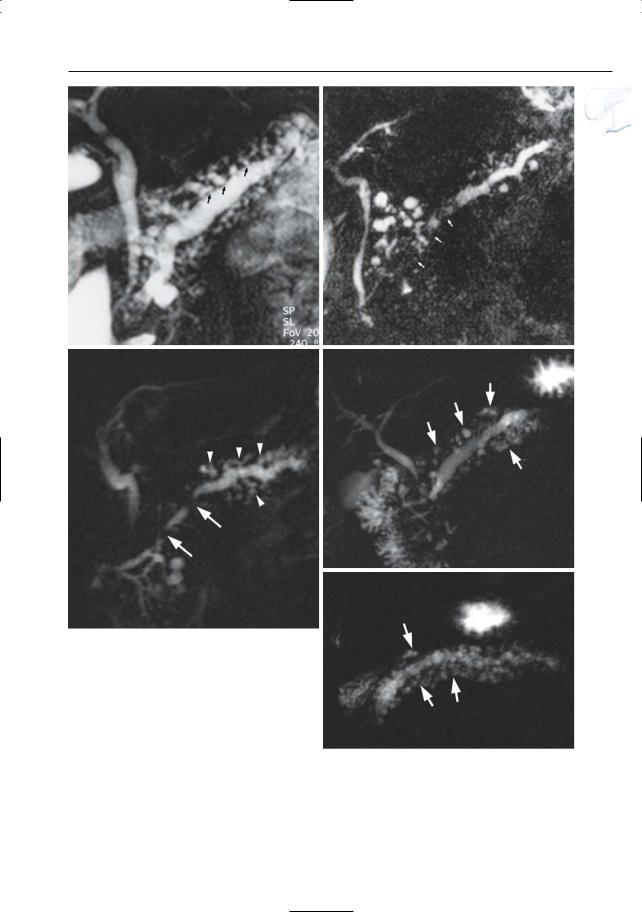
Fig. 163 a, b. a Projective MR image showing nonuniform dilation of the side branches with “nipping” (arrows). b Projective image showing more discrete dilation of the side branches. In this patient, the image is dominated by the presence of multiple small intrapancreatic pseudocysts. Also note the presence of intraductal stones (arrows) and an “hourglass” configuration of the bile duct. c Different patient. Projective image showing typical findings in advanced chronic pancreatitis: dilatation of the pancreatic duct with two stenoses (arrows), and dilatation of the side branches with “nipping” and “clubbing” (arrowheads). d, e. Projective images in different patient showing diffuse dilation of the main pancreatic duct and of several side branches, with “nipping” and “clubbing”
b
d
e

328 6.2 Benign Nontraumatic Abnormalities
#164 Atypical Ductal Changes
in Chronic Pancreatitis
KEY FACTS
Marked Diffuse Dilation of Ducts
●May be caused by distal intraductal stone, orificial stenosis, etc.
●Differential diagnosis:
–Small papillary tumor (see # 130, 132)
–Dysfunction of the vaterian sphincter complex (see # 127)
–Intraductal mucinous neoplasm (see
# 199)
Focal Narrowing
●Focal narrowing of the main pancreatic duct may be the only sign on MRCP in patients with chronic pancreatitis (Fig. 164a)
●Differential diagnosis:
–Acute pancreatitis
–Duct-destructive chronic pancreatitis (see # 169)
–Neoplasm (usually shows more irregular narrowing)
–Rare causes: e.g., granulomatous disease
●Clues in the differential diagnosis with pancreatic cancer: see # 167
Double Duct Sign (Fig. 164 b – d)
●Narrowing of the pancreatic duct and the adjacent segment of the common bile duct
●Although this sign is more classically seen in malignancy, it may also be
observed in patients with chronic pancreatitis (differential diagnosis: more gradual narrowing, kinking, etc.; see
# 167)
References
Ponette E,Brys P,Van Steenbergen W (1994) Chronic pancreatitis: endoscopic retrograde cholangiopancreatography. In: Baert AL (ed) Radiology of the pancreas. Springer, Berlin Heidelberg New York, pp 106–115
6 Pancreatic Ducts 329
a |
b |
c |
d |
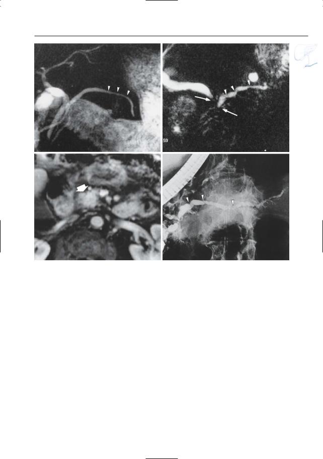
Fig. 164. a Projective MR image showing ductal narrowing in the pancreatic body (arrowheads), without associated abnormalities. b–d Double duct sign in chronic pancreatitis. b Projective MR image showing double duct sign (arrows), possibly indicating a pancreatic tumor.Also note the multiple short stenoses of the pancreatic duct (arrowheads) and
pseudocysts. c Contrast-enhanced T1-weighted image showing normal enhancement of the pancreatic head (arrow): no signs of a tumor. d ERCP confirming the presence of multiple stenoses (arrowheads). Chronic pancreatitis was confirmed by biopsy and follow-up

330 6.2 Benign Nontraumatic Abnormalities
#165 Complications of Chronic
Pancreatitis (1): Pseudocyst
Related topic: # 156 (pseudocyst in acute pancreatitis)
KEY FACTS: DISEASE
●Definition, classification, etc.: see # 156
●Can occur in chronic pancreatitis with or without superimposed acute pancreatitis
●Treatment: surgical or endoscopic drainage
KEY FACTS: MRI
●Signal intensity: although most pseudocysts exhibit fluid intensity, the signal intensity may be variable if blood degradation products are present within the pseudocyst
●Uncomplicated pseudocyst: differential diagnosis:
–Simple cyst
–Mucinous neoplasm (see # 198): may be indistinguishable from pseudocysts on MRCP; clinical history, puncture with aspiration, and/or ERCP may provide important clues
●Differential diagnosis of complicated (hemorrhagic) pseudocyst:
–Phlegmon (may also contain hemorrhage)
–Hemorrhagic tumor (diagnostic clues: associated signs of chronic pancreatitis, secondary signs of malignancy, presence and degree of upstream ductal dilation and atrophy, visualization of solid components)
●Note: Small intrapancreatic pseudocysts are very common in chronic pancreatitis; similar small fluid collections may be seen in:
–Acute pancreatitis
–Pancreatic carcinoma (more focal)
References
Ahearne PM, Baillie JM, Cotton PB, Baker ME, Meyers WC, Pappas TN (1992) An endoscopic retrograde cholangiopancreatography (ERCP)-based algorithm for the management of pancreatic pseudocysts. Am J Surg 163 : 111–115
Sand JA, Seppannen SK, Nordback IH (1997) Intracystic hemorrhage in pancreatic pseudocysts: initial experiences of a treatment protocol. Pancreas 14 : 187–191
van Sonnenberg E,Wittich GR, Casola G et al. (1989) Percutaneous drainage of infected and noninfected pancreatic pseudocysts: experience in 101 cases. Radiology 170 : 757–761
6 Pancreatic Ducts 331
b
a
d
c |
e |
Fig. 165 a, b. Projective image (a) showing typical findings in advanced chronic pancreatitis: dilatation of the main pancreatic duct, prominent dilatation of the side branches with “nipping” (arrowheads) and a pseudocyst in the pancreatic head (arrow). Axial T2-weighted HASTE image (b) showing the fluid collection in the pancreatic head: pseudocyst (arrow). c Different patient. Projective
image showing two small pseudocysts in the pancreatic head and several peripancreatic pseudocysts extending towards the stomach (arrows). d, e Different patient. Axial T1 weighted FLASH image (d) and T2-weighted HASTE image (e) showing a large hemorrhagic pancreatic pseudocyst with fluid–fluid level (small arrow)
332 6.2 Benign Nontraumatic Abnormalities
#166 Complications
of Chronic Pancreatitis (2):
Other Complications
Related topics: # 74 (common bile duct stones complicated by fistula), # 157 (vascular complications in acute pancreatitis)
KEY FACTS: DISEASE
Vascular Complications
●See # 157
●Types:
–Pseudoaneurysm
(encountered in 6%–10% of patients)
–Venous thrombosis
Fistula
●Pancreatic fluid may penetrate several organs/cavities and create fistulas:
–Pleura: pathway usually via the lesser sac to the aortic or esophageal hiatus
–Peritoneum (pancreatic ascites): via the lesser sac and foramen of Winslow
–Portal vein
●May develop de novo or secondary to rupture of a pseudocyst
a
KEY FACTS: MRI
●Vascular complications: see # 157 (Fig. 166d–f)
●Fistula: usually best seen on coronal heavily T2-weighted cross-sectional images; both the rupture of the pancreatic duct and the connection with the pleura may be demonstrated by MRCP (Fig. 166a–c)
References
Fulcher AS, Capps GW, Turner MA (1999) Thoracopancreatic fistula: clinical and imaging findings. J Comput Assist Tomogr 23 : 181–187
Maule WF, Reber HA (1985) Diagnosis and management of pancreatic pseudocysts, pancreatic ascites, and pancreatic fistulas. In: Go VLM, Gardner JD, Brooks FP et al. (eds) The exocrine pancreas: biology, pathobiology and diseases. Raven, New York, pp 601–610
Miller FH, Keppke AL, Wadhwa A et al. (2004) MRI of pancreatitis and its complications: part 2, chronic pancreatitis. AJR Am J Roentgenol 183 : 1645–1652
Sankaran S, Sugawa C,Walt AJ (1979) Value of endoscopic retrograde pancreatography in pancreatic ascites. Surg Gynecol Obstet 148 : 185–192
Vujic I (1989) Vascular complications of pancreatitis. Radiol Clin North Am 27 : 81–91
b
Fig. 166. a Heavily T2-weighted axial image (a) showing communication (arrows) between the distal pancreatic duct and a retroperitoneal collec-
tion. Coronal images (b, c) showing connection between the retroperitoneal collection and the right pleural space (arrowheads)
6 Pancreatic Ducts 333
c
e
Fig. 166 d–f. Axial T2-weighted HASTE image (d) showing a small heterogeneous lesion in the pancreatic head (arrow). e Axial contrast-enhanced T1weighted VIBE image obtained in the arterial phase showing bright contrast enhancement of the anterior portion of this lesion (arrow): pseudoaneurysm. f MIP image showing the small pseudoaneurysm (arrow) adjacent to the gastroduodenal artery
(arrowhead)
d
f

334 6.2 Benign Nontraumatic Abnormalities
#167 Chronic Pancreatitis
with Focal Inflammatory Mass
KEY FACTS: DISEASE
●A focal inflammatory mass is seen in up to 30% of patients with chronic pancreatitis (up to 49% in some series; Luetmer et al. 1989), most commonly in the pancreatic head
●Etiology:
–Superimposed acute pancreatitis
–Possibly a specific chronic inflammatory process
●Symptoms: patients with chronic pancreatitis and pancreatic head enlargement commonly have severe pain
KEY FACTS: MRI
!Together with the segmental type of chronic pancreatitis (see # 164), this entity commonly poses problems in the differential diagnosis with pancreatic carcinoma. The following features suggest chronic pancreatitis:
●On projective images:
–Dilation of the main pancreatic duct distal to the site of stenosis (i.e., downstream)
–Abnormal side branches distal to the site of stenosis
–Intraductal stones
–Multiple pseudocysts at different locations
–Side branches: nipping, clubbing, distorted appearance
–Signs of attraction of the common bile duct (suggesting fibrosis; see #78)
–Smooth tapering of the common bile duct
–Duct-penetrating sign (Ichikawa et al. 2001)
●On cross-sectional images:
–CT: calcifications within the mass
–Visualization of dilated side branches traversing the suspect area (Fig. 167a–d) (in malignant tumors, obstructed side branches are seen adjacent to, but not within the mass; see Fig. 187a)
–No atrophy or uniform atrophy of the gland
–Focal mass with relatively high signal intensity on non-fat-suppressed T1weighted images (most reliable sign but not always present) (Fig. 167k–n)
–Focal mass not presenting a a “black hole” on early-phase dynamic con- trast-enhanced images (Fig. 167k–n, compare to Fig. 1 and Fig 186a–f)
References
Büchler M, Malfertheiner M, Friess H, Senn T, Beger HG (1990) Chronic pancreatitis with inflammatory mass in the head of the pancreas: a specific entity? In: Beger HG (ed) Chronic pancreatitis. Springer, Berlin Heidelberg New York, pp 41–46
Ichikawa T, Sou H, Araki T et al. (2001) Duct-pene- trating sign at MRCP: usefulness for differentiating inflammatory pancreatic mass from pancreatic carcinomas. Radiology 221 : 107–116
Ishihara T, Yamaguchi T, Tsuyuguchi T, Hayasake A, Saisho H (1996) Characteristic findings of ERCP in the diagnosis of chronic pancreatitis with inflammatory mass. Nippon Shokakibyo Gakkai Zasshi 93 : 725–731
Luetmer PH, Stephens DH,Ward EM (1989) Chronic pancreatitis: reassessment with current CT. Radiology 171 : 353–357
6 Pancreatic Ducts 335
a |
b |
c |
d |
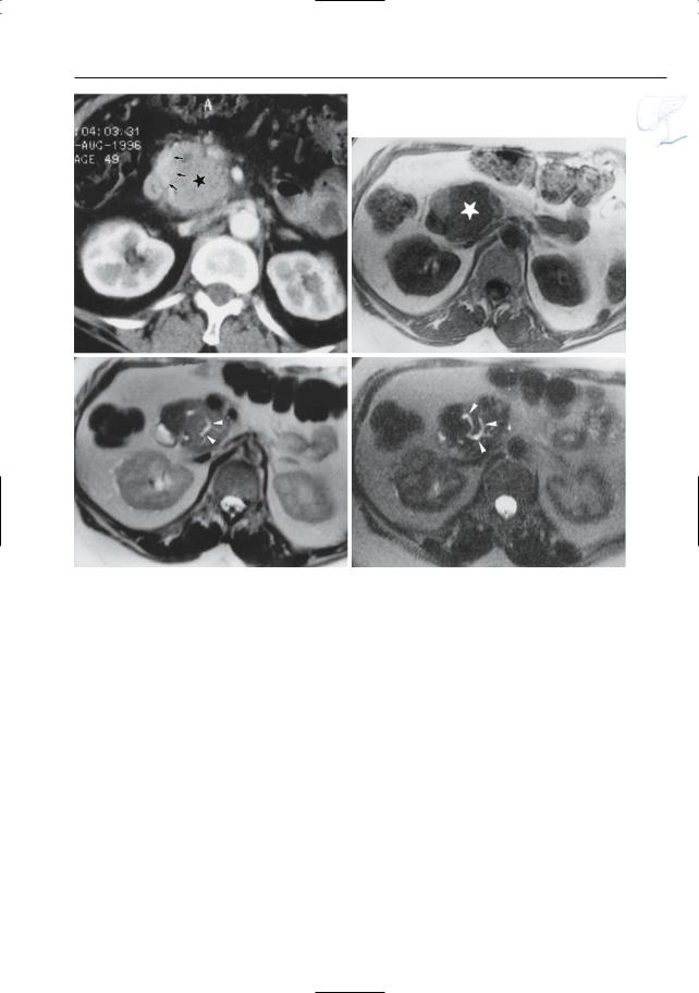
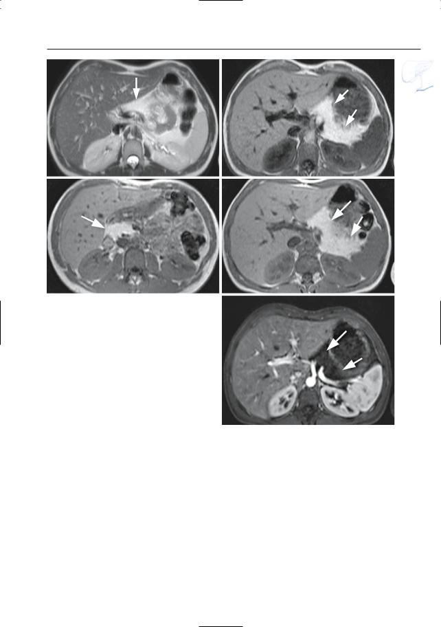
Fig. 167 a–d. Patient with chronic pancreatitis. a CT image showing enlargement and decreased iodine uptake of the pancreatic head (asterisk): Tumor? Note the normal enhancement more laterally (arrows). b T1-weighted MR image showing
the enlarged part of the pancreatic head as markedly hypointense (asterisk), which is still consistent with a tumor. c Moderately and d heavily T2weighted images showing dilated side branches (arrowheads): this rules out a tumor.
336 6.2 Benign Nontraumatic Abnormalities
e |
f |
g |
h |
i |
j |
Fig. 167 e–j. T1-(e) and T2-(f)weighted images showing a focal lesion in the pancreatic tail that appears as a T1-hypointense and T2-hyperintense mass (arrow). Axial contrast-enhanced T1-weighted VIBE images obtained in the arterial (g) and venous (h) phase showing this mass to be hypovascular
(arrow). Axial CT images (i, j) also confirm the presence of a small hypointense mass in the pancreatic tail (arrow). Needle biopsy and lack of evolution over a period of 9 months proved this mass to be a focal inflammatory mass.
6 Pancreatic Ducts 337
k |
l |
m |
n |
Fig. 167 k–n. k Projective image showing a doubleduct sign (arrow). l Axial T1-weighted image showing an enlarged pancreatic head (arrow) with small hypointense areas at the duodenal groove. m Axial contrast-enhanced T1-weighted VIBE image obtained in the arterial phase showing decreased/delayed uptake of contrast medium (arrow). n Image obtained in the venous phase sho-
wing late enhancement of this mass (arrow). The enhancement features together with the relatively high signal intensity at T1-weighted images favour the diagnosis of a focal inflammatory mass. Because of increasing biochemical abnormalities, surgery was performed. Final diagnosis: groove pancreatitis, no tumor

338 6.2 Benign Nontraumatic Abnormalities
#168 Chronic Obstructive
Pancreatitis
Related topic: # 194 (adenocarcinoma with retro-obstructive pancreatitis)
KEY FACTS: DISEASE
●Pancreatitis related to (caused by) obstruction of the main pancreatic duct
●Histology: evenly distributed chronic parenchymal inflammation with preservation of the ductal epithelium. Intraductal stones are uncommon (except as a causal factor)
●Cause: either benign (e.g., papillary stenosis) or malignant
●Complications: endocrine and/or exo-
crine insufficiency may develop if less than 40% of the parenchyma lies beyond the obstruction
●Therapy: removal of obstructive component
KEY FACTS: MRI
(FIG. 168, SEE ALSO FIG. 182 B)
●Typical features:
–Uniform dilation of the main pancreatic duct
–Commonly relative sparing or mild uniform dilation of side branches
–No intraluminal filling defects (except as a causal factor)
–Intraparenchymal pseudocysts may be seen
–Diminished signal intensity of parenchyma on T1-weighted MRI (fibrosis)
●Note: Chronic obstructive pancreatitis is commonly seen in patients with ductal adenocarcinoma. This may cause problems in determining the exact proximal extent of the tumor (both tumor and pancreatitis are hypointense on T2-weighted images). In such cases, tumor delineation may be improved by obtaining contrastenhanced scans (see # 1a–d, 15, 194)
References
Freeny PC (1989) Classification of pancreatitis. Radiol Clin North Am 27 : 1–3
6 Pancreatic Ducts 339
a |
b |
Fig. 168 a, b. a Projective image showing dilatation of the pancreatic duct in the body/tail secondary due to an intraductal stone (arrow). b Axial CT im-
age confirming the presence of a stone in the pancreatic head (arrow)

340 6.2 Benign Nontraumatic Abnormalities
|
|
|
|
|
|
KEY FACTS: MRI (FIG. 169) |
|
||
|
|
#169 Nonalcoholic |
|
|
|||||
|
|
|
|
|
|
|
|||
|
|
|
Duct-Destructive Chronic |
|
● |
Focal or more diffuse mass-like enlarge- |
|
||
|
|
|
Pancreatitis (auto-immune |
|
|
||||
|
|
|
pancreatitis) |
|
|
ment of the pancreas |
|
||
|
|
|
|
|
|
||||
|
|
|
|
|
|
● |
Signal intensity: |
|
|
|
|
|
|
|
|
|
|||
|
|
KEY FACTS: DISEASE (ECTORS ET AL. 1997) |
|
– |
T1: hypointense |
|
|||
|
|
|
|
|
|
|
– |
T2: variable, depending on the histo- |
|
|
|
● Probably identical to chronic sclerosing |
|
|
logic composition (fibrosis versus |
|
|||
|
|
pancreatitis |
|
|
|
|
active inflammation) |
|
|
|
|
● Type of chronic pancreatitis with spe- |
● |
Other features: |
|
||||
|
|
cific histologic features |
|
– |
Hypovascular |
|
|||
|
|
● Histology: |
periductal inflammation |
|
– |
Causes narrowing of the main pan- |
|
||
|
|
causing duct obstruction and focal duct |
|
|
creatic duct (regular or irregular); |
|
|||
|
|
destruction. |
The infiltrate consists |
|
|
narrowing of the CBD may be associ- |
|
||
|
|
mainly of lymphocytes and is associated |
|
|
ated |
|
|||
|
|
with periductal and interlobular fibrosis |
|
– Sometimes multiple biliary stenoses |
|
||||
|
|
● Incidence: not known, probably not rare |
|
– |
Mimics tumor |
|
|||
|
|
|
! |
||||||
|
|
● No characteristic age or sex distribution |
● |
Features that distinguish nonalcoholic |
|||||
|
|
● Causes: probably auto-immune related |
|
duct-destructive chronic pancreatitis |
|
||||
|
|
● Associated diseases: auto-immune or |
|
from pancreatic cancer: |
|
||||
|
|
related diseases such as Sjögren’s syn- |
|
– |
No extrapancreatic spread |
|
|||
|
|
drome, primary sclerosing cholangitis, |
|
– |
Less pronounced ductal dilation/par- |
|
|||
|
|
and chronic ulcerative colitis |
|
|
enchymal atrophy proximal to the |
|
|||
|
|
● Symptoms (variable): |
|
|
lesion |
|
|||
|
|
– |
No symptoms |
|
– Sometimes smooth rim of contrast |
|
|||
|
|
– |
Obstructive jaundice |
|
|
enhancement and/or signal intensity |
|
||
|
|
– |
Signs of acute pancreatitis |
|
|
alteration at the anterior and poste- |
|
||
|
|
● Remission has been observed after |
|
|
rior border of pancreatic parenchyma |
|
|||
|
|
administration of steroids |
|
|
(pathognomonic finding) |
|
|||
6 Pancreatic Ducts 341
References
Bogomoletz W (1997) Duct destructive chronic pancreatitis. A new insight into the pathology of idiopathic non-alcoholic chronic pancreatitis. Gut 41 : 272–273
Ectors N, Maillet B, Aerts R et al. (1997) Non-alco- holic duct destructive chronic pancreatitis. Gut 41 : 263–268
a
Fig. 169 a–c. a Fat-suppressed T1-weighted image showing a hypointense mass in the pancreatic tail (arrowheads). b T2-weighted image showing the mass as hyperintense (arrowheads). c ERCP showing narrowing of the pancreatic duct in the pancreatic body/tail (arrows). Note that,despite its large size, the mass is relatively well defined and limited to the confines of the pancreas.
Irie H, Honda H, Baba S et al. (1998) Auto-immune pancreatitis: CT and MR characteristics. AIR; 170 : 1323–1327
Sahani DV, Kalva SP, Farrell J et al. (2004) Autoimmune pancreatitis: imaging features. Radiology 233 : 345–352
Van Hoe L, Gryspeerdt S, Ectors N et al. (1998) Nonalcoholic duct destructive chronic pancreatitis: imaging findings. AJR Am J Roentgenol 170 : 643–647
b
c
342 6.2 Benign Nontraumatic Abnormalities
d |
e |
f |
g |
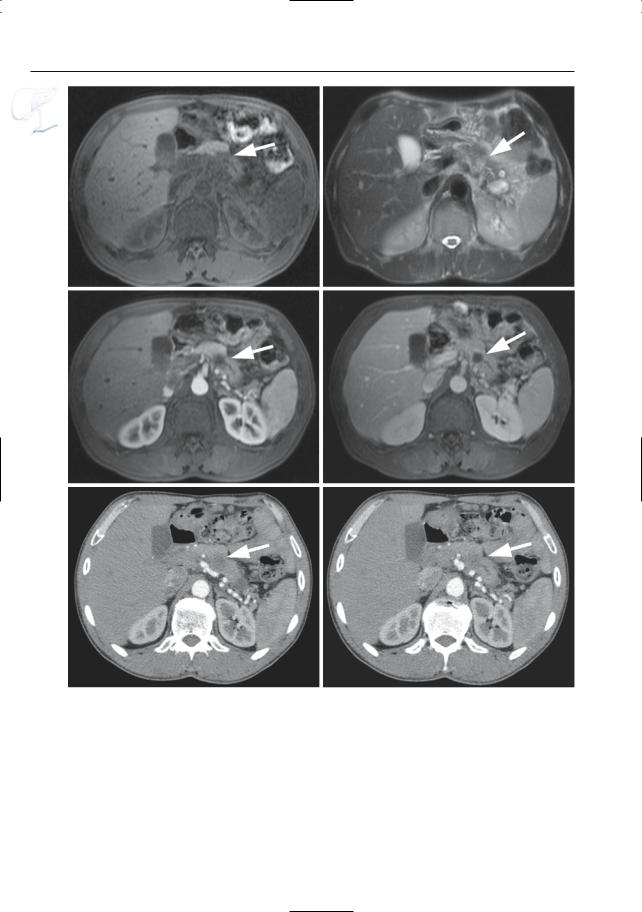
Fig. 169. (continued) d–k Axial T2-weighted HASTE images (d, e) and axial T1-weighted image (f) showing subtle enlargement of the pancreatic body/tail (arrows) and focal enlargement of the uncinate process (arrow), with abnormally low signal intensity of the affected parenchyma on the T1-weighted image. g, h Axial contrast-enhanced T1-weighted VIBE images obtained in the pancreatic phase show decreased enhancement (arrows). Note normally
h enhancing lateral part of the pancreatic head (arrowhead)
6 Pancreatic Ducts 343
i |
j |
k |
l |
m |
n |
Fig. 169. (continued) i Axial CT image in the venous phase confirming the enlargement of the pancreatic body/tail with decreased iodine uptake (arrows). j Projective image showing subtle dilatation of the pancreatic duct in the body/tail (arrows), with narrowing in the head/neck. k Axial T1-weighted image after 1 month steroid therapy shows an important regression of the findings (arrows) confirming the diagnosis of autoimmune pancreatitis. l–n Singleslice MRCP image obtained in a different patient shows severe narrowing of the CBD and proximal
biliary dilatation. m Axial T2-weighted image shows bizarre appearance of pancreatic parenchyma with presence of peripheral (ventral and dorsal) hypointense bands (arrows). n Axial contrast-enhanced T1weighted image shows poor uptake of contrast material in this region (arrows). The patient was known with primary sclerosing cholangitis and also had a severe stenosis at the hepatic hilum [arrowheads in (l). At the time of admission, a rapid clinical deterioration was observed. However, spectacular improvement was noted after steroid administration

344 6.2 Benign Nontraumatic Abnormalities
#170 Hereditary Pancreatitis
KEY FACTS: DISEASE
●Form of chronic pancreatitis with autosomal dominant inheritance
●Cause: mutation of the cationic trypsinogen gene
●Clinical onset: usually between 2 and 12 years (mean, 7 years)
●Symptoms: attacks of acute abdominal pain
●Complications:
–Exocrine and/or endocrine insufficiency (10%–30%)
–Pseudocyst formation (17%)
–Thrombosis of splenic vein (5%)
KEY FACTS: MRI (FIG. 170)
●Typical features:
–Moderate to marked dilation of the main pancreatic duct (commonly more than 1–2 cm)
–Typically large intraductal calculi
●Other features:
–Signs of acute pancreatitis
–Signs of complications (see also # 165, 166)
●Differential diagnosis: other types of chronic pancreatitis (age is often the most important clue)
References
Niederau C, Luthen R (1997) Hereditary pancreatitis is due to a mutation of the cationic trypsinogen gene. Z Gastroenterol 35 : 363–365
Perrault J (1994) Hereditary pancreatitis. Gastroenterology Clin North Am 23 : 743–752
Rohrmann CA, Surawicz CM, Hutchinson D, Silverstein FE, White TT, Marchioro TL (1981) The diagnosis of hereditary pancreatitis by pancreatography. Gastrointest Endosc 27 168–173
6 Pancreatic Ducts 345
a |
b |
Fig. 170. a A 13-year-old boy. Projective image showing dilation of the pancreatic duct with large intraductal concretions (arrowheads). b, c An 18-year-old girl with chronic pancreatitis. b Projective image showing dilatation of the pancreatic duct (arrowheads) with a focal stenosis in the pancreatic body (arrow). c Axial T1-weighted image showing decrea-
sed signal intensity of the pancreatic tissue due to c pancreatic inflammation (arrows)

346 6.2 Benign Nontraumatic Abnormalities
#171 Simple True Cyst
Related topics: #31 (hepatic cysts), # 172 (cysts in von Hippel-Lindau disease)
KEY FACTS: DISEASE
●Definition: cyst lined by an epithelial layer
●Histology: clear cystic fluid; inner surface lined with ductal cells
●Incidence: less common than pseudocysts
●Types:
–Congenital cysts (associated with polycystic disease in other organs)
–Acquired cysts: retention cysts (arising from obstruction of pancreatic duct); parasitic cysts
●Usually solitary; if multiple, underlying disease is common (see # 172)
●Usually asymptomatic; jaundice is possible
KEY FACTS: MRI
●T1 hypointense, T2 hyperintense
●Homogeneous, no solid components, unilocular
●May compress the common bile duct or pancreatic duct (rare) (Fig. 171)
●Differential diagnosis (may be difficult):
–Features suggesting pseudocyst: clinical history; connection with pancreatic duct; associated signs of chronic pancreatitis; multiple cystic lesions
–Features suggesting cystic tumor (see
# 175, 198): peripheral rim of solid tissue; irregular contours; mural nodules
–Lymphangioma (see # 173)
References
Heindryckx E,Van Steenbergen W,Van Vaerenbergh W, Van Hoe L, Vanbeckevoort D, Ectors N, Baert AL. Solitary true cyst of the pancreas: case report. Eur Radiol (in press)
Kim YH, Saini S, Sahani D, Hahn PF, Mueller P, Auh Y (2005) Imaging diagnosis of cystic pancreatic lesions: pseudocyst versus nonpseudocyst. RadioGraphics 25 : 671–685
Mao C, Greenwood S, Wagner S, Howard JM (1992) Solitary true cyst of the pancreas in an adult. Int J Pancreatol 12 : 181–186
Sperti C, Pasquali C, Constantino V, Perasole A, Liessi G, Pedrazzoli S (1995) Solitary true cyst of the pancreas in adults. Int J Pancreatol 18 : 161–67 Zhang X,Mitchell DG,Dohke M,Holland GA,Parker L (2002) Pancreatic cysts: depiction on singleshot fast spin-echo MR images. Radiology 223 :
547–553
6 Pancreatic Ducts 347
a |
b |
Fig. 171 a–c. Projective images (a, b) showing a large cystic structure in the pancreatic head, displacing the common bile duct (arrows). Note the nor-
mal aspect of the pancreatic duct in a (arrowhead). |
|
c CT image confirming the lack of solid components |
c |
within the cyst (arrow) |

348 6.2 Benign Nontraumatic Abnormalities
#172 Cysts in Von Hippel-Lindau
Disease
Related topic: # 171 (simple true cyst)
KEY FACTS: DISEASE
●Von Hippel-Lindau disease: autosomal disorder associated with abnormal tissue growth in different organs (e.g., retinal angiomatosis, hemangioblastoma of the central nervous system, renal cell carcinoma, adrenal pheochromocytoma)
●Incidence: pancreatic cysts are found in 72% of patients with von Hippel-Lindau disease
●Number of cysts: varies from a few cysts to polycystic transformation of the entire organ
●Other pancreatic abnormalities seen with increased frequency in von HippelLindau disease: serous cystadenoma, islet cell tumor, adenocarcinoma
●Other diseases associated with the presence of (multiple) pancreatic cysts:
–Adult polycystic kidney disease
–Cystic fibrosis (see # 174)
KEY FACTS: MRI
●Pancreatic cysts are easily recognized (Fig. 172)
References
Horton WA, Wong V, Eldridge R (1976) Von HippelLindau disease: clinical and pathological manifestations in nine families with 50 affected members. Arch Intern Med 136 : 769–777
Hough DM,Stephens DH,Johnson CD,Binkovitz LA (1994) Pancreatic lesions in von Hippel-Lindau disease: prevalence,clinical significance,and CTfindings. AJR Am J Roentgenol 162 : 1091–1094
Neumann HP, Dinkel E, Brambs H et al. (1991) Pancreatic lesions in the von Hippel Lindau syndrome. Gastroenterology 101 : 465–471
6 Pancreatic Ducts 349
a |
b |
c |
d |
Fig. 172. a, b Axial T2-weighted image and b pro- |
Disease. Axial T2-weighted image (TE 360) (c) and |
jective image showing multiple cysts throughout the |
projective image (d) showing multiple cysts scatte- |
pancreas. c, d Other patient with Von Hippel-Lindau |
red throughout the pancreas |

350 6.2 Benign Nontraumatic Abnormalities
#173 Lymphangioma
KEY FACTS: DISEASE
●Rare benign “tumor,” actually representing ectasia of lymphatic channels
●Most common in children
●Location:
–Neck and axilla (95%)
–Numerous other locations have been reported
●May contain serous fluid or chyle
●Abdominal lymphangiomas may also contain fat if lymphatic flow is received from the mesentery
KEY FACTS: MRI
●Fluid-containing lesion without characteristic features
–Unior multilocular, round or elongated
–May contain fat or calcifications
–Located within or adjacent to the pancreas
●Differential diagnosis: simple cyst, pseudocyst, mucinous cystic neoplasm
References
Khandelwal M, Lichtenstein GR, Morris JB, Furth EE, Long WB (1995) Abdominal lymphangioma masquerading as a pancreatic cystic neoplasm. J Clin Gastroenterol 20 : 142–144
Pandolfo I,Scribano E,Gaeta M,Fiumara F,Longo M (1985) Cystic lymphangioma of the pancreas: CT demonstration. J Comp Assist Tomogr 9 : 209– 210
Salimi Z, Fishbein M, Wolverson MK, Johnson FE (1991) Pancreatic lymphangioma: CT, MRI, and angiographic features. Gastrointest Radiol 16 : 248–250
6 Pancreatic Ducts 351
a
c
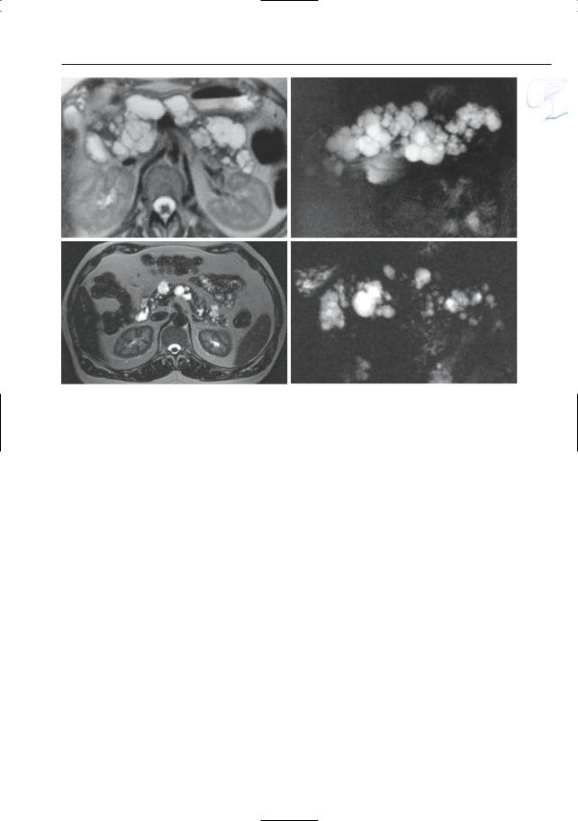
Fig. 173 a–e. a, b Axial T2-weighted HASTE image (TE 60 and 360) and (c) axial T1-weighted image showing a large fluid-containing mass anterior to the pancreas (large arrow) with internal debris (arrowheads) causing posterior displacement of the residual pancreatic parenchyma (small arrows). d, e Axial contrast-enhanced T1-weighted VIBE images obtained in the pancreatic phase showing the large non-enhancing cyst-like mass anterior (large arrow) and the severely compressed enhancing pancreatic tissue posteriorly (small arrows). Pathology confirmed the diagnosis of a large cystic lymphangioma
b
d
e

352 6.2 Benign Nontraumatic Abnormalities
#174 Cystic Fibrosis
KEY FACTS: DISEASE
●Autosomal recessive multisystem disorder with widespread abnormalities of mucus-secreting exocrine glands
●Incidence: 1 in 1500
●Pancreatic disease (pathophysiology):
–Decreased secretion of chloride, sodium, and water, followed by precipitation of relatively insoluble proteins and obstruction of small pancreatic ducts
–Progressive dilation of the acini and ducts with secondary fibrosis, atrophy, lipomatous degeneration, and/or cyst formation
●Morphologic types:
–Diffuse fatty pancreas
–Combination of fat and fibrotic parenchyma residuals (liposclerosis)
–Atrophy and fibrosis
–Numerous cysts (pancreatic cystosis)
●Clinical features: acute pancreatitis, diabetes mellitus, cirrhosis, exocrine pancreatic insufficiency, obstructive lung disease, chronic pulmonary infection
●Prognosis: mainly determined by the progression of pulmonary obstruction and infection
KEY FACTS: MRI
●MRI findings may reflect the various stages of the disease. Appearance on T1weighted images:
–Mottled hyperintensity (partial fatty replacement)
–Lobulated hyperintense pancreas (nearly complete fatty replacement)
–Diffuse hyperintense pancreas (complete fatty replacement) (Fig. 174)
–Atrophic or hypotrophic pancreas with normal signal intensity
–Multiple hypointense areas corresponding to cysts (probably rare)
●Differential diagnosis depends on the stage: chronic pancreatitis (atrophy and fibrosis), von Hippel-Lindau disease (multiple pancreatic cysts)
References
Ferrozzi F, Bova D, Campodonico F et al. (1996) Cystic fibrosis: MR assessment of pancreatic damage. Radiology 198 : 875–879
Hernanz-Schulman M, Teele RL, Perez-Atayde et al. (1986) Pancreatic cystosis in cystic fibrosis. Radiology 158 : 629–631
King LJ, Scurr ED, Murugan N et al. (2000) Hepatobiliary and pancreatic manifestations of cystic fibrosis: MR imaging appearances. Radiographics 20 : 767–777
6 Pancreatic Ducts 353
a
c
Fig. 174 a–e. Axial T2-weighted HASTE image (a) and axial T1-weighted images (b–d) showing an pancreatic atrophy with diffuse fat replacement of the normal pancreatic tissue (arrows). e Axial con- trast-enhanced fat-suppressed T1-weighted VIBE image obtained in the pancreatic phase showing low signal intensity of pancreatic parenchyma, as expected (arrows)
b
d
e
354 6.2 Benign Nontraumatic Abnormalities
#175 Microcystic Serous
Cystadenoma
Related topics: # 176 (macrocystic serous cystadenoma), # 198 (mucinous cystic neoplasm)
KEY FACTS: DISEASE
●Glycogen-containing cystic tumor, virtually always benign
●Histology: small cysts containing proteinous fluid separated by thin connective tissue septa; the cysts are lined with a monolayer of epithelial cells
●± 50% of all cystic pancreatic neoplasms
●Ratio of women to men: 4 : 1
●Most commonly observed in middleaged and elderly women
●Diagnosis: the cytoplasm of the cuboidal cells stains darkly (red) with periodic acid–Schiff (PAS). This positivity for PAS
a
c
disappears after pretreatment with amylase, thus identifying glycogen
●Can occur anywhere in the pancreas
●Predisposing factor: von Hippel-Lindau disease
KEY FACTS: MRI
●Well-defined lesion containing multiple small (< 2 cm) cysts (Fig. 175)
●Contrast-enhanced scans usually show enhancement of septa and peripheral
part of tumor (Fig. 175) |
|
|
! |
||
● Central calcifications (33%): usually not |
||
observed on MRI scans |
|
|
● The cysts never communicate with the |
|
|
pancreatic ducts (differential diagnosis |
|
|
with pseudocyst) |
|
|
● Features distinguishing this entity from |
|
|
mucinous neoplasms (# 198): |
|
|
– Large number of cysts |
|
|
– Small size of the cysts (< 2 cm) |
|
|
– Rich arterial blood supply |
|
b
Fig. 175. a–c Gadolinium-enhanced T1-weighted image (a) showing multiseptated cystic lesion in the pancreatic neck (arrows). Note enhancement of septa and small size of individual cysts. b T2weighted axial image and c projective image also showing multiple small cysts and thin septa
(arrows)
6 Pancreatic Ducts 355
d |
e |
f |
g |
Fig. 175 d– g. (continued) Different patient. Projective image (d) and T2-weighted HASTE image (e) showing a multiseptated cystic lesion in the pancreatic tail containing multiple small cysts (arrows). f Axial T1-weighted image showing this lesion to be
References
Compagno J, Oertel J (1978) Microcystic adenomas of the pancreas (glycogen-rich cystadenomas): a clinicopathologic study of 34 cases. Am J Clin Pathol 69 : 289–298
diffusely hypointense due to the multiple cysts (arrows). g Axial contrast-enhanced T1-weighted VIBE image obtained in the portal venous phase showing septal enhancement (arrows). Note the small size of the individual cysts
Kim YH, Saini S, Sahani D, Hahn PF, Mueller P, Auh Y (2005) Imaging diagnosis of cystic pancreatic lesions: pseudocyst versus nonpseudocyst. RadioGraphics 25 : 671–685
Procacci C, Graziani R, Bicego E et al. (1997) Serous cystadenoma of the pancreas: report of 30 cases with emphasis on the imaging findings. J Comp Assist Tomogr 21 : 373–382

356 6.2 Benign Nontraumatic Abnormalities
#176 Macrocystic Serous
Cystadenoma
Related topics: # 175 (microcystic serous cystadenoma), # 198 (mucinous cystic neoplasm)
KEY FACTS: DISEASE
●Macrocystic variant of serous cystadenoma (cysts may measure up to 8 cm, often with one large dominant cyst and several smaller daughter cysts)
●Histology: similar to the microcystic variant
●Extremely rare
●Only benign cases reported
KEY FACTS: MRI (FIG. 176)
●Identical to MR features of mucinous cystic tumor (see # 198)
References
Lewandrowski K, Warshaw A, Comptom C (1992) Macrocystic cystadenoma of the pancreas: a morphologic variant differing from microcystic adenoma. Hum Pathol 23 : 871–875
Mori K, Takeyama S, Hirosawa H et al. (1995) A case of macroscopic serous cystadenoma of the pancreas. Int J Pancreatol 17 : 91–93
6 Pancreatic Ducts 357
a |
b |
Fig. 176. a Coronal T2-weighted image showing cystic lesion with multiple septa (arrows) and several large cystic components. b Different patient. T2weighted HASTE image (TE 60) showing a large
multiseptated cystic lesion in the pancreatic head containing innumerable cysts. Note the presence of several larger cystic components (arrows)

358 6.2 Benign Nontraumatic Abnormalities
#177 Benign Neuroendocrine
Tumors
Related topic: # 197 (malignant neuroendocrine tumors)
KEY FACTS: DISEASE
●Synonym: islet cell tumors
●Histology: tumors composed of neuroendocrine cells characterized by the expression of neuroendocrine markers
●Incidence: 0.4%–1.5% of autopsies
●Nearly 85% of endocrine pancreatic tumors are functioning tumors
●The incidence of malignancy is correlated with:
–Histologic subtype (see # 197): insulinomas are usually benign (> 90%); all other subtypes are more likely to be malignant
–Size: the smaller the tumor, the less likely malignant behavior is (Buetow et al. 1995)
●Predisposing factor: type-1 multiple endocrine neoplasia (MEN)
KEY FACTS: MRI
●Signal intensity:
–T1: nearly invariably hypointense
–T2: variable, most commonly hyperintense, sometimes (partially) cystic (Fig. 177a–f and g–j)
●On dynamic contrast-enhanced scans, most endocrine tumors enhance more than the surrounding pancreatic parenchyma (rich arterial blood supply)
●The conspicuity on contrast-enhanced scans is variable and depends on:
–The “baseline” intensity of the tumor and surrounding parenchyma
–The degree of vascularization of both tissues
–The imaging parameters (e.g., delay between the injection and the scan) (Fig. 177n–q)
●Aspect on contrast-enhanced scans
–Hyperintense (common)
–Hypointense with hyperintense enhancing rim (common)
–Isointense (hypointense tumors not uncommonly become invisible after intravenous administration of contrast agent) (Fig. 177n–q)
–Hypointense
●Note: Benign functioning tumors (particularly insulinomas and gastrinomas) are usually small (< 2 cm in the large majority of cases); detection requires an optimized technique and special attention
●Note: Gastrinomas associated with type- 1 multiple endocrine neoplasia syndrome are even smaller (< 1 cm), commonly multicentric, and predominantly located in the wall of the duodenum
●Differential diagnosis of hypervascular pancreatic tumors:
–Primary: endocrine tumors; serous cystic tumor (enhancement of periphery and septa); solid and papillary cystic neoplasm (rare)
–Secondary: metastases from angiosarcoma, melanoma, carcinoid, renal adenocarcinoma, adrenal carcinoma, thyroid carcinoma, etc.
References
Buetow PC, Parrino TV, Buck JL et al. (1995) Islet cell tumors of the pancreas: pathologic-imaging correlation among size, necrosis and cysts, calcification, malignant behaviour, and functional status. AJR Am J Roentgenol 165 : 1175–1179
Kraus BB, Ros PR (1994) Insulinoma: diagnosis with fat-suppressed MR imaging. AJR Am J Roentgenol 162 : 69–70
Pavone P, Mitchell DG, Leonetti F et al. (1993) Pancreatic beta-cell tumors: MRI. J Comput Assist Tomogr 17 : 403–407
Sheth S, Hruban RK, Fishman EK (2002) Helical CT of islet cell tumors of the pancreas: typical and atypical manifestations. AJR Am J Roentgenol 179 : 725–730
Van Hoe L, Vanbeckevoort D, Baert AL (1997) Endocrine tumors of the pancreas: computed tomography and magnetic resonance imaging. Chir Gastroenterol 13 : 301–306
!
!
6 Pancreatic Ducts 359
a |
b |
c |
d |
e |
f |
Fig. 177 a–f.

360 6.2 Benign Nontraumatic Abnormalities
g |
h |
i |
j |
k |
l |
Fig. 177 g–m. (continued) |
m |
|
6 Pancreatic Ducts 361
n |
o |
p |
q |
Fig. 177 a–f. Axial T2-weighted HASTE images |
veals a hypointense nodule at this location (arrow). |
(a, b) (TE 60 and 360) and projective image (c) |
m Axial contrast-enhanced T1-weighted VIBE im- |
showing a tick-walled cystic lesion in the pancreat- |
age obtained in the pancreatic phase showing this |
ic body (arrow). Axial pre- (d) and post-contrast |
mass to be isointense to the surrounding normal |
(e, f) T1-weighted images show a strong enhance- |
pancreatic tissue (arrow), this feature points to the |
ment of the anterior wall of this cystic lesion (ar- |
strong enhancement of the lesion. n–q Patient with |
row). Final diagnosis: cystic neuroendocrine tumor |
insulinoma. n T1-weighted image showing small hy- |
without obvious signs of malignancy. g–j Patient |
pointense lesion in the pancreatic neck (arrow). |
with MEN-I syndrome and multiple neuroen- |
o The lesion is isointense on the T2-weighted image. |
docrine pancreatic tumors. g, h Axial T2-weighted |
p Contrast-enhanced T1-weighted image obtained |
HASTE images and (i, j) axial contrast-enhanced T1- |
in the pancreatic phase fails to reveal the lesion. |
weighted VIBE images obtained in the pancreatic |
q The lesion becomes hypointense again on images |
phase show two partially cystic lesions in the pan- |
obtained in the portal venous phase (arrow). These |
creatic tail. k–m Different patient. Axial T2-weight- |
features point to strong enhancement in the early |
ed HASTE image (k) showing an anterior bulge in |
phase and early washout of contrast medium |
the pancreatic tail. Axial T1-weighted image (l) re- |
|

362 6.2 Benign Nontraumatic Abnormalities
|
|
|
|
|
|
KEY FACTS: MRI (FIG. 178) |
|
|
|
|
|
#178 Schwannoma |
|
|
|
|
|
||
|
|
|
|
|
|
|
|
||
|
|
KEY FACTS: DISEASE |
|
|
|
● Signal intensity: typically high |
signal |
|
|
|
|
|
|
|
intensity on T2-weighted images (cystic/ |
|
|||
|
|
|
|
|
|
|
|||
|
|
|
|
|
|
|
|||
|
|
● Tumor derived from the lining cells of |
necrotic component, myxoid material) |
|
|||||
|
|
● Usually strong enhancement |
|
|
|
||||
|
|
the nerve sheath (Schwann cells) |
|
|
|
||||
|
|
● Clues for differential diagnosis |
with |
|
|||||
|
|
● Synonyms: neurinoma, neurilemmoma |
! |
||||||
|
|
other retroperitoneal tumors: |
|
|
|||||
|
|
● Pathology: |
|
|
|
|
|
||
|
|
|
|
|
– High signal intensity (T2) |
|
|
||
|
|
– Typically well encapsulated, rounded |
|
|
|
||||
|
|
– Rounded form, sharp delineation |
|
||||||
|
|
– Cystic degeneration, necrosis, and |
|
||||||
|
|
|
|
|
|
||||
|
|
hemorrhage common |
|
|
|
|
|
|
|
|
|
– Consists of Antoni A areas (high cellu- |
|
|
|
|
|||
|
|
References |
|
|
|
||||
|
|
larity) and Antoni B areas (myxoid |
|
|
|
||||
|
|
|
|
|
|
||||
|
|
substance) |
|
|
|
Cerofolini E,Landi A(1991) MR of benign peripheral |
|
||
|
|
● To be distinguished from neurofibroma |
nerve sheath tumors. J Comput Assist Tomogr |
|
|||||
|
|
(proliferation of |
Schwann |
cells and |
15 : 593–597 |
|
|
|
|
|
|
fibroblasts; associated with |
Reckling- |
Kim SH, Choi BI, Han MC, Kim YI (1992) Retroperi- |
|
||||
|
|
toneal neurilemoma: CT and MR findings. AJR |
|
||||||
|
|
hausen’s disease, i.e., neurofibromatosis I) |
|
||||||
|
|
Am J Roentgenol 159 : 1923–1026 |
|
|
|
||||
|
|
● Incidence: 5%–10% of tumors of the |
Urban BA, Fishman EK, Hruban RH, Cameron JL |
|
|||||
|
|
central nervous |
system; rare in the |
(1992) CT findings in cystic schwannoma of the |
|
||||
|
|
abdomen |
|
|
|
pancreas. J Comp Assist Tomogr 16 : 492–493 |
|
||
|
|
|
|
|
|
|
|
|
|
●Age peak: 30–60 years
●Usually no symptoms
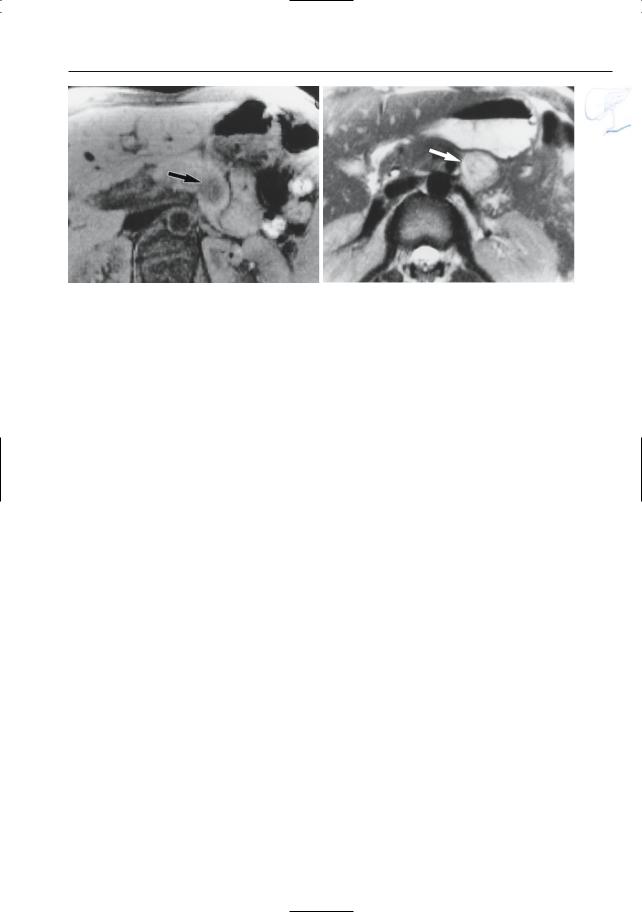
6 Pancreatic Ducts 363
a |
b |
Fig. 178 a, b. a T1-weighted image showing a |
showing the tumor as a markedly hyperintense mass |
hypointense lesion apparently surrounded by pan- |
(arrow). The tumor originated from the mesenteric |
creatic parenchyma (arrow). b T2-weighted image |
root adjacent to the pancreas |

364 6.2 Benign Nontraumatic Abnormalities
#179 Granulomatous Disease
of the Pancreas
KEY FACTS: DISEASE
●Granulomatous involvement of the pancreas may (rarely) be seen in sarcoidosis, tuberculosis, fungal diseases, cat scratch disease, and lymphoma
●Sarcoidosis:
–Granulomatous disease of unknown etiology
–Lung and lymph nodes most commonly affected
–Other organs sometimes involved:eyes, liver, skin, salivary glands, spleen, pericardium, peritoneum
–Diagnosis: bronchoalveolar lavage (BAL)
KEY FACTS: MRI
●Common findings in abdominal sarcoidosis:
–Enlarged lymph nodes
–Hepatosplenomegaly
–Focal lesions in liver and spleen
●Uncommon findings:
–Focal lesion(s) in the pancreas with or without displacement of the pancreatic duct (Fig. 179)
–Narrowing of the distal common bile duct (Toda et al. 1994)
–Ascites (peritoneal involvement/congestive heart failure)
●Differential diagnosis: although rare, sarcoidosis should be considered in the differential diagnosis of focal pancreatic masses not typical of adenocarcinoma
References
Bonhomme A, Dhadamus A, De Bie P, Van Hoe L, Baert AL (1997) Pancreatic involvement in systemic sarcoidosis: CT findings. J Belge Radiol 80 : 116–117
Caldwell JH, Evans WE (1978) Granuloma (sarcoid?) of the pancreas. Am J Gastroenterol 69 : 320–322 Purdy DJ, Levine EJ, Forsthoefel KJ, Fromkes JJ (1994) Periampullary pseudotumor secondary to granulomatous disease. Am J Gastroenterol 89 :
2087–2088
Toda K,Souda S,Yoshikawa Y,Momiyama T,Ohshima M (1994) Narrowing of the distral common bile duct and the portal vein secondary to pancreatic sarcoidosis.Am J Gastroenterol 89 : 1259–1261

6 Pancreatic Ducts 365
a |
b |
Fig. 179 a, b. Patient with sarcoidosis. a Projective MR image showing apparent narrowing of the pancreatic duct in the pancreatic body and neck (arrows). b ERCP image showing focal narrowing (arrow). Overestimation of the length of the steno-
sis by MRCP is related to the lack of distention (no retrograde injection of contrast material; see #23). Also note increased background signal intensity in a, caused by pancreatitis after ERCP

366 6.2 Benign Nontraumatic Abnormalities
#180 Hemochromatosis
KEY FACTS: DISEASE
●Excess iron deposition in various parenchymal organs leading to organ damage
●Autosomal recessive inheritance
●Organs especially affected: liver, pancreas, heart
●Basic disorder: mucosal defect in the intestinal wall resulting in increased absorption of intestinal iron
●Symptoms: hyperpigmentation (90%), hepatomegaly (90%), arthralgia (50%), diabetes (30%), hypogonadism
●Complications:
–Periportal fibrosis and cirrhosis
–Hepatocellular carcinoma (14%–30%)
●Differential diagnosis: secondary hemochromatosis (hemosiderosis) (Fig.180c):
–Iron overload due to blood transfusions
–Storage of iron mainly in the reticuloendothelial system (liver, spleen)
–Little clinical significance
KEY FACTS: MRI
●Pancreatic parenchyma exhibits very low signal intensity on T1and T2-weighted images
●Always associated with low signal intensity of liver tissue
References
Runge VM, Clanton JA, Smith FW et al. (1983) NMR of iron and copper disease states. AJR Am J Roentgenol 141 : 943–948
Yoon DY, Choi BI, Han JK, Park MO, Suh SJ (1994) MR findings in secondary hemochromatosis: transfusional versus erythropoietic. J Comput Asist Tomogr 18 : 416–419
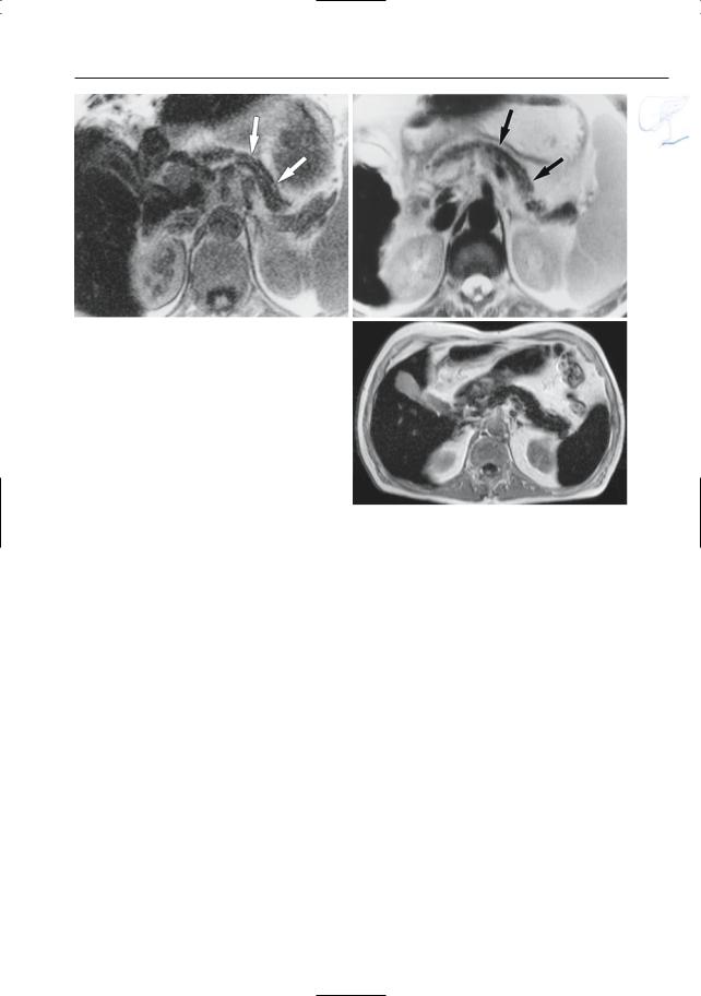
6 Pancreatic Ducts 367
a
Fig. 180 a, b. a T1and b T2-weighted images show an atrophic and markedly hypointense pancreas (arrows). Also note the markedly hypointense aspect of the liver parenchyma. c Patient with history of multiple blood transfusions and hemosiderosis. Axial T2-weighted HASTE image shows low signal intensity of liver, spleen, and pancreas, due to secondary hemosiderosis. The low signal intensity of the pancreatic parenchyma observed in this case is rather atypical for hemosiderosis
b
c

368 6.3 Traumatic, Postoperative, and Iatrogenic Abnormalities
6.3Traumatic, Postoperative, and Iatrogenic Abnormalities
#181 Pancreatic Duct Injury
Related topics: #48 (sequelae of direct liver trauma), # 113 (blunt gallbladder trauma)
KEY FACTS: DISEASE
KEY FACTS: MRI (FULCHER ET AL. 2000)
●MRI usually plays no role in the acute setting, but may be used for follow-up purposes
●(Continued) leakage, strictures, pseudocysts, atrophy, and/or ductal dilation may be shown
●Relatively uncommon: 1%–12% of all abdominal injuries
●Cause: blunt or penetrating injury
●Classification of pancreatic trauma:
–Grade 1: contusion
–Grade 2: small laceration
–Grade 3: laceration with rupture of the main pancreatic duct
–Grade 4: crush injury
●Most common location of laceration: pancreatic neck (compression against the first lumbar vertebra)
●Associated disease: other abdominal injuries in 90%
●Complications of grade 3 injuries:
–Fistula
–Pseudocyst
–Stricture with or without recurrent pancreatitis
–Focal necrosis
–Abscess formation
References
Carr ND, Cairns SJ, Lees WR, Russell RC (1989) Late complications of pancreatic trauma. Br J Surg 76 : 1244–1246
Fulcher AS, Turner MA, Yelon JA et al. (2000) Magnetic resonance cholangiopancreatography (MRCP) in the assessment of pancreatic duct trauma and its sequelae: preliminary findings. J Trauma 48 : 1001–1007
Van Hoe L, Gryspeerdt S, Oyen R, Baert AL (1996) Retroperitoneal trauma. In: Heller M (ed) Radiology of trauma. Springer, Berlin Heidelberg New York, pp 163–192
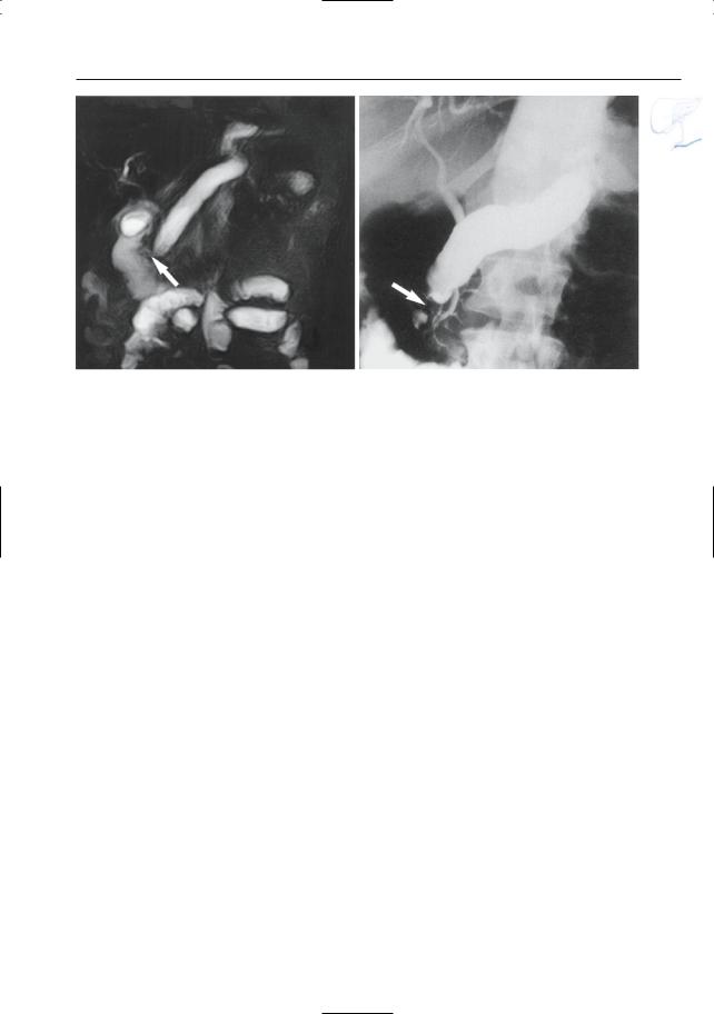
6 Pancreatic Ducts 369
a |
b |
Fig. 181 a, b. a Projective MR image and b ERCP image showing marked dilation of the pancreatic duct secondary to ductal stenosis in the pancreatic
head (arrow). The patient suffered a severe blunt trauma to the pancreas as a child

370 6.3 Traumatic, Postoperative, and Iatrogenic Abnormalities
#182 Complications of Pancreatic
Transplantation (1):
Pancreatitis
Related topic: # 149 (postoperative anatomy: after pancreatic transplantation)
KEY FACTS: DISEASE
●Postoperative pancreatitis commonly occurs immediately after surgery and usually subsides within 4 weeks
●Incidence: 1.8%
●Causes of persistent pancreatitis (acute/ relapsing/chronic):
–Urinary reflux
–Stenosis/obstruction of the pancreatic duct or anastomosis
●Complications:
–Fistula
–Ascites
–Pseudocyst
KEY FACTS: MRI
●Classical MR features of acute pancreatitis (see # 151–157)
–Diffuse parenchymal edema
–Peripancreatic fluid
● Features of chronic pancreatitis: see
# 160–168 (Fig. 182a, b)
●Pancreatic duct dilation may be observed if obstruction or reflux is present
●Signs of complications: pseudocysts, fistula, etc.
●Note: The differentiation between pancreatitis and rejection (see # 183) may be difficult. Features more or less specific for pancreatitis are:
– Ductal dilation
– Large fluid collections/ascites
– Focal areas of necrosis
References
Moudry-Munns K, Gruessner A, Sutherland DE (1994) Analysis of United States pancreas transplant registration data. J Transplant Coordination 4 : 18–22
Yuh WT, Hunsicker LG, Nghiem DD et al. (1989) Pancreatic transplants: evaluation with MR imaging. Radiology 170 : 171–177
6 Pancreatic Ducts 371
a |
b |
Fig. 182. a Projective image showing long segmental stenosis of the pancreatic duct (arrowheads). Also note the slight dilation of the pancreatic duct in the tail, with several dilated side branches (arrows). It remained unclear in this patient whether the stenosis was the primary lesion or a secondary lesion in
preexisting chronic pancreatitis. b Projective MR image showing a dilated pancreatic duct (arrowheads), a small fluid exudate, and a large pseudocyst (asterisk): chronic obstructive pancreatitis secondary to an anastomotic stricture (see also # 168)

372 6.3 Traumatic, Postoperative, and Iatrogenic Abnormalities
#183 Complications of Pancreatic
Transplantation (2):
Other Complications
Related topic: # 149 (postoperative anatomy: after pancreatic transplantation)
KEY FACTS: DISEASE
●Major causes of graft loss include:
–Rejection (35%)
–Vascular thrombosis (12%–20%)
–Sepsis (8%–22%)
●Other complications:
–Hemorrhage
–Infarction
–Pseudoaneurysm (may be related to biopsy)
–Anastomotic leak
KEY FACTS: MRI
●Acute rejection:
–Usually large edematous graft (as in pancreatitis)
–Dynamic contrast-enhanced MRI shows delayed parenchymal enhancement (probably not specific)
●Chronic rejection:
–Small graft
–Low signal intensity on T1and T2weighted images (fibrosis)
●Hematoma: signal intensity usually characteristic of blood (see #51)
●Abscess (Fig. 183):
–Typical fluid collection with thick, irregular wall
–Air–fluid level or multiple air bubbles may be seen
–Differential diagnosis with pseudocyst, loculated ascites, and urinoma may be difficult
●Secretin-augmented MRCP and MR perfusion measurements have been proposed to differentiate between functional and dysfunctional grafts (Heverhagen et al. 2004)
References
Fernandez MP, Bernardino ME, Neylan JF, Olson RA (1991) Diagnosis of pancreatic transplant dysfunction: value of gadopentetate dimeglumineenhanced MR imaging. AJR Am J Roentgenol 156 : 1171–1176
Snider JF, Hunter DW, Kuni CC, Castenada-Zuniga WR, Letourneau JG (1991) Pancreatic transplantation: radiologic evaluation of vascular complications. Radiology 178 : 749–753
Heverhagen JT,Wagner HJ, Ebel H et al. (2004) Pancreatic transplants: noninvasive evaluation with secretin-augmented MR pancreatography and MR perfusion measurements – preliminary results. Radiology 233 : 273–280
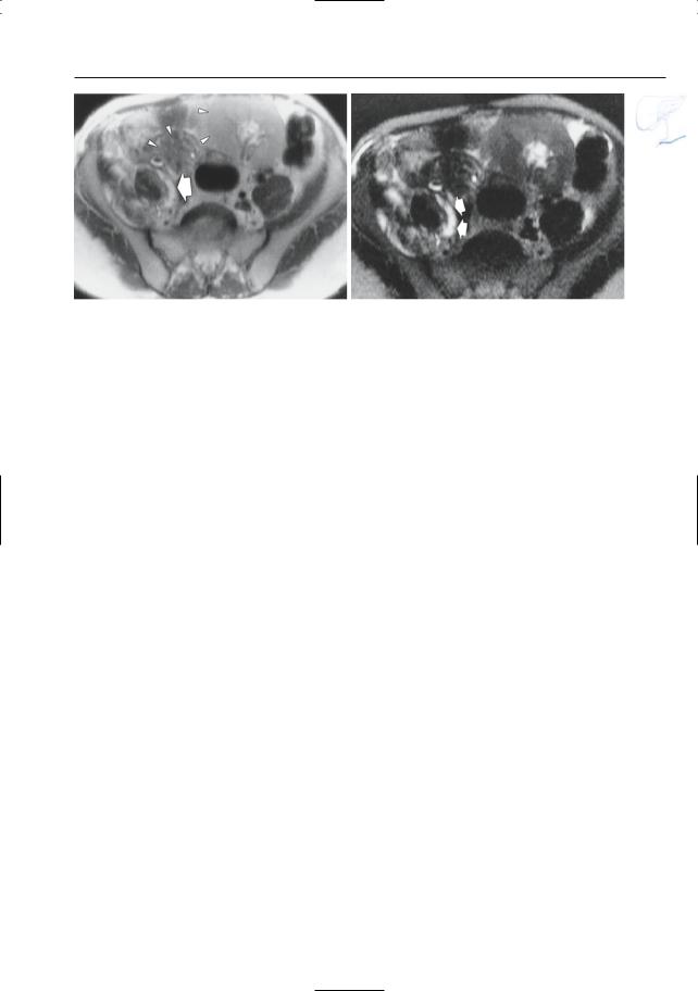
6 Pancreatic Ducts 373
a |
b |
Fig. 183 a, b. a Moderately T2-weighted image showing transplanted pancreas and kidney (arrowheads). Also note the small hyperintense area more dorsally (arrows). b Heavily T2-weighted image
confirming the presence of fluid in the psoas compartment (arrows). The final diagnosis was psoas abscess
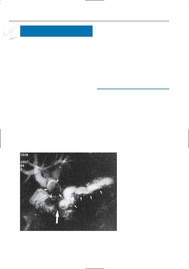
374 6.3 Traumatic, Postoperative, and Iatrogenic Abnormalities
#184 Complications of Partial
Pancreatic Resection
Related topic: # 148 (postoperative anatomy: after partial resection)
KEY FACTS: DISEASE
●Surgical procedures: see # 148
●Main complications:
–Anastomotic leak
–Anastomotic stenosis
–Tumor recurrence
–Intraductal lithiasis
KEY FACTS: MRI
●Specific features depend on the type of disease (e.g., stricture, fluid collection)
●Note: Adequate visualization of the normal and pathologic anastomosis requires sufficient filling of the draining jejunal loop
●Note: Once early postoperative edema has disappeared, anastomotic leaks and stenoses as well as recurrent tumor are usually easily recognized
References
Lepanto L, Gianfelice D, Déry R, Dagenais M, Lapointe R, Roy A (1994) Postoperative changes, complications, and recurrent disease after Whipple’s operation: CT features. AJR Am J Roentgenol 163 : 841–846
Trerotola SO, Jones B, Crist DW, Cameron JL (1989) Pylorus-preserving Whipple pancreaticoduodenectomy: postoperative evaluation. Radiology 171 : 735–738
Fig. 184. Patient who had undergone a Whipple operation and subsequently presented with jaundice. Projective MR image showing marked obstructive dilation of the pancreatic duct, secondary to an anastomotic stricture (arrows). The intraheptratic ducts are also dilated and contain a large stone
(arrowheads)