


180 3.4 Malignant Tumors
3.4 Malignant Tumors
#87 Extrahepatic Cholangio-
carcinoma (1): Klatskin Tumor
Related topics: #54,55 (intrahepatic cholangiocarcinoma), #88 (extrahepatic cholangiocarcinoma: distal duct type)
KEY FACTS: DISEASE
●Cholangiocarcinoma involving the proximal common hepatic duct and sometimes the bifurcation (= hilar cholangiocarcinoma)
●Histology: varies from well-differenti- ated adenocarcinoma to anaplastic carcinoma
●Incidence: 70% of cholangiocarcinomas
●Age peak: sixth to seventh decade
●Growth pattern:
–Focal stenotic
–Diffuse sclerosing
–Polypoid (least common)
●Classification: see Fig. 87a (Deviere et al. 1988)
●Predisposing factors:
–Inflammatory bowel disease
–Primary sclerosing cholangitis (see #41–44)
–Caroli’s disease (see #30)
–Choledochal cysts (see #68)
–Clonorchis sinensis infection
●Symptoms: painless jaundice, cholangitis, weight loss
●Spread:
–Most are locally infiltrative
–Invasion of portal vein unusual
–Lymphatic spread: most often in common bile duct nodes
–Infiltration of the liver (23%)
–Peritoneal seeding (9%)
–Hematogenous: extremely rare
KEY FACTS: MRI (FIG. 87 b – f)
● Projective images:
– Usually stenotic lesion (usually 1–3 cm in length, may be longer; contour may be smooth or irregular)
–Nodular intraluminal components not uncommon
–Differential diagnosis: trauma, bile duct surgery, compression by metastatic lymph nodes, lymphoma, hepatocellular carcinoma,or gallbladder carcinoma
●Cross-sectional images:
–Mass located at the liver hilum causing intrahepatic bile duct dilation
–Signal intensity on T2-weighted images: well-differentiated and mucincontaining types are hyperintense; the scirrhous type is moderately hyperintense (or more or less isointense)

3 Extrahepatic Bile Duct 181
–Enhancement pattern: heterogeneous progressive enhancement
–Other features: lobar atrophy, segmental portal vein occlusion without caliber expansion of the vein, hilar lymph node enlargement
–Small (<1–2 cm) and infiltrating tumors are usually not clearly visible
!● Note: In the absence of surgical antecedents, strictures involving the region of the porta hepatis or the midportion of the extrahepatic duct are almost invariably malignant, regardless of their appearance
References
Deviere J, Baize M, De Toeuf J, Cremer M (1988) Long-term follow-up of patients with hilar malignant stricture treated by endoscopic biliary drainage. Gastrointest Endosc 34 : 95–101
Guthrie JA, Ward J, Robinson PJ (1996) Hilar cholangiocarcinomas: T2-weighted spin echo and gadolinium-enhanced FLASH MR imaging. Radiology 201 : 347–351
Nichols DA, MacCarty RL, Gaffey TA (1983) Cholangiographic evaluation of bile duct carcinoma. AJR Am J Roentgenol 141 : 1291–1294
Park MS, Kim TK, Kim KW et al. (2004) Differentiation of extrahepatic bile duct cholangiocarcinoma from benign stricture: findings at MRCP versus ERCP. Radiology 233 : 234–240
Soyer P, Bluemke DA, Reichle R et al. (1995) Imaging of intrahepatic cholangiocarcinoma. 2. Hilar cholangiocarcinoma. AJR Am J Roentgenol 165 : 1433–1436
Yeh TS, Jan YY, Tseng JH et al. (2000) Malignant perihilar biliary obstruction: magnetic resonance cholangiopancreatographic findings. Am J Gastroenterol 95 : 432–440
182 3.4 Malignant Tumors
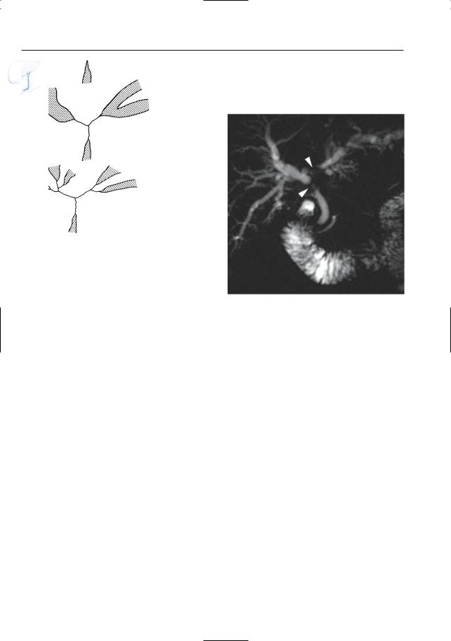
a
Fig. 87. a Classification of malignant hilar stenoses (Bismuth). Type I: tumors involving the proximal common hepatic duct, sparing the bifurcation; type II: tumors of the bifurcation involving the more distal hepatic ducts; type III: tumors of the bifurcation extending up to and involving secondary hepatic ducts. b–f Projective image (b) and corresponding ERCP image (c) showing a long stricture of the proximal common hepatic duct and bifurcation (arrow-
b
heads) with secondary intrahepatic bile duct dilatation: Klatskin tumor type II. d T1-weighted FLASH image showing a hypointense soft tissue mass in the liver hilum (arrow). e Axial contrast-enhanced T1weighted VIBE image obtained in the arterial phase showing only slight peripheral enhancement (arrow). f Same image obtained after 10-min interval showing the typical delayed central enhancement, characteristic for a cholangiocarcinoma (arrow)
3 Extrahepatic Bile Duct 183
c |
d |
e |
f |
Fig. 87. (continued)

184 3.4 Malignant Tumors
#88 Extrahepatic Cholangio-
carcinoma (2): Distal Duct Type
Related topics: #54, 55 (intrahepatic cholangiocarcinoma), #87 (Klatskin tumor)
KEY FACTS: DISEASE
●Definition: cholangiocarcinoma located below the proximal part of the common hepatic duct (usually within the midextrahepatic biliary tree)
●Macroscopic appearance: mass encasing the bile duct or rounded intraluminal mass
●Usually small
●Incidence: 10% of cholangiocarcinomas
●Predisposing disease: see #87
●Better prognosis than Klatskin tumor (higher probability of surgical cure; Nichols et al. 1983)
KEY FACTS: MRI (FIG. 88)
●Projective images:
–Usually focal stenosis
–Rarely polypoid mass
●Cross-sectional images:
–Mass lesion (if sufficiently large)
–T1: hypointense; T2: variable, usually moderately hyperintense
–With or without enlarged lymph nodes
References
Kim JH, Kim MJ, Chung JJ et al. (2002) Differential diagnosis of periampullary carcinomas at MR imaging. Radiographics 22 : 1335–1352
Nichols DA, MacCarty RL, Gaffey TA (1983) Cholangiographic evaluation of bile duct carcinoma. AJR Am J Roentgenol 141 : 1291–1294

3 Extrahepatic Bile Duct 185
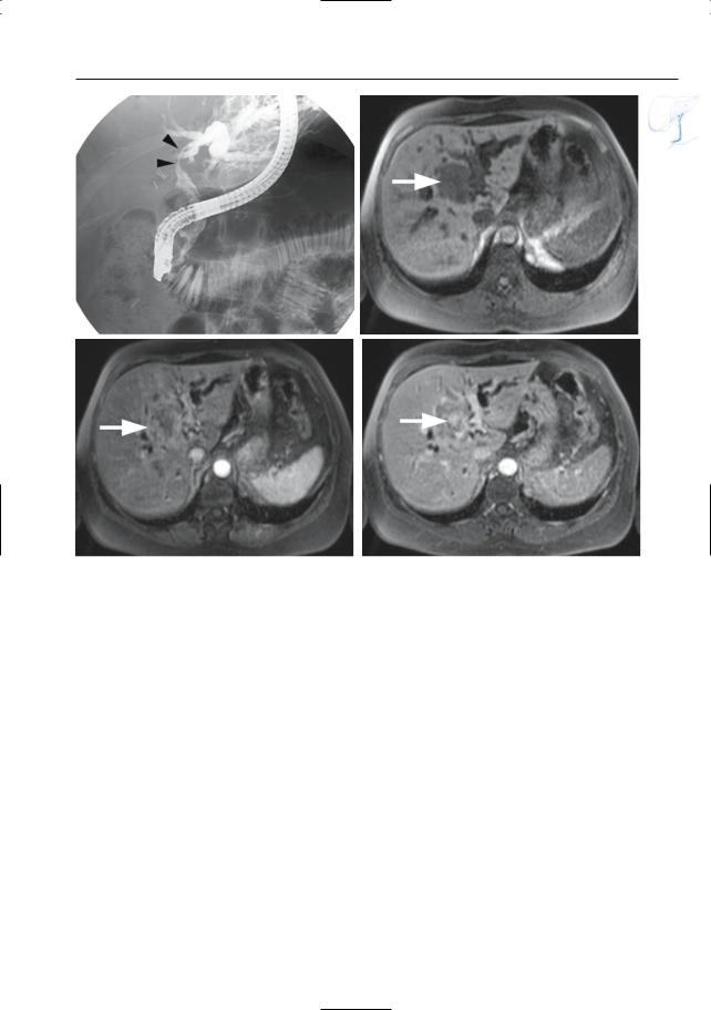
b
a
d
c |
e |
Fig. 88. a, b Projective image (a) showing marked bile duct dilatation with an abrupt stenosis in the distal common bile duct (arrow). b Axial contrastenhanced T1-weighted VIBE image obtained in the venous phase showing a small hypo-intense lesion in the distal common bile duct, corresponding to a small cholangiocarcinoma (arrow) c–e Different
patient. Projective image (c) showing marked bile duct dilatation with an abrupt stenosis in the distal common bile duct (arrow). d, e Coronal and axial T2-weighted HASTE images (TE 60) showing a slightly hyperintense infiltrating mass in the pancreatic head (arrow). Final diagnosis: cholangiocarcinoma

186 3.4 Malignant Tumors
#89 Bile Duct Involvement
by Gallbladder Carcinoma
Related topics: # 115–118 (gallbladder carcinoma)
KEY FACTS: DISEASE (SEE ALSO # 115 – 118)
●Incidence of biliary tree stenosis/ obstruction in gallbladder carcinoma: 50%
●Mechanisms of involvement of bile duct:
–Gallbladder carcinomas have a propensity for direct centripetal extrahepatic spread
–Bile duct compression/invasion by metastatic lymph nodes
KEY FACTS: MRI (FIG. 89)
●Cholangiographic appearance: indistinguishable from Klatskin tumors
●Characteristic features (see # 115–118):
–Focal or diffuse thickening of the gallbladder wall
–Abnormal gallbladder bed
–Sometimes large mass directly invading the liver
References
Lane J, Buck JL, Zeman RK (1989) Primary carcinoma of the gallbladder: a pictorial essay. Radiographics 9 : 209–228
Schwartz LH, Black J, Fong Y et al. (2002) Gallbladder carcinoma: findings at MR imaging with MR cholangiopancreatography. J Comput Assist Tomogr 26 : 405–410
Silvis S, Rohrmann C, Ansel H (eds) (1995) Endoscopic retrograde cholangiopancreatography. Igaku-Shoin, New York, p 287
Thorsen MK, Quiroz F, Lawson TL, Smith DF, Foley WD, Stewart ET (1984) Primary biliary carcinoma: CT evaluation. Radiology 152 : 479–483

3 Extrahepatic Bile Duct 187
b
a
d
c |
e |
Fig. 89. a, b Patient who had previously undergone “routine” cholecystectomy. Histology revealed gallbladder carcinoma with positive section margins. a Projective MR image obtained 3 weeks after surgery showing narrowing of the proximal common hepatic duct (arrow). b Axial T2-weighted image obtained at the level of the stenosis showing a small hypointense soft tissue mass around the narrowed bile duct
(arrowheads), representing infiltrating tumor. c–e Different patient. Projective image (c) showing severe narrowing of the proximal common hepatic bile duct (arrow) with secondary intrahepatic bile duct dilatation. d, e Axial T2-weighted HASTE image (TE 60) showing an infiltrating hyperintense mass originating from the gallbladder (arrow) and invading the adjacent bile duct (arrowhead)
188 3.4 Malignant Tumors
#90 Bile Duct Involvement
by Pancreatic Carcinoma
Related topics: # 133 (vaterian sphincter complex, invasion by pancreatic carcinoma), # 185–195 (pancreatic carcinoma)
KEY FACTS: DISEASE
●Pancreatic carcinoma commonly occurs in the pancreatic head (60%–70%)
●Secondary involvement of the biliary tree is common
KEY FACTS: MRI (FIG. 90)
●Stenosis:
–Abrupt transition with or without “shouldering”
–Median length: several centimeters
–Usually irregular contours
●May also become manifest as obstruction
●Location:
–Usually intrapancreatic segment of the common bile duct
–Midsegment of the common bile duct: in pancreatic tumors extending cranially
●Note: The diagnosis of pancreatic carcinoma can be made with confidence if:
–Cross-sectional images show a mass lesion located in the pancreatic head
–Both the common bile duct and the pancreatic duct show typical irregular narrowing with abrupt transition (typical “double duct” sign)
References
Baert AL, Rigauts H, Marchal G (1994) Ductal adenocarcinoma. In: Baert AL (ed) Radiology of the pancreas. Springer, Berlin Heidelberg New York, pp 129–172
Freeny PC (1989) Radiological diagnosis and staging of pancreatic ductal adenocarcinoma. Radiol Clin North Am 27 : 121–128
Kim JH, Kim MJ, Chung JJ et al. (2002) Differential diagnosis of periampullary carcinomas at MR imaging. Radiographics 22 : 1335–1352
3 Extrahepatic Bile Duct 189
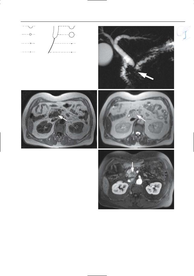
a
c
Fig. 90. a Typical patterns of ductal narrowing in chronic pancreatitis (left) and pancreatic adenocarcinoma (right). In adenocarcinoma, an abrupt transition is usually seen. (From Rohrman and Baron 1989, with permission). b–e Projective image (b) showing severe, abrupt narrowing of the distal common bile duct and pancreatic duct (“double duct sign”) (arrow). T1-weighted FLASH image (c) and T2weighted HASTE image (TE 60) (d) showing a markedly T1 hypointense and T2 hyperintense mass in the pancreatic head (arrow).e Contrast-enhanced T1weighted VIBE image obtained in the arterial phase showing this mass to be hypointense compared with the surrounding normal pancreatic tissue (arrow). These are typical characteristics of a pancreatic adenocarcinoma
b
d
e

190 3.4 Malignant Tumors
#91 Bile Duct Involvement
by Other Extrabiliary
Neoplasms
Related topics: #60 (intrahepatic bile ducts, metastases), # 196 (secondary pancreatic tumors)
KEY FACTS: DISEASE
●Incidence: at least 10% of tumors involving the bile ducts
●Causes:
–Metastatic disease (usually lymph node metastases in pericholedochal lymphatics)
–Lymphoma
KEY FACTS: MRI (FIG. 91)
●Projective images show a varying appearance:
–Smooth eccentric compression
–Smooth or irregular concentric narrowing
–Polypoid intraluminal mass (metastases represent ± 10% of polypoid bile duct tumors; Ponette et al. 1994)
●Cross-sectional images usually enable identification of enlarged lymph nodes
References
Ponette E, Biebau G, Gelin J et al. (1994) Imaging of polypoid endoluminal growing bile duct tumors. J Belge Radiol 77 : 157–161
3 Extrahepatic Bile Duct 191
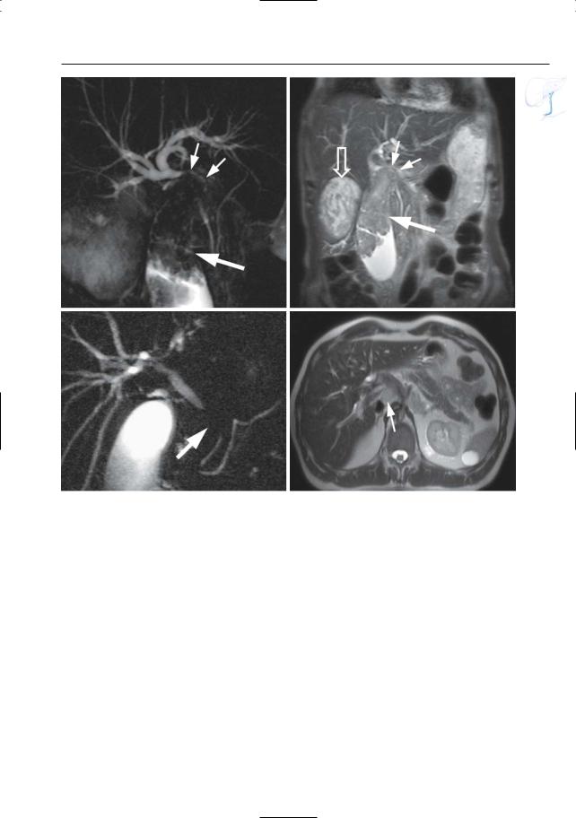
a |
b |
c |
d |
Fig. 91. a, b Patient with a known malignant melanoma. Projective image (a) and coronal T2-weight- ed HASTE image (b) showing a distended gallbladder filled with solid tissue (large arrow): gallbladder metastasis. Secondary invasion of the proximal common hepatic duct is also observed (small arrows). Note the partially necrotic liver metastasis adjacent
to the gallbladder (open arrow). c, d Patient with known lymphoma. c Projective image showing a medial deviation and severe narrowing of the distal common bile duct (arrow). d T2-weighted HASTE image showing a large retroperitoneal adenopathy adjacent to the common bile duct, causing extrinsic compression and stenosis (arrow)