

Книги по МРТ КТ на английском языке / MRI for Orthopaedic Surgeons Khanna ed 2010
.pdf130 II Upper Extremity
graphic evidence of a fractured scaphoid. However, when a patient continues to have tenderness in the anatomic snu - box or over the scaphoid tubercle at follow-up examination, other advanced imaging modalities often are used for the detection of a radiographically occult scaphoid fracture. Recent comparative studies have sought to determine the superior modality for confirming the diagnosis: bone scintigraphy, CT, MRI, or ultrasound.3–6 MRI has consistently outperformed other methods in the accurate early detection of scaphoid fracture and other radiographically indeterminate carpal injuries.7–9 In addition, the use of early MRI in the management of clinically suspected scaphoid fractures may
prove to be more cost-e ective than follow-up conventional radiography.10,11
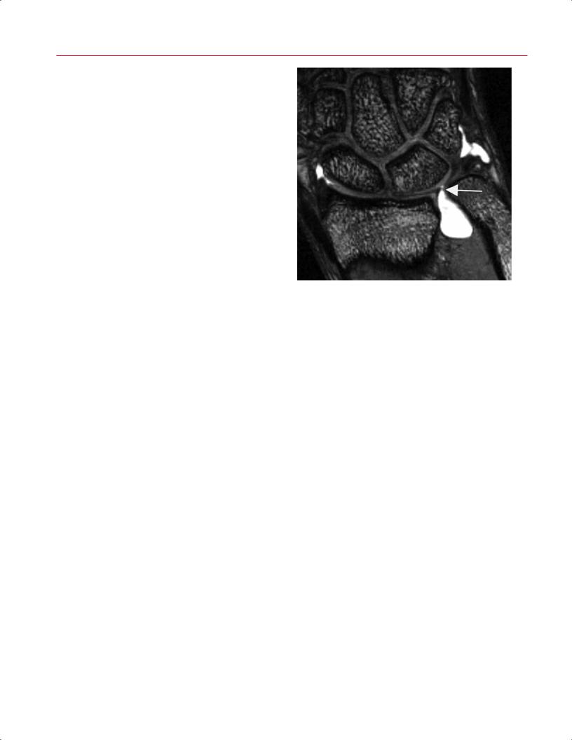
A fracture may be identified on T1-weighted images as a distinct line of low signal intensity that contrasts with the surrounding high-signal marrow (Fig. 6.1A). Fat-suppressed, T2-weighted images typically show high-signal marrow edema surrounding the fracture line (Fig. 6.1B). A scaphoid humpback deformity may also be shown on sagittal images (Fig. 6.1C).
MRI has proven to be sensitive in the detection of occult fractures in the distal radius, distal ulna, and other carpal bones (Fig. 6.2)12,13 and can also di erentiate fractures from
A B
|
Fig. 6.1 Scaphoid fracture/deformity. (A) A coronal T1-weighted image of the wrist showing a frac- |
|
ture through the scaphoid waist (arrow), seen as a discrete line of low signal intensity. (B) A coronal |
|
fat-suppressed T2-weighted image showing the same fracture as in (A), with focal marrow edema |
C |
in the scaphoid waist consistent with fracture (arrow). (C) A sagittal gradient-echo image showing |
a humpback deformity of the scaphoid. |

6 The Wrist and Hand 131
Fig. 6.2 A coronal T1-weighted image of the wrist showing a radiographically occult capitate fracture (arrows).
bone bruises (contusions), which show marrow edema without a discrete fracture line. MRI is also sensitive for diagnosing subtle physeal injuries in children (Fig. 6.3) and for evaluating physeal bars and growth arrest.14 Radiographically occult fractures of the metacarpals and phalanges are uncommon.
pisotriquetral joint injury) by history and physical examination. Historically, three-compartment wrist arthrography was the imaging test of choice for evaluation of the TFCC. However, this modality su ers from unacceptably low sensitivity and specificity and lacks the ability to determine lesion size, location, and acuity.17,18 Although wrist arthroscopy remains the standard for the diagnosis of TFCC pathology, advances in MRI and MR arthrography are increasing the reliability in accurate diagnosis of the size, location, and character of TFCC tears before wrist arthroscopy. In the most specialized centers, the sensitivity and accuracy of MRI for detecting TFCC tears approaches 100%, using arthroscopy as the standard diagnostic modality.19
The normal TFCC is a low signal intensity structure shaped like a bowtie in the coronal plane. The articular disc is readily apparent on coronal and sagittal images. Advances in imaging techniques have enabled MRI to visualize some other individual components of the TFCC, such as the radioulnar ligaments and the ECU subsheath. T2-weighted images may help to di erentiate between degenerative and traumatic tears of the TFCC, with traumatic tears showing increased signal intensity in adjacent tissues. The excellent anatomic
TFCC Injury
The TFCC encompasses the major stabilizing structures of the ulnar aspect of the wrist, spanning the distal radioulnar joint and consisting of the following components15:
•Fibrocartilaginous articular disc
•Dorsal and volar radioulnar ligaments
•Ulnolunate ligament
•Ulnotriquetral ligament
•UCL
•ECU subsheath
•A variable meniscus homologue
In general, tears of the TFCC are classified as degenerative or traumatic.16 Degenerative central perforations of the TFCC are common and often asymptomatic. On the other hand, acute injuries to the TFCC frequently lead to ulnar-side wrist pain and dysfunction. It is di cult to distinguish this condition from other causes of ulnar-side wrist pain (such as occult fractures, lunotriquetral ligament injury, ECU tendinopathy or subluxation, distal radioulnar joint injury, or
Fig. 6.3 A sagittal STIR image of the wrist showing di use marrow edema in the distal radius metaphysis adjacent to the growth plate, which is widened along its volar aspect (arrow).
132 II Upper Extremity
A 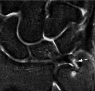 B
B
Fig. 6.4 Peripheral TFCC tear. (A) A coronal fat-suppressed T2weighted image showing a tear of the peripheral portion of the TFCC near its ulnar attachment (arrow). (B) A coronal STIR image showing
detail a orded by MRI also helps in determining specific lesion location, with degenerative tears typically occurring in the thinner central portion and traumatic tears most often found in the periphery of the TFCC (Fig. 6.4). Nonetheless, all TFCC attachments must be carefully evaluated in all three planes. Ulnar attachment tears have been consistently more di cult to diagnose.20 Concomitant pathology of the ECU tendon, lunotriquetral ligament, and surrounding osseous structures must be pursued. MR arthrography may prove superior to MRI in detecting TFCC lesions.2,21 A TFCC tear is often shown by the presence of gadolinium contrast within the tear itself and/or in the distal radioulnar joint. Strong evidence exists that observer experience is critical for the accurate detection of TFCC pathology.22
Interosseous Ligament Injury
The scapholunate and lunotriquetral ligaments are the most commonly injured wrist ligaments secondary to acute trauma. These C-shaped ligaments vary in thickness throughout their courses. MRI has supplanted stress radiography and conventional wrist arthrography as the study of choice for suspected carpal ligament injury. Compared with arthroscopic findings, conventional MRI is highly specific but still has an overall low sensitivity for the detection of tears of the scapholunate ligament,23 although the sensitivity seems to be improving with technologic advances in imaging techniques. Disruption of the interosseous ligaments is best evaluated on coronal gra- dient-echo (Fig. 6.5) and fat-suppressed T2-weighted images.
a perforation of the peripheral portion of the TFCC near its radial attachment (arrow).
Carpal ligaments show low signal intensity on most MR images. However, the scapholunate ligament normally shows a more heterogeneous appearance on coronal images, which may lead to the misdiagnosis of a tear. Distinct linear areas of high signal intensity equal to that of fluid, especially in the thick dorsal portion of the ligament, usually indicate clinically significant tears, which may lead to carpal instability if not treated adequately. Alternative findings in ligament tears include nonvisualization of the ligament or morphologic distortion.24 MR arthrography may be more accurate than conventional MRI for the detection of tears of the scapholunate ligament when compared with arthroscopic findings.25 MR arthrography is most useful for the detection of tears of the lunotriquetral ligament, which although rarely an isolated injury, often accompanies an injury to the TFCC. The assessment of lunotriquetral ligament pathology by MRI techniques is still met with skepticism in the literature.26,27
Extrinsic Carpal Ligament Injury
The extrinsic carpal ligaments are a complex assortment of dorsal and volar structures that lie in close relation to the wrist joint capsule. The volar extrinsic ligaments are stronger and more robust than their dorsal counterparts. Isolated acute injuries to the extrinsic ligaments are rare. Typically, disruptions of these ligaments are part of a constellation of acute bone and soft-tissue injury about the wrist joint. Although MRI can distinguish the extrinsic ligament complexes with multiple image planes or 3D reconstructions
6 The Wrist and Hand 133
A 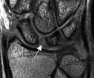 B
B
Fig. 6.5 Scapholunate ligament tear. (A) A coronal gradient-echo image of the wrist showing partial stretching of the scapholunate ligament with concurrent widening of the scapholunate interval
(Fig. 6.6), the role of MRI in this clinical setting is more often to exclude occult fracture, disruption of the intrinsic carpal ligaments, or injury to the TFCC. If diagnosed, these conditions may require a change in treatment algorithm.
Thumb UCL Injury
The UCL of the MCP joint is the primary stabilizer of the ulnar aspect of the thumb. Rupture of this ligament is common in skiers and results from forced abduction of the thumb. A
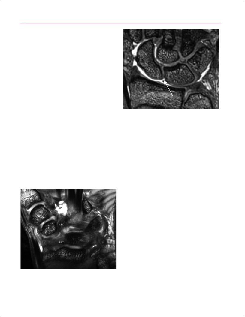
Fig. 6.6 A coronal STIR image of the wrist showing the extrinsic ligaments comprising part of the volar wrist capsule, including the radioscaphocapitate (RSC) and radiolunotriquetral (RLT) ligaments.
(arrow). (B) A coronal gradient-echo image of the wrist showing a small perforation in the membranous portion of the scapholunate ligament (arrow).
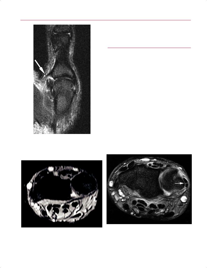
Stener lesion describes displacement of the torn ligament to a position superficial to the aponeurosis of the adductor pollicis muscle, a conformation that inhibits adequate ligament healing. Although clinical presentation and stress radiography may be enough for the diagnosis of a torn UCL, MRI may detect a Stener lesion and help guide the surgical treatment of acute injuries (Fig. 6.7).28,29
Tendon Injuries
Tendon pathology may occur as a result of acute trauma, chronic overuse, or inflammatory arthropathies such as RA. The spectrum of pathology ranges from tendon thickening and various degrees of tendon or tendon sheath inflammation to frank tears. In patients with traumatic injury, partial or complete rupture of a hand or wrist tendon may be di - cult to diagnose clinically. Although tenderness and localized swelling are often present, the degree of flexion or extension loss may be equivocal. The use of MRI and ultrasound in this setting is evolving. Occasionally, ruptures of the extensor wrist tendons can be detected by MRI (Fig. 6.8). On T2-weighted sequences, partial tendon tears typically show increased intrasubstance signal intensity (Fig. 6.9), whereas complete tears show separation of the tendon ends with fluid tracking between its margins. Timely and accurate diagnosis has important clinical implications because early surgical repair provides the best outcome for zone II flexor tendon tears comprising >50% of their width.30 Similarly, identifying the location of the retracted tendon end after a zone I flexor digitorum profundus avulsion injury (Jersey finger) is important for determining the appropriate treatment. Advances in image quality with sophisticated systems and MR
134 II Upper Extremity
Fig. 6.7 A coronal oblique T2-weighted image of the thumb showing a complete rupture of the UCL at the MCP joint with increased fluid emanating from the joint (arrow).
tenography, in which contrast material is injected within the tendon sheath, have led to the detection of flexor pulley system injuries (see Fig. 2.21) and concomitant bowstringing deformities of the flexor tendons.31
■ Degenerative Conditions
The wrist and hand are a ected by various degenerative conditions, although the usefulness of MRI may be limited to the evaluation of ulnar impaction syndrome, tendon disorders, and carpal instability in select patients. Other common degenerative conditions in the hand and wrist, such as first carpometacarpal joint and radiocarpal joint osteoarthritis, are adequately imaged with conventional radiography.
Ulnar Impaction Syndrome
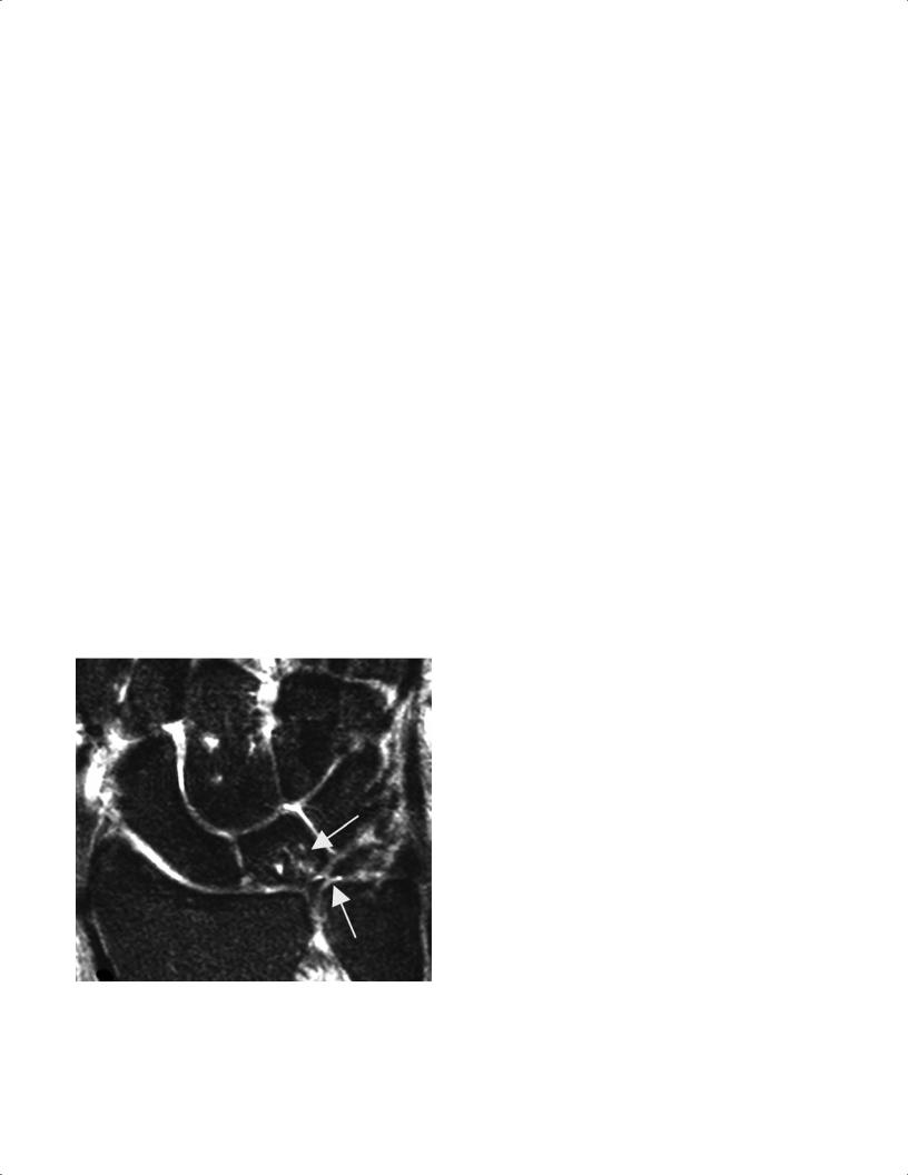
Ulnar variance relates to the relative lengths of the radius and ulna. In patients with ulnar positive variance, the relatively long ulna may abut the proximal carpal row when the wrist is ulnarly deviated, leading to ulnar-side wrist pain from excessive loading across the ulnar aspect of the wrist. Chronic impaction may lead to subchondral sclerosis or cyst formation of the proximal and ulnar portions of the lunate, the radial portion of the triquetrum, and/or the distal and radial portions of the ulnar head. Focal defects of the articular cartilage or increased marrow signal on T2-weighted images may be visualized (Fig. 6.10). These findings can be di eren-
Fig. 6.8 An axial STIR image of the wrist showing absence of the extensor pollicis longus tendon in the third extensor compartment, indicative of complete rupture (arrow).
Fig. 6.9 An axial fat-suppressed T2-weighted image at the level of the distal radioulnar joint, showing thickening of the ECU tendon and a high signal intensity split within the tendon, indicating a partial tear (arrow).
|
6 The Wrist and Hand 135 |
||
tiated from Kienböck disease because the signal changes are |
dinous T2-weighted signal intensity (Fig. 6.11). Similar find- |
|
|
focal rather than di use, and the ulnar variance is usually |
ings are described for other commonly a ected tendons, |
||
positive rather than negative or neutral. MRI may also detect |
including the ECU, extensor carpi radialis longus, extensor |
||
accompanying degenerative TFCC or lunotriquetral ligament |
pollicis longus, flexor carpi radialis, and flexor carpi ulnaris |
||
tears.32 |
(Fig. 6.12). Tendon subluxation or dislocation from chronic |
||
|
overuse may be detected on axial images. MRI may help to |
||
Tendon Disorders |
di erentiate chronic tenosynovitis and soft-tissue masses |
||
such as giant cell tumor of the tendon sheath. |
|||
|
|||
Pain and diminished function of the wrist and hand may |
The clinician should keep in mind that when a tendon |
||
be caused by various forms of tendon pathology, rang- |
lies at approximately 55 degrees relative to the direction |
||
ing from tendinosis to tenosynovitis to partial or complete |
of the external magnetic field during scanning, artifactual |
||
tears. Although these conditions may result from simple |
increased intratendinous signal intensity may be observed |
||
overuse, other etiologies include inflammatory arthropa- |
on some imaging sequences secondary to the magic angle |
||
thies, indolent infections, PVNS, gout, sarcoidosis, or amy- |
phenomenon, not to true pathology.34,35 This phenomenon |
||
loidosis.33 Although tendon pathology is often self-limiting, |
is recognized easily because the signal disappears and the |
||
MRI is excellent for assessing the spectrum of tendon pa- |
tendon appears normal on heavily T2-weighted images. This |
||
thology. MRI may reveal tendon thickening or thinning |
phenomenon also is seen in the shoulder where the pres- |
||
in patients with tendinosis or tendinitis. T2-weighted im- |
ence of the magic angle e ect may be mistaken for rotator |
||
ages may reveal distended tendon sheaths filled with high |
cu tendinopathy (see chapter 4). |
||
signal intensity fluid in patients with tenosynovitis (Fig. |
Given that the clinical findings are typically diagnostic, |
||
6.10). Occasionally, tiny foci of low signal intensity, called |
MRI is not necessary for the diagnosis of stenosing tenosyno- |
||
rice bodies, are seen within the fluid in patients with chronic |
vitis of the digits (trigger finger), although it may have a role |
||
tenosynovitis. |
in evaluating patients with postoperative recurrence or in |
||
As an example, de Quervain tenosynovitis is a common |
ruling out other causes of symptoms, such as tendon sheath |
||
inflammatory disorder of the first dorsal compartment. A |
tumor, rheumatoid synovitis, or volar plate pathology. |
||
spectrum of MRI findings may be seen, including thickening |
|
|
|
of the abductor pollicis longus and extensor pollicis brevis |
Carpal Instability |
||
tendons, adjacent fluid collections, and increased intraten- |
The most common pattern of carpal instability is dorsal in- |
||
|
|||
|
tercalated segment instability, often associated with disrup- |
||
|
tion of the scapholunate and volar extrinsic carpal ligaments. |
||
|
Volar intercalated segment instability from lunotriquetral |
||
|
ligament deficiency is far less common. Clinically, carpal in- |
||
|
stabilities usually present as chronic wrist pain months to |
||
|
years after traumatic injury or secondary to inflammatory |
||
|
arthropathy. Physical examination findings are often equivo- |
||
|
cal, and standard lateral wrist radiographs may not always |
||
|
show abnormal carpal alignment. In patients with static car- |
||
|
pal instability, the malalignment will be evident on sagittal |
||
|
images, and MRI may also detect associated abnormalities of |
||
|
the stabilizing carpal ligaments, such as laxity, thickening, |
||
|
retraction, edema, or absence. MR arthrography and thin- |
||
|
section coronal gradient-echo images are the most useful for |
||
|
identifying the pathology of these structures. Perforations |
||
|
of the central portions of the interosseous ligaments may be |
||
|
asymptomatic and consistent with normal aging.36 |
||
|
|
|
|
|
■ Infectious Conditions |
||
Fig. 6.10 A coronal STIR image of the wrist showing focal edema and cystic changes at the proximal ulnar aspect of the lunate consistent with ulnar impaction syndrome (top arrow). A small perforation of the central portion of the TFCC is also shown (bottom arrow).
Soft-tissue abscesses within the hand or wrist typically show intermediate to low signal intensity on T1-weighted images and high signal intensity on T2-weighted images. A thick, enhancing wall is usually seen after the administra-
136 II Upper Extremity
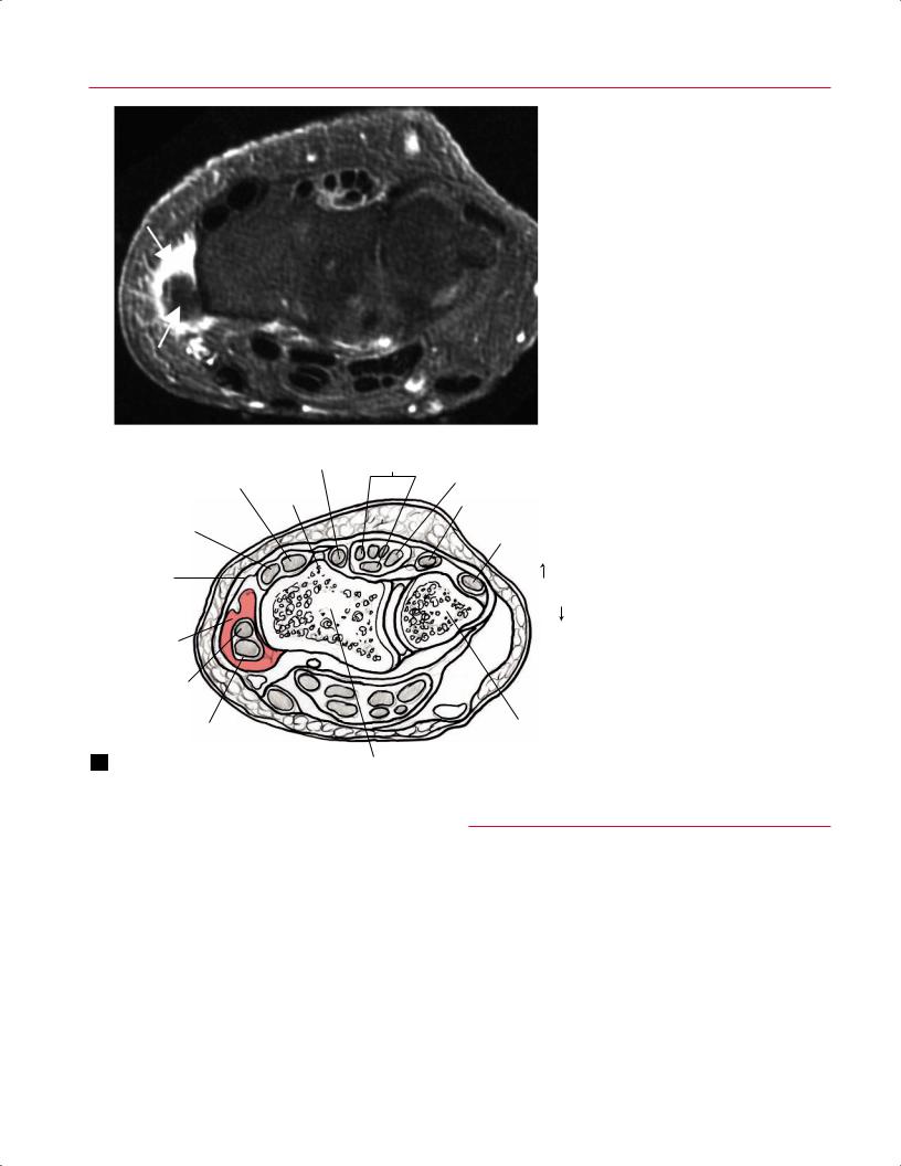
A
Extensor pollicis Extensor carpi longus tendon radialis brevis
tendon |
Lister’s |
|
tubercle |
||
Extensor carpi |
||
|
||
radialis longus |
|
|
tendon |
|
|
Extensor |
|
|
retinaculum |
|
|
Edema |
|
|
Extensor pollicis |
|
|
brevis tendon |
|
|
Abductor pollicis |
|
|
longus tendon |
|
B
Fig. 6.11 De Quervain tenosynovitis. An axial fat-sup- pressed T2-weighted image (A) and artist’s sketch
(B) showing thickening of the first compartment tendons and surrounding edema (arrows on A).
Extensor digitorum communis tendons
Extensor indicis tendon
Extensor digiti minimi tendon
Extensor carpi ulnaris tendon
Dorsal
Palmar
Ulna
Radius
tion of intravenous contrast. MRI can evaluate the extent of soft-tissue involvement and the presence or absence of accompanying osteomyelitis. An emergent MRI may be necessary in select patients with suspected necrotizing fasciitis, where T2-weighted images may help confirm the diagnosis with increased signal intensity seen in the deep fascia, although this imaging finding is nonspecific. Given the need for urgent treatment of this diagnosis, the clinician should also carefully evaluate for the presence of crepitus within the soft tissues and rapidly spreading erythema and induration. Prompt treatment of infectious processes in critically ill patients should never be delayed while awaiting an MRI study.
■ Other Pathologic Conditions
MRI has proven useful in the evaluation of other pathologic conditions a ecting the wrist and hand, such as osteonecrosis, RA, compression neuropathies, and soft-tissue masses.
Kienböck Disease
Idiopathic osteonecrosis of the lunate is known as Kienböck disease. This diagnosis must be considered in patients with subacute or chronic dorsal wrist pain of unknown etiology. A strong association with negative ulnar variance exists, and the etiology of the disorder may stem from chronic micro-

6 The Wrist and Hand 137
•Type 1 variant, di use involvement of the scaphoid
•Type 2 variant, only partial necrosis
The recognition of two distinct patterns has implications for treatment, and the clinical outcome may be more favorable for patients with type 2 involvement.38
Fig. 6.12 Axial fat-suppressed T2-weighted image showing fluid between the first and second extensor compartments consistent with intersection syndrome from chronic overuse (arrow).
trauma secondary to increased shear stress on the lunate. Lichtman et al37 defined four stages of Kienböck disease:
•Stage I: normal conventional radiographs, changes in the lunate on MRI
•Stage II: lunate sclerosis on conventional radiographs
•Stage III: lunate collapse
•Stage IV: pancarpal arthritis
Early diagnosis by MRI is critical because both nonoperative measures and surgical intervention are most successful in the initial stages of the disease, long before progression to lunate collapse or pancarpal arthritis.
In the first stage of Kienböck disease, MRI shows distinct changes in the lunate. On T1-weighted images, decreased lunate vascularity leads to di use low signal intensity of the bone marrow compared with the surrounding carpal bones (Fig. 6.13). This di use involvement distinguishes Kienböck disease from focal T1-weighted signal loss secondary to ulnar impaction syndrome, fracture, or tumor. The findings on T2-weighted images have been correlated with prognosis, showing low signal intensity with complete marrow fibrosis and increased signal intensity with lunate revascularization. When stage I Kienböck disease is diagnosed, patients are typically immobilized for up to 3 months, and follow-up MRI examinations are used to monitor the success of nonoperative treatment.
MRI findings are similar in idiopathic osteonecrosis of the scaphoid, also known as Preiser disease, and other carpal bones. One study supports the possibility of two patterns of Preiser disease based on disparate MRI patterns of vascular impairment38:
Posttraumatic Scaphoid Osteonecrosis
The tenuous retrograde blood supply of the scaphoid often results in delayed union, nonunion, and/or osteonecrosis of the proximal pole. These complications are minimized with the early diagnosis of a scaphoid fracture and the implementation of appropriate treatment. In patients with scaphoid nonunion, determination of bone viability is critical to subsequent management. Vascularity of the proximal fragment has been shown to be an important determinant of outcome after surgical reconstruction of a scaphoid nonunion.39 Patients with proximal pole osteonecrosis may require vascularized bone grafting. Visual inspection of intraoperative punctate bleeding typically corresponds with viability of the proximal pole. Because conventional radiographs are unreliable for detecting bone vascularity, MRI with intravenous gadolinium contrast can be used to assess the degree of osteonecrosis preoperatively. Osteonecrosis is indicated by low signal intensity on T1-weighted images, in stark contrast to the high signal intensity of the marrow of the normal surrounding carpal bones (Fig. 6.14). T2-weighted images also show low signal intensity.
Fig. 6.13 Coronal T1-weighted image showing complete loss of normal marrow signal within the lunate, indicating necrosis (Kienböck disease).
138 II Upper Extremity
Fig. 6.14 A coronal T1-weighted image showing loss of normal marrow signal in the proximal pole of the scaphoid (arrow), indicative of posttraumatic osteonecrosis.
ture so that other diagnoses (systemic lupus erythematosus, scleroderma, polymyositis, dermatomyositis, Behçet disease, or mixed connective disease) are excluded.43 Advanced techniques have been developed to quantify the volume of a ected synovium and bone erosion with MRI. These methods may prove useful in predicting disease progression and monitoring the response to treatment.44
Compression Neuropathies
The nerve branches in the hand and wrist most frequently a ected by compression syndromes are the median nerve in the carpal tunnel and the ulnar nerve in Guyon’s canal. The superficial sensory branch of the radial nerve is a ected much less commonly. Compression neuropathies may be triggered by intrinsic or extrinsic sources. Most patients are diagnosed by history and physical examination findings, which are then corroborated by electrodiagnostic testing. MRI can serve as a useful adjunctive diagnostic modality in the evaluation of compression neuropathies. Some patients present with atypical or equivocal clinical features or nerve conduction study results, and an MRI examination may be indicated for the investigation of unusual etiologies, such as nerve sheath tumors or extrinsic space-occupying lesions. Another indication for MRI is patients with recurrent symptoms after surgical intervention, in whom incomplete
RA
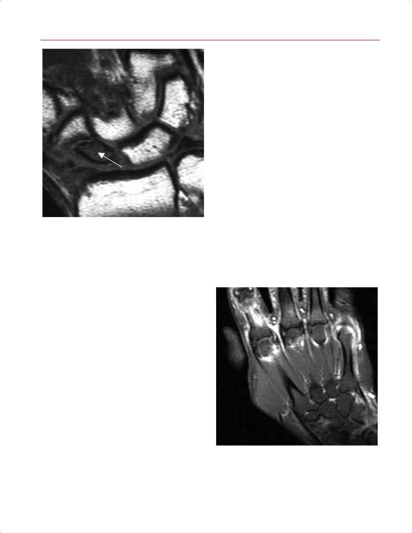
RA, the most common form of inflammatory arthritis, is characterized by inflammation of the synovial lining of joints and tendon sheaths. It frequently a ects the hand and wrist early in the disease process. Although RA may become a severely disabling condition, recent clinical trials unequivocally support early intervention with aggressive pharmacologic therapy for the prevention of rapid joint erosion and the improvement of long-term outcomes.40 The importance of timely and accurate diagnosis is implicit within this strategy. For many patients, this diagnosis is achieved only by history, physical examination, laboratory data, and conventional radiographic findings; a more expeditious diagnosis of early RA, however, may be provided with MRI.
Several reports have shown the value of MRI in detecting bone erosion and active synovitis before changes are visible on conventional radiographs.41,42 To investigate a possible diagnosis of RA via MRI, contrast-enhanced images are obtained after an intravenous injection of contrast material. Intense bilateral contrast enhancement in the wrist, MCP, or proximal IP joints suggests active synovitis and is indicative of inflammatory arthritis (Fig. 6.15). Additionally, adjacent bone edema or frank erosions can be detected (Fig. 6.16). Because these imaging findings are nonspecific, the clinician must take into account the entire clinical pic-
Fig. 6.15 A coronal fat-suppressed postgadolinium T1-weighted image showing di use high signal enhancement surrounding the MCP joints, consistent with synovitis from early RA. Conventional radiographs were normal.
6 The Wrist and Hand 139
Fig. 6.16 A sagittal gradient-echo image of the wrist showing multiple subchondral cysts and periarticular erosions in the setting of advanced RA.
decompression, excessive scarring, hematoma formation, or another space-occupying lesion may be detected and for whom additional treatment can be initiated accordingly (Fig. 6.17).
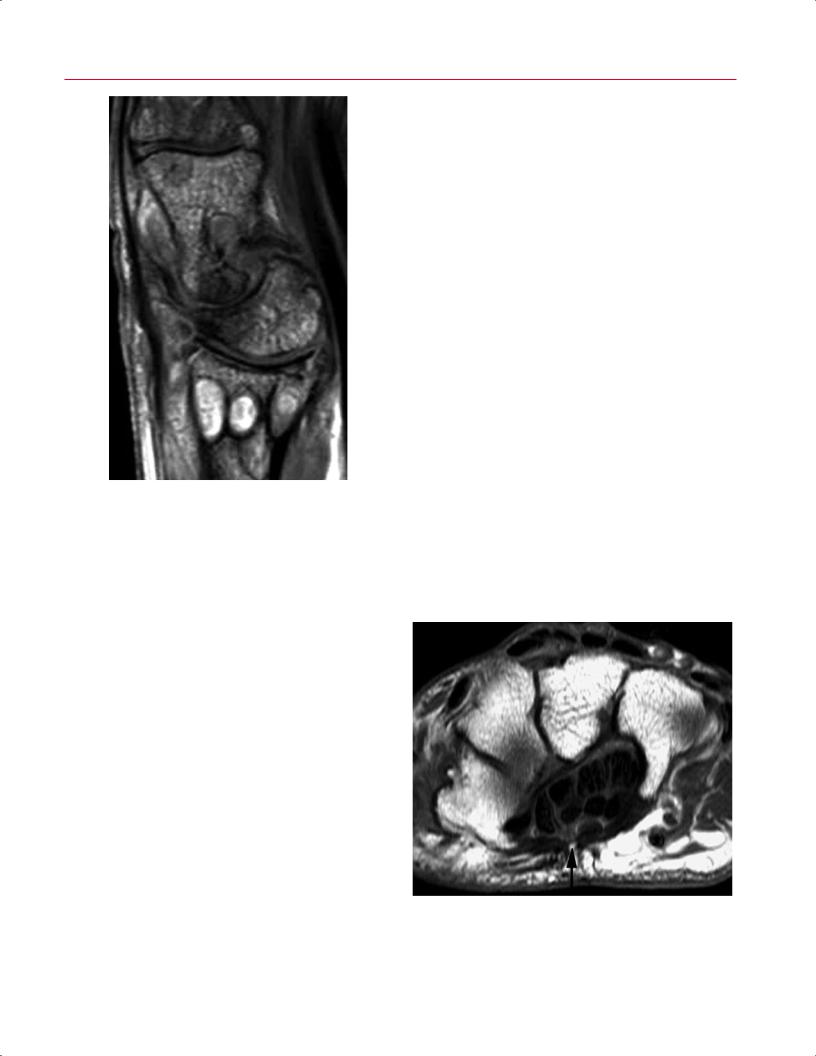
In the evaluation of the median nerve at the carpal tunnel, T1-weighted and T2-weighted axial images are obtained to screen for obvious anatomic anomalies or pathologic findings. The median nerve is examined for changes in size, shape, or signal intensity. A predominant finding in patients with carpal tunnel syndrome is increased signal within the median nerve on T2-weighted images, which is related to chronic compression or ischemia. Other detectable abnormalities include bowing of the flexor retinaculum, deep palmar bursitis, tenosynovitis, or the presence of a spaceoccupying soft-tissue mass. Intraneural lesions usually are well delineated. Although dynamic and motion studies of the median nerve in the carpal tunnel have been described, they are rarely used.45
In the presence of ulnar nerve compression at the wrist, MRI may elucidate the cause when other diagnostic methods are unrevealing. Several etiologies include posttraumatic neuritis, occult fractures, pisotriquetral arthropathy, anatomic variants or ganglion cysts, and other soft-tissue
masses (Fig. 6.18). Again, axial images are best for evaluating the ulnar nerve in Guyon’s canal. The information gained from MRI may help guide treatment decisions, with surgical decompression reserved for those patients in whom an extrinsic source of compression is found.
In chronic cases of any nerve compression syndrome, MRI may reveal patterns of muscle atrophy consistent with denervation of the a ected nerve. Denervated muscles typically show increased signal intensity on T1-weighted images, representing fatty replacement in the more chronic setting, or on fat-suppressed T2-weighted sequences, representing muscle edema in the more acute to subacute phases.
Soft-Tissue Masses
Soft-tissue masses occur commonly in the wrist and hand. Many of these lesions, such as ganglia, can be managed reliably without the use of advanced imaging. Nevertheless, MRI remains the standard for evaluating soft-tissue masses of unclear clinical diagnoses. The primary role of MRI is in di erentiating determinate from indeterminate lesions (see Chapter 15).46 Determinate lesions are lesions that may be diagnosed on MRI by a radiologist or clinician with a high degree of certainty based on the imaging characteristics. Indeterminate lesions are lesions for which the radiologist or clinician is unable to provide a definitive diagnosis and which therefore warrant a needle or open biopsy. Intravenous contrast may help in distinguishing solid and cystic lesions.
Benign solid lesions of the wrist and hand with characteristic MRI features include hemangioma, giant cell tumor of
Fig. 6.17 A postoperative axial T1-weighted image at the level of the carpal tunnel showing incomplete release of the transverse carpal ligament (arrow).