

Книги по МРТ КТ на английском языке / MRI for Orthopaedic Surgeons Khanna ed 2010
.pdf50 I Initial Concepts
A
Femur
|
|
Semimembranosus m. |
Biceps femoris m. |
||||
|
|
|
|
|
|
|
|
|
|
|
|
|
|
|
PCL |
|
Patella |
|
|
|
|
|
|
|
Trochlear groove |
|
|
|
|
|
Popliteal v. |
|
cartilage |
|
|
|
|
|
|
|
|
|
|
|
|
|
|
|
Patellar tendon |
|
|
|
|
|
Popliteal a. |
|
|
|
|
|
|
|
|
|
Infrapatellar fat pad (Hoffa) |
|
|
|
|
|
ACL |
|
|
|
|
|
|
|
|
|
Transverse ligament |
|
|
|
|
Gastrocnemius |
|
|
|
|
|
|
|||
|
|
|
|
|
m. |
||
|
|
|
|
|
|
||
|
Anterior horn of |
|
|
|
|
Posterior horn |
|
|
|
|
|
|
of medial |
||
|
medial meniscus |
|
|
|
|
||
|
|
|
|
|
meniscus |
||
|
|
|
|
|
|
||
|
|
|
|
|
|
|
Soleus m. |
B |
Deep infrapatellar bursa |
|
|
|
Popliteus m. |
||
|
Tibia |
|
|
|
|
|
|
|
|
|
|
|
|
|
|
Fig. 2.43 A sagittal proton-density image (A) and artist’s sketch (B) of the knee at the level of the intercondylar notch showing the ACL and PCL. The transverse ligament is seen in close association with
Menisci
The medial and lateral menisci are uniformly low signal intensity, triangle-shaped structures (when imaged in crosssection) under the femoral condyles. The menisci increase in thickness toward the periphery of the joint. At the edge of the joint, near the capsule, the menisci can be seen as thick, dark bands running parallel to the articular surface.
the anterior horn of the medial meniscus. Also shown are the patellofemoral cartilage and the patellar and quadriceps tendons.
The transverse ligament can be seen as a tiny dark circle or triangle just superior to the anterior aspect of the tibial surface in midsagittal images. The transverse ligament also can be seen just superior to the anterior horns of the menisci, separated from the menisci in some images by a thin line of high signal intensity (Fig. 2.44). Clinicians unaware of this normal appearance may interpret the hyperintense line

2 Normal MRI Anatomy of the Musculoskeletal System 51
Fig. 2.44 A sagittal proton-density image of the knee showing the entire course of the PCL.
|
|
|
Extensor hallucis |
Anterior |
Tibialis anterior |
||||
|
Anterior |
longus tendon |
tibial v. |
||||||
|
tendon |
||||||||
|
|
|
|
||||||
|
tibial a. |
|
|
|
|
|
|
|
|
|
Extensor |
|
|
|
|
|
|
|
|
|
digitorum |
|
|
|
|
|
|
|
|
|
longus |
|
|
|
|
|
|
|
|
|
tendon |
|
|
|
|
|
|
|
|
|
Anterior |
|
|
|
|
|
Tibialis |
||
|
inferior |
|
|
|
|
|
posterior |
||
|
tibiofibular |
|
|
|
|
|
tendon |
||
|
lig. |
|
|
|
|
|
Flexor |
||
|
|
|
|
|
|
|
|
||
|
Fibula |
|
|
|
|
|
digitorum |
||
|
|
|
|
|
|
longus |
|||
|
|
|
|
|
|
|
|
||
|
Posterior |
|
|
|
|
|
tendon |
||
|
inferior |
|
|
|
|
|
|
Posterior |
|
|
|
|
|
|
|
|
|||
|
tibiofibular |
|
|
|
|||||
|
|
|
|
tibial a. |
|||||
|
lig. |
|
|
|
|
|
|||
|
|
|
|
|
|
|
|
||
|
Peroneus |
|
|
|
|
|
|
|
|
|
longus |
|
|
|
|
|
Posterior |
||
|
tendon |
|
|
|
|
|
|||
|
|
|
|
|
|
tibial v. |
|||
|
|
|
|
|
|
|
|
||
|
|
|
|
|
|
|
|
FHL |
|
|
|
|
|
|
|
|
|
tendon |
|
A |
|
|
|
|
|
|
|
|
|
|
|
Achilles tendon |
|
|
|
B |
|||
|
|
|
|
|
|
||||
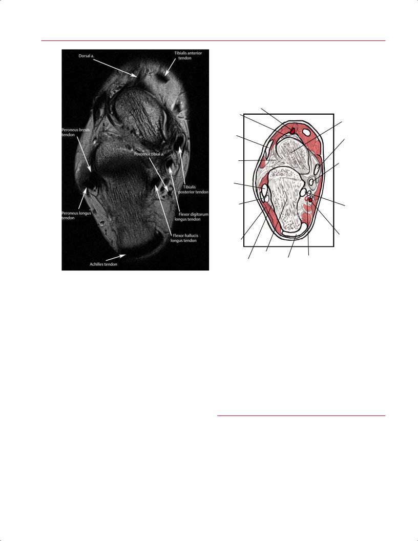
Fig. 2.45 An axial T2-weighted image (A) and artist’s sketch (B) of the right ankle at the level of the tibial plafond. The tibialis posterior, flexor digitorum longus, and FHL tendons are seen posterior to the
medial malleolus. The normal concave appearance of the Achilles tendon is seen in the midline posteriorly. The peroneus longus and brevis tendons are shown posterior to the lateral malleolus.
52 I Initial Concepts
A
|
|
Tibialis anterior |
|
|
||
Dorsal a. |
|
tendon |
|
|
||
|
|
|
|
|
|
|
|
|
|
|
|
|
|
Navicular |
|
|
|
|
|
|
Extensor |
|
|
|
|
Talus |
|
|
|
|
|
Tibialis |
||
digitorum |
|
|
|
|
||
longus |
|
|
|
|
posterior |
|
tendon |
|
|
|
|
tendon |
|
Extensor |
|
|
|
|
Flexor |
|
retinaculum |
|
|
|
|
digitorum |
|
roots |
|
|
|
|
longus |
|
|
|
|
|
|
tendon |
|
Peroneus |
|
|
|
|
Sustenta- |
|
brevis |
|
|
|
|
||
tendon |
|
|
|
|
culum tali |
|
Peroneus |
|
|
|
|
FHL |
|
longus |
|
|
|
|
||
|
|
|
|
tendon |
||
tendon |
|
|
|
|
||
|
|
|
|
|
|
|
Peroneal |
|
|
|
|
Posterior |
|
|
|
|
|
tibial a. |
||
retinaculum |
|
|
|
|
|
|
Calcaneus |
|
|
|
|
|
|
Calcaneo |
Achilles |
|
Posterior |
|
|
|
tendon |
|
tibial v. |
|
B |
||
fibular lig. |
|
|
||||
Fig. 2.46 An axial T2-weighted image (A) and artist’s sketch (B) of the right ankle at the level of the calcaneus, showing the posterior tibialis, flexor digitorum longus, and FHL tendons in their expected
between the menisci and the transverse ligament as pathologic, leading to a mistaken diagnosis of a meniscal tear.
The so called magic-angle phenomenon can cause the false diagnosis of a lateral meniscus tear. Specifically, the upsloping portion of the posterior horn of the lateral meniscus may have increased signal on short-TE images secondary to this phenomenon.13
Extensor Mechanism
The structures of the extensor mechanism of the knee (the patella, patella tendon, quadriceps tendon, and medial and lateral retinaculum) are well seen on midsagittal images (Fig. 2.43). The fat pad is just posterior to the patellar tendon. The patellar and femoral bone marrow signal should be uniform without edema. The signal of the patellar tendon should be uniformly dark. The quadriceps tendon may have a laminated appearance reflecting its multiple tendon composition. There should be no areas of focal thickening of the tendons. The cartilage surfaces of the patella are particularly thick (up to 1 cm). There should be no areas of signal change, fibrillation, or thinning; the presence of these findings is indicative of chondromalacia.
locations from anteromedial to posterolateral, respectively. The peroneus longus and brevis tendons are seen coursing along the lateral aspect of the calcaneus.
Neurovascular Structures
The popliteal artery courses several millimeters posterior to the knee joint. The popliteal vein is just posterior and lateral to the artery (Fig. 2.43) and has a large cross-section. The tibial nerve runs just posterior and lateral to the vein. The common peroneal nerve arises from the tibial nerve above the joint line and may be seen coursing inferiorly and laterally toward the fibular head. The relationships between these structures are easily identified on coronal images.
■ Foot/Ankle
Axial Images
Axial images are extremely helpful in identifying normal and abnormal anatomy of the ankle (Figs. 2.45 and 2.46) and are usually evaluated from superior to inferior (proximal to distal). These images are acquired perpendicular to the plane of the tibia and fibula. The tibia and fibula expand distally to
|
|
|
|
|
|
|
|
|
2 Normal MRI Anatomy of the Musculoskeletal System |
|
53 |
Interosseous |
|
Metatarsals |
|
Interosseous |
form the tibial plafond, medial malleolus, and lateral malleo- |
|
|||||
metatarsal ligs. |
|
|
lig. |
lus. Axial images also show the distal tibiofibular joint and |
|
||||||
|
|
|
|
|
|
|
|
|
|||
Tarsometa- |
|
|
|
|
|
|
|
ankle syndesmosis, with the fibula located laterally and pos- |
|
||
|
|
|
|
|
|
|
|
||||
tarsal joint |
|
|
|
|
|
|
|
teriorly in the syndesmotic notch. More inferiorly, the talar |
|
||
|
|
|
|
|
|
|
|
Tarsometa- |
dome is visualized, followed sequentially by the calcaneus |
|
|
Cuneiforms |
|
|
|
|
|
|
|
and the midfoot osseous structures (Fig. 2.47). The navicular |
|
||
|
|
|
|
|
|
|
tarsal joint |
|
|||
Cuneo- |
|
|
|
|
|
|
bone can be seen on axial images and can be evaluated in this |
|
|||
|
|
|
|
|
|
|
|
||||
|
|
|
|
|
|
|
plane. More distally, the tarsometatarsal joints and metatar- |
|
|||
navicular joint |
|
|
|
|
|
|
|
|
|||
|
|
|
|
|
|
|
sals are seen. These structures are not all in the same axial |
|
|||
|
|
|
|
|
|
|
|
Cuboid |
|
||
|
|
|
|
|
|
|
|
plane because of the transverse arch of the foot. Therefore, |
|
||
Navicular |
|
|
|
|
|
|
|
|
|||
|
|
|
|
|
|
|
|
|
they must be examined on sequential images, which is fa- |
|
|
Talonavicular |
|
|
|
|
|
|
Calcaneceo- |
cilitated by correlation with the coronal and sagittal images. |
|
||
|
|
|
|
|
|
cuboid joint |
|
||||
joint |
|
|
|
|
|
|
The forefoot structures (Fig. 2.48) are usually best evaluated |
|
|||
|
|
|
|
|
|
|
|
||||
|
|
|
|
|
|
|
|
Calcaneus |
with specialized planes for the hallux and separate planes |
|
|
|
|
|
|
|
|
|
|
for the lesser metatarsals. |
|
||
|
|
|
|
|
|
|
|
|
|
||
|
|
|
|
|
|
|
|
|
On axial images, most tendon structures in the ankle |
|
|
|
|
|
|
|
|
|
|
|
are seen in cross-section. Therefore, it is important to iden- |
|
|
Interosseous |
|
Subtalar joint |
|
|
tify and follow each tendon from image to image along its |
|
|||||
talocalcaneal lig. |
|
|
|
|
|
|
|
course. These tendons should be dark on all sequences. At |
|
||
|
|
|
|
|
|
|
|
|
|
||
Fig. 2.47 Illustration of an axial section through the osseous struc- |
the level of the ankle joint, the tibialis anterior and extensor |
|
|||||||||
tures of the right foot showing the tarsometatarsal and intertarsal |
hallucis longus tendons are the most anteromedial struc- |
|
|||||||||
joints and the interosseous ligaments. |
|
|
tures, anterior to the neurovascular bundle. The extensor |
|
|||||||
digitorum longus tendon (Fig. 2.49) has a more muscular, broad appearance at the level of the ankle joint and lies more laterally than does the extensor hallucis longus. The most
|
|
|
|
|
|
|
|
|
|
|
|
|
|
|
Distal |
|
|
|
|
|
|
|
|
|
|
|
|
|
|
|
|
|
|||
|
|
|
|
|
|
|
|
|
|
|
|
|
|
|
|
|
|
|
|
|
|
|
|
|
|
|
|
|
|
|
|
||||
|
|
|
|
|
|
|
|
|
|
|
|
|
|
|
|
|
|
|
|
|
|
|
|
|
|
|
|
|
|
|
|||||
|
|
|
|
|
|
|
|
|
|
|
|
Phalanges |
|
|
|
|
|
|
|
|
|
|
|
|
|
|
|
||||||||
|
|
|
|
|
|
|
|
|
|
|
|
|
|
Middle |
|
|
|
|
|
|
|
|
|
|
|
|
|
Distal |
|||||||
|
|
|
|
|
|
|
|
|
|
|
|
|
|
|
|
|
|
|
|
|
|
|
|
|
|
|
|||||||||
|
|
|
|
|
|
|
|
|
|
|
|
|
|
|
|
|
|
|
|
|
|
|
|
|
|
|
|
|
|
|
|
|
Middle |
||
|
|
|
|
|
|
|
|
|
|
|
|
|
|
|
|
|
|
|
|
|
|
|
|
|
|
|
|
|
|
|
|
|
|||
|
|
|
|
|
|
|
|
|
|
|
|
|
|
|
|
|
|
|
|
|
|
|
|
|
|
|
|
|
|
|
|
|
Proximal |
||
|
|
|
|
|
|
|
|
|
|
|
|
|
Metatarsals |
|
|
|
|
|
|
|
|
|
|
|
|||||||||||
|
|
|
|
|
|
|
|
|
|
|
|
|
|
|
|
|
|
|
|
|
|
|
|
||||||||||||
|
|
|
|
|
|
|
|
|
|
|
|
|
|
|
|
|
|
|
|
|
|
|
|
|
|
|
|||||||||
|
|
|
|
|
|
|
|
|
|
|
|
|
|
|
|
|
|
|
|
|
|
|
|
|
|
|
|||||||||
|
|
|
|
|
|
1 |
|
|
|
|
|
|
|
|
|
|
|
|
|
1 |
|
|
|
|
|
|
|
|
|
||||||
|
|
|
|
|
|
|
|
|
|
|
|
|
|
|
|
|
|
|
|
|
|
|
|
|
|
|
|
|
|
|
|
|
|||
4 |
3 |
2 |
|
|
|
|
|
|
|
|
|
|
|
|
|
|
|
|
2 |
3 |
4 |
|
|
|
|
|
|
||||||||
|
|
|
|
|
|
|
|
|
|
|
|
|
|
|
|
|
|
|
|
|
|
||||||||||||||
5 |
|
|
|
|
|
|
|
|
|
|
|
|
|
|
|
|
|
|
1 |
|
|
|
|
|
|
|
|
|
|||||||
|
|
|
|
|
|
|
|
|
|
|
|
Cuneiform |
2 |
3 |
5 |
|
|
|
|
|
|
||||||||||||||
|
|
|
|
|
|
|
|
|
|
|
|
|
|
|
|
|
|
|
|
|
|
|
|
||||||||||||
|
|
|
|
|
|
|
|
|
|
|
|
|
Navicular |
|
|
|
|
|
|
|
|
|
|
|
|
|
Cuboid |
||||||||
|
|
|
|
|
|
|
|
|
|
|
|
|
|
|
|
|
|
|
|
|
|
|
|
|
|
||||||||||
|
|
|
|
|
|
|
|
|
|
|
|
|
|
|
|
|
|
|
|
|
|
|
|
|
|
|
|
|
|
|
|||||
|
|
|
|
|
|
|
|
|
|
|
|
Talus |
|
|
|
Head |
|
|
|
|
|
|
|
|
|
|
|
|
|
|
|||||
|
|
|
|
|
|
|
|
|
|
|
|
|
|
|
|
|
|
|
|
|
|
|
|
|
|
|
|
|
|||||||
|
|
Calcaneus |
|
|
|
|
|
|
|
|
|
|
|
|
Neck |
|
|
|
|
|
|
|
|
|
|
|
|
|
|
||||||
|
|
|
|
|
|
|
|
|
|
|
|
|
|
|
|
|
|
|
|
|
|
|
|
|
|
|
|
||||||||
|
|
|
|
|
|
|
|
|
|
|
|
|
|
|
|
|
|
|
|
|
|
|
|
|
|
|
|
|
|
|
|
|
|
||
|
|
|
|
|
|
|
|
|
|
|
|
|
|
|
|
Trochlea |
|
|
|
|
|
|
|
|
|
|
|
|
|
|
|||||
|
|
|
|
|
|
|
|
|
|
|
|
|
|
|
|
|
|
|
|
|
|
|
|
|
|
|
|
|
|
||||||
|
|
|
|
|
|
|
|
|
|
|
|
|
|
|
|
|
|
|
|
|
|
|
|
|
|||||||||||
|
|
|
|
|
|
|
|
|
|
|
Sustentaculum |
|
|
|
|
|
|
|
|
|
|
|
|
Calcaneus |
B |
||||||||||
|
|
|
|
|
|
|
|
|
|
|
|
|
|
|
|
|
|
|
|
|
|
|
|
||||||||||||
|
|
|
|
|
|
FHL |
tali |
|
|
|
|
|
|
|
|
|
|
|
|
|
|
|
|
|
|
|
|
|
|
||||||
|
A |
|
|
|
|
|
|
|
|
|
|
|
|
|
|
|
|
|
|
|
|
|
|
|
|
|
|
|
|
|
|||||
|
|
|
groove |
|
|
|
|
|
|
|
|
|
|
|
|
|
|
|
|
|
|
|
|
|
|
|
|
|
|||||||
|
|
|
|
|
|
|
|
|
|
|
|
|
|
|
|
|
|
|
|
|
|
|
|
|
|
|
|
|
|
|
|||||
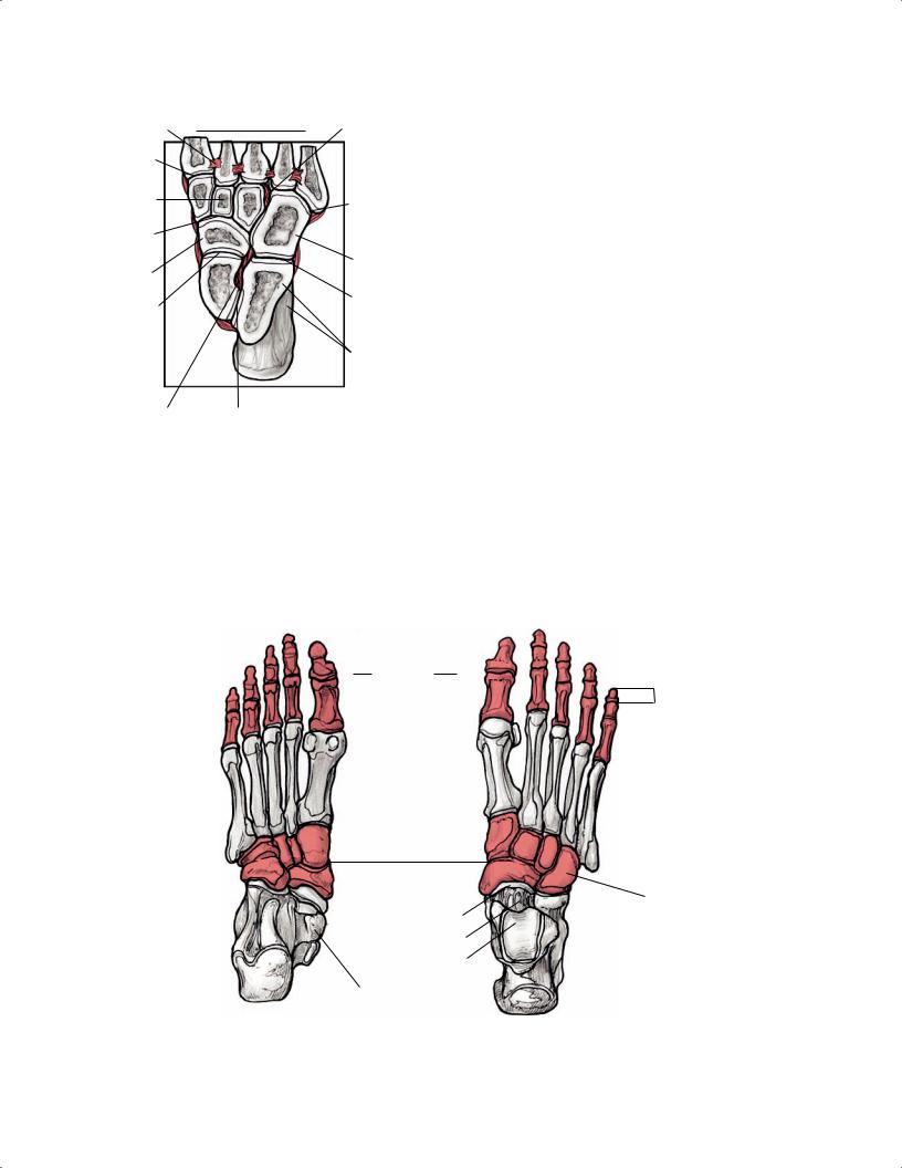
Fig. 2.48 Illustrations of the osseous structures of the plantar (A) and dorsal (B) surface views of the right foot. |
|||||||||||||||||||||||||||||||||||
|
|
Extensor digitorum |
|
|
longus m. |
|
|
Tibialis anterior m. |
|
|
Superior extensor |
|
|
retinaculum |
|
|
Extensor digitorum longus |
Sural n. |
tendon sheath |
|
|
|
Lateral malleolar bursa |
Peroneus brevis |
Inferior extensor retinaculum |
|
tendon |
|
|
Calcaneal tendon |
|
Extensor hallucis longus tendon |
|
Extensor digitorum brevis m. |
|
|
||
|
|
|
|
|
Extensor digitorum longus tendon |
Superior |
|
|
peroneal retinaculum |
|
|
|
|
Abductor digiti |
Inferior |
minimi m. |
|
|
||
peroneal retinaculum |
|
|
Peroneus longus
Peroneus brevis
Abductor digiti m. Peroneus tertius m. tendon
minimi m.
Extensor digitorum Peroneus brevis longus m.
tendon
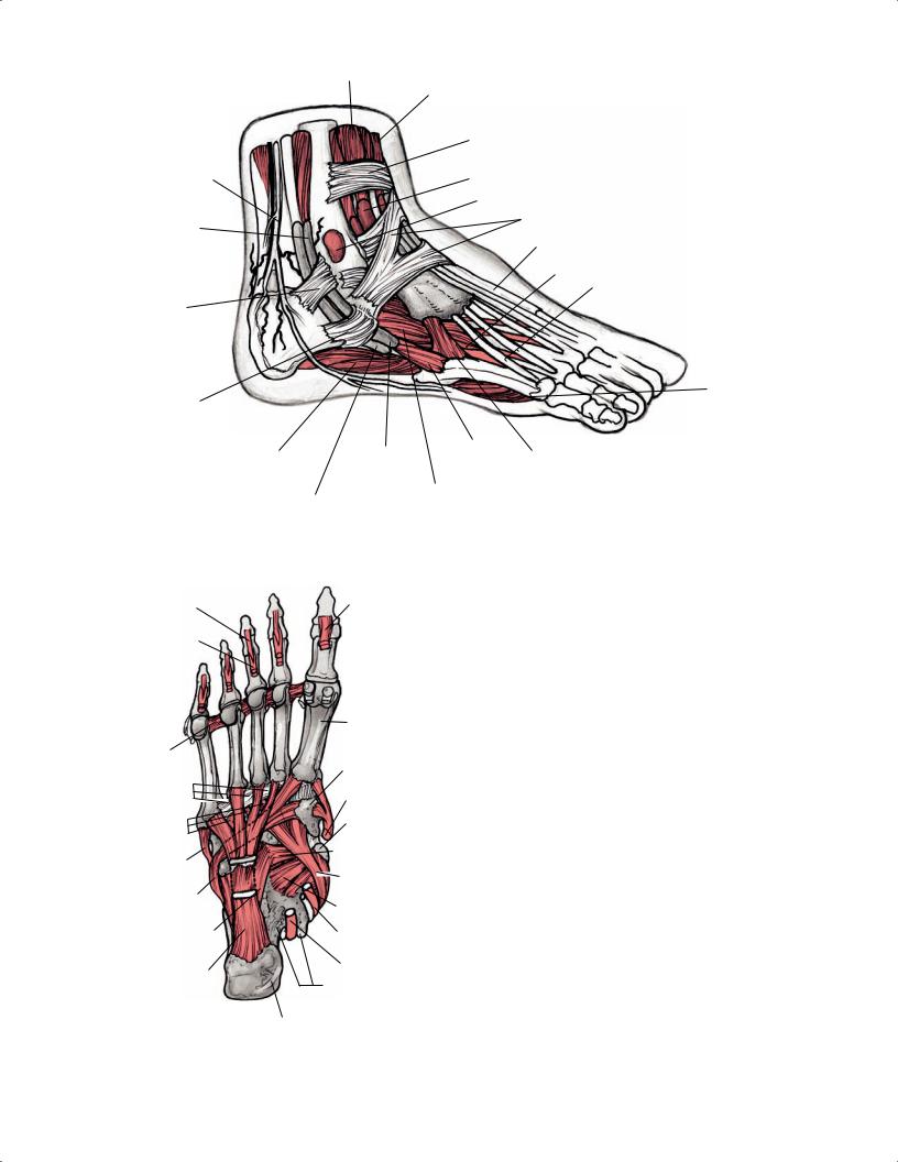
Fig. 2.49 A 3D illustration of the ankle and foot from the lateral aspect showing the musculotendinous units passing deep to the superior and inferior extensor retinacula.
Flexor digitorum |
|
longus tendons |
FHL tendon |
Flexor digitorum brevis tendons
|
First metatarsal |
|
Deep transverse |
Medial |
|
metatarsal ligs. |
cuneiform |
|
Plantar |
Tibialis |
|
metatarsal ligs. |
anterior tendon |
|
|
||
Plantar |
Tuberosity of |
|
tarsometa- |
navicular |
|
tarsal ligs. |
|
|
|
Cuboideonavicular |
|
Peroneus |
lig. |
|
brevis tendon |
Tibialis |
|
|
||
Peroneus |
posterior tendon |
|
Plantar |
||
longus tendon |
||
calcaneonavicular |
||
|
||
|
lig. |
|
Calcaneocuboboid |
Flexor digitorum |
|
lig. |
longus tendon |
|
Long plantar lig. |
FHL tendon |
|
|
||
|
Sustentaculum tali |
Calcaneus
Fig. 2.50 An axial 3D illustration of the plantar aspect of the right foot showing the tarsometatarsal and intertarsal ligaments, as well as the tendons crossing the ankle into the foot.
anterolateral structure is the peroneus tertius tendon (variably present), which can sometimes be di cult to visualize because of its small size.
The posterior tibialis tendon is the most obvious tendon structure just posterior to the medial malleolus. The flexor digitorum longus tendon is just posterior to the posterior tibialis tendon and medial to the posterior neurovascular bundle. It is approximately one third the diameter of the posterior tibialis tendon. Lateral and deep to the posterior neurovascular bundle is the FHL, which has a broad muscular cross-section at the level of the ankle joint. This tendon can be followed further distally in its sheath between the posteromedial and posterolateral processes of the talus. These three tendons can be identified not only by their appearance and insertions but also by their predictable relationships to each other as they pass posterior to the medial malleolus.
The peroneus brevis and longus (Figs. 2.50 and 2.51) are easily identified posterior to the fibula. The peroneus brevis lies directly against the fibula, and the longus is more superficial at the level of the ankle joint. The overlying superior peroneal retinaculum should be assessed for integrity.
The Achilles tendon is the most posterior tendinous structure in the ankle. At its insertion on the calcaneus, it has an oblong appearance on axial images and should appear concave more proximally on the anterior surface.
|
|
|
2 Normal MRI Anatomy of the Musculoskeletal System |
55 |
|
Peroneus longus |
|
|
|
|
|
tendon |
Peroneus |
|
|
|
|
|
brevis tendon |
|
|
|
|
|
|
|
Fibula |
|
|
|
|
|
Anterior talofibular lig. |
|
|
Gastrocnemius |
|
|
|
|
|
m. |
|
|
Dorsal talonavicular lig. |
|
|
|
|
|
|
||
|
|
|
Interosseous talocalcaneal ligs. |
|
|
Superior peroneal |
|
|
Dorsal cuneonavicular ligs. |
|
|
retinaculum |
|
|
|
||
|
|
|
|
|
|
Achilles |
|
|
1 |
|
|
tendon |
|
|
|
|
|
|
|
2 |
|
|
|
|
|
|
|
|
|
Calcaneus |
|
|
3 |
|
|
|
|
4 |
|
|
|
|
|
|
|
|
|
|
|
|
5 |
|
|
Inferior peroneal |
Dorsal |
Dorsal |
Dorsal |
|
|
calcanocuboid |
cuneocuboid |
metatarsal ligs. |
|
||
retinaculum |
|
||||
lig. |
ligs. |
|
|
|
|
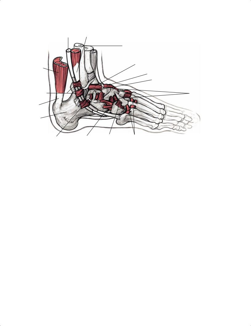
Fig. 2.51 A 3D illustration of the ankle and foot from the lateral aspect showing the deep ligaments and the peroneus longus and brevis tendons passing posterior to the lateral malleolus, deep to the peroneal retinaculum.
The anterior and posterior tibiofibular ligaments, along with the dense interosseous tibiofibular ligament, constitute the syndesmotic complex between the tibia and fibula. The anterior and posterior tibiofibular ligaments are identified easily as low signal intensity ligamentous connections between the distal tibia and fibula at the level of the ankle joint.
Of the lateral ligamentous complex structures, the anterior and posterior talofibular ligaments are best seen on axial images. They appear as dark, thin, ligamentous bands extending distally from the fibula anteriorly and posteriorly to insert on the talus. The anterior band attaches to the neck of the talus, and the posterior band attaches to the posterior lateral process of the talus. However, the structures of the lateral ligamentous complex do not all run in the same plane, and thus, the calcaneofibular ligament component of the lateral ligamentous complex is best seen on the coronal images, whereas the anterior and posterior talofibular ligaments are best seen on axial images.
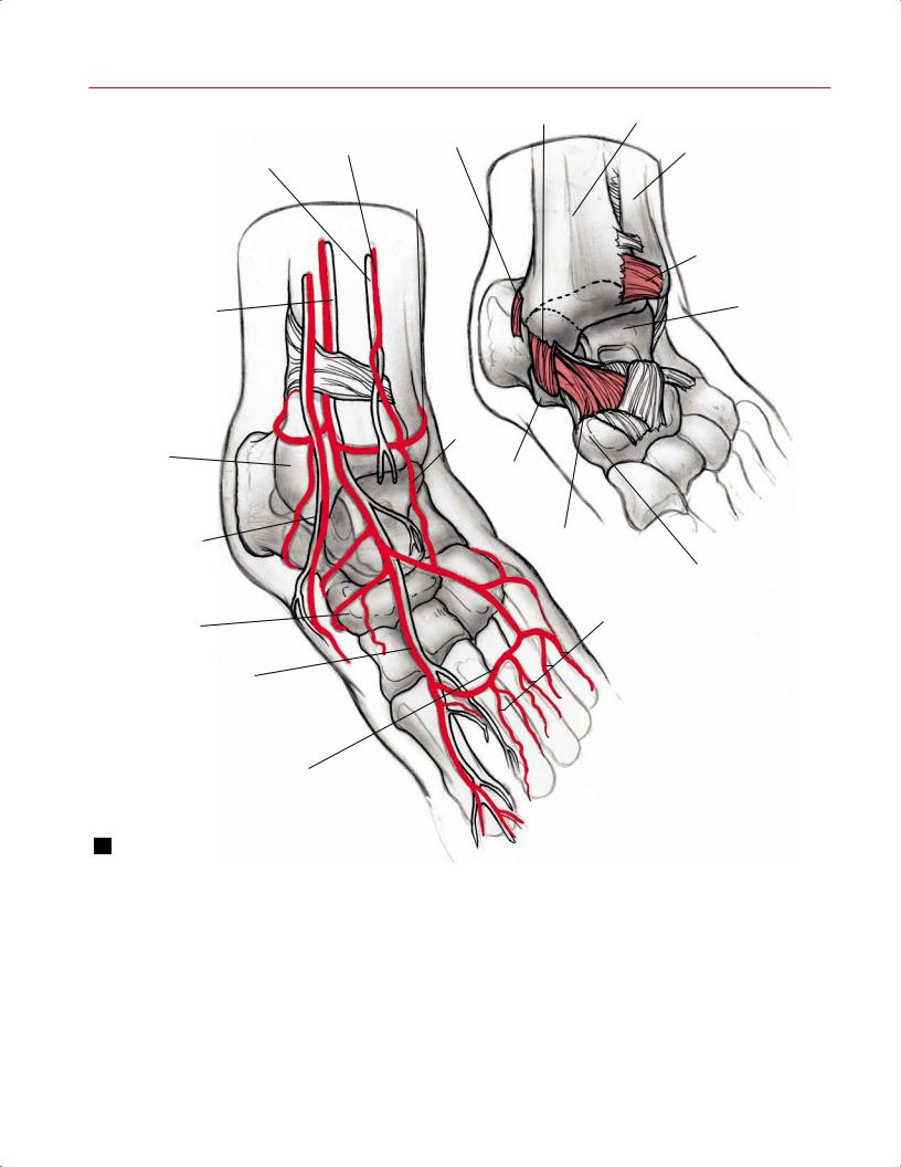
The anterior neurovascular bundle (anterior tibial artery, anterior tibial vein, and deep peroneal nerve) is slightly lateral and deep to the extensor hallucis longus (Fig. 2.52). The posterior neurovascular bundle (posterior tibial artery, vein, and nerve) lies between the flexor digitorum longus and FHL and deep to the flexor retinaculum.
Coronal Images
Coronal images of the ankle are obtained in the plane of the tibia (Figs. 2.53 and 2.54). The tibial plafond, medial and lateral malleoli, and talar dome are well visualized on protondensity and T2-weighted coronal images. Such images are useful in diagnosing osseous pathology such as occult fractures, stress fractures, OCDs, degenerative arthritis, osseous infections, and neoplastic processes.
The ankle structures identified on coronal images are the medial and lateral ligament complexes. The deltoid ligament forms the MCL complex of the ankle and consists of superficial and deep layers. The tibionavicular and tibiocalcaneal ligaments constitute the superficial layer, and the tibiotalar ligament constitutes the deep layer (Fig. 2.55). The tibionavicular ligament originates from the medial malleolus and attaches to the tuberosity of the navicular. The tibiocalcaneal ligament is seen as a thick, dark band extending from the medial malleolus inferiorly to the sustentaculum tali of the calcaneus. The tibiotalar ligament extends from the medial malleolus to the entire nonarticular medial aspect of the talus.
The lateral ligamentous complex of the ankle consists of the calcaneofibular, anterior talofibular, and posterior talofibular ligaments. Of these three structures, the calcaneofibular ligament is best seen on coronal images, extending from
56 I Initial Concepts
|
|
Tibiocalcaneal lig. |
Tibia |
|
Superficial |
Posterior tibiotalar lig. |
|
|
Peroneal a. |
Fibula |
|
|
peroneal n. |
||
|
|
|
|
|
|
Lateral |
|
|
|
malleolar a. |
|
|
|
|
Anterior talofibular |
Anterior tibial a. |
|
|
lig. |
|
|
||
Deep peroneal n. |
|
Talus |
|
|
|
||
Medial |
|
|
Lateral |
||
malleolar a. |
|
||||
|
tarsal a. |
||||
|
|
|
|
||
Medial |
|
|
|
||
malleolus |
|
Sustentaculum |
|||
|
|
|
|
||
Saphenous v. |
|
|
tali |
||
|
|
|
|
||
|
|
|
|
||
|
|
|
|
|
|
|
|
|
|
Deltoid lig. |
B |
Saphenous n. |
|
|
|||
|
|
|
|||
|
|
|
|
Navicular |
|
|
|
|
|
Dorsal |
|
Navicular |
|
metatarsal aa. |
|||
|
|
|
|||
Dorsalis pedis a. |
|
|
|
||
Arcuate a.
A
Fig. 2.52 Anterior oblique illustrations of the left foot and ankle showing the neurovascular (A) and ligamentous (B) structures.
the tip of the lateral malleolus to the lateral wall of the calcaneus. Despite the fact that many tendinous structures can be di cult to identify in the coronal plane, the posterior tibial tendon and the peroneus longus and brevis tendons can be identified inferior to the medial and lateral malleoli, respectively. The posterior tibial tendon should be seen as a lon-
gitudinal dark signal structure just posterior to the medial malleolus. The FHL can be identified at its broad origin from the posterior two thirds of the fibula. The peroneus longus and brevis tendons are seen as longitudinal dark signal structures on most coronal images, passing just posterior to the lateral malleolus.
|
|
|
Tibia |
|
|
|
|
|
|
Anterior tibiofibular lig. |
Talar dome cartilage |
||||||
|
|
|
|
|
|
|||
|
Anterior |
|
|
|
Deltoid lig. |
|||
|
|
|
|
|
|
|||
|
talofibular lig. |
|
|
|
|
|
||
|
|
|
|
|
|
|
Tibialis |
|
|
|
|
|
|
|
|
posterior |
|
|
|
|
|
|
|
|
tendon |
|
|
|
|
|
|
|
|
Interosseous |
|
|
Fat in sinus |
|
|
|
talocal- |
|||
|
|
|
|
|||||
|
|
|
|
caneal lig. |
||||
|
tarsi |
|
|
|
|
|
Sustenta- |
|
|
|
|
|
|
|
|
||
|
|
|
|
|
|
|
culum tali |
|
|
Calcaneus |
|
|
|
Flexor |
|||
|
|
|
|
digitorum |
||||
|
|
|
|
|
|
|
||
|
|
|
|
|
|
|
longus tendon |
|
|
|
|
|
|
|
|
Abductor |
|
|
Abductor |
|
|
|
hallucis m. |
|||
|
|
|
FHL tendon |
|||||
|
digiti minimi m. |
|
|
|||||
|
|
|
|
|
|
|||
|
|
Long plantar |
Flexor digitorum |
|||||
|
|
lig. |
||||||
|
|
brevis m. |
|
|
|
|||
|
|
|
|
|
|
|
||
|
|
|
|
|
|
|||
A |
|
Quadratus plantae m. |
|
|
B |
|||
|
|
|
|
|||||
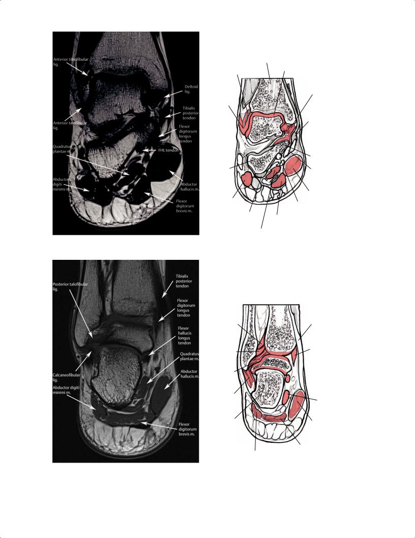
Fig. 2.53 |
A coronal T1-weighted image (A) and artist’s sketch (B) of the right ankle at the level of the tibial plafond and talus. |
|||||||
A
Fig. 2.54 A coronal T1-weighted image (A) and artist’s sketch (B) of the right ankle through the posterior aspect of the joint showing the posterior tibialis, flexor digitorum longus, and FHL tendons coursing
Fibula |
Tibia |
|||||||
|
|
|
|
|
|
Tibialis |
||
Posterior inferior |
|
|
|
|
||||
|
|
|
|
posterior |
||||
tibiofibular |
|
|
|
|
||||
|
|
|
|
tendon |
||||
lig. |
|
|
|
|
||||
|
|
|
|
|
|
|
||
|
|
|
|
|
|
Flexor |
||
Transverse |
|
|
|
|
digitorum |
|||
|
|
|
|
longus |
||||
tibiofibular |
|
|
|
|
tendon |
|||
lig. |
|
|
|
|
|
|
|
|
Posterior |
|
|
|
|
Talus |
|||
|
|
|
|
|||||
|
|
|
|
|
FHL |
|||
talofibular |
|
|
|
|
|
|||
lig. |
|
|
|
|
|
tendon |
||
Calcaneo- |
|
|
|
|
|
|
|
|
fibular lig. |
|
|
|
|
Quadratus |
|||
|
|
|
|
|
|
plantae m. |
||
Calcaneus |
|
|
|
|
Abductor |
|||
|
|
|
|
|
|
|||
|
|
|
|
|
|
hallucis m. |
||
|
|
|
|
|
Flexor digitorum |
|||
Abductor digiti |
|
|
|
brevis m. |
|
|||
|
|
|
B |
|||||
minimi m. |
|
|
|
|
|
|
||
posterior to the medial malleolus in their expected locations from anteromedial to posterolateral, respectively.

Tibia
Fibula
Medial malleolus
Posterior tibiofular lig.
Posterior tibiotalar part of deltoid lig.
Posterior talofibular lig.
Tibiocalcaneal part
of deltoid lig.
Calcaneofibular lig.
Talus
Posterior talocalcaneal lig.
 Calcaneal tendon
Medial talocalcaneal lig.
Calcaneus
Fig. 2.55 A posteroanterior 3D illustration of the right ankle showing the posterior ligamentous structures, including the tibiocalcaneal and posterior tibiotalar parts of the deltoid ligament.
The osseous structures of the foot can also be evaluated on the coronal images. The hindfoot, including the talar body, calcaneus, and subtalar joint, can be examined for occult talocalcaneal coalitions or arthritic processes. The talocalcaneal ligaments and soft-tissue contents of the sinus tarsi can be examined on T2-weighted images or fat-suppressed proton-density images for evidence of increased signal secondary to ligament injury or soft-tissue impingement that supports a diagnosis of sinus tarsi syndrome. Coronal imaging of the midfoot also shows the talonavicular and calcaneocuboid joints, the navicular body and cuboid, and the tarsometatarsal joints. Coronal images through the midfoot are useful for diagnosing navicular or cuboid stress fractures, arthritis of the midfoot joints, or soft-tissue injuries of the tarsometatarsal (Lisfranc) complex. These images can show the plantar tarsometatarsal ligament complex and facilitate evaluation of tendinitis or tearing of the extensor and flexor tendons. Coronal imaging of the forefoot osseous structures includes the following:
•Metatarsals
•Phalanges
•MTP and IP joints
•Sesamoids
The flexor and extensor tendons can be visualized at the forefoot, particularly the extensor hallucis longus tendon and prominent flexor tendons at the first MTP level. The capsuloligamentous structures (plantar plate) of the MTP joints are also visualized in the coronal plane, allowing for diagnosis of MTP sprain injuries, such as the common turf-toe injury at the first MTP joint.
Sagittal Images
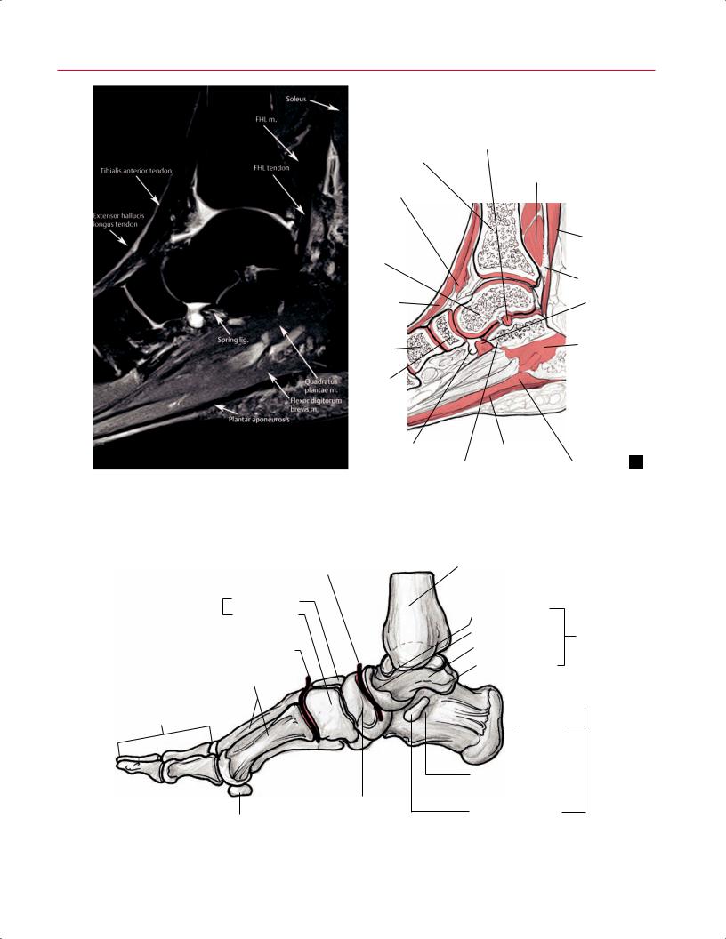
Like coronal images, sagittal images allow most structures to be visualized longitudinally along their courses (Fig. 2.56). The osseous structures of the ankle and foot can be visualized in sequence from medial to lateral (Fig. 2.57). The tibial plafond, talar dome, and lateral malleolus can be inspected for arthritic, degenerative, neoplastic, infectious, or traumatic conditions. Sagittal imaging shows the midfoot bones (particularly the talar head, navicular, and cuboid) and can permit sequential examination of the cuneiforms
2 Normal MRI Anatomy of the Musculoskeletal System 59
Interosseous talocalcaneal lig.
FHL m.
Soleus m.
FHL tendon
Spring lig.
Quadratus plantae m.
Plantar aponeurosis
A |
Sustentaculum tali |
Flexor digitorum brevis m. |
B |
|
Fig. 2.56 A sagittal proton-density fat-suppressed image (A) and artist’s sketch (B) of the medial aspect of the ankle at the level of the navicular.
Transverse tarsal joint
Cuneiforms
Intermediate
Medial
Tarsometatarsal joint
Metatarsals
Phalanges
Navicular
Sesamoid
Fig. 2.57 An illustration of the osseous structures of the right foot from a medial view.
Tibia
Head
Neck
Talus
Tochlea
Posterior process
Calcaneus
Tuberosity
Groove for FHL tendon
Sustentaculum tali