

Книги по МРТ КТ на английском языке / MRI for Orthopaedic Surgeons Khanna ed 2010
.pdf120 II Upper Extremity
A 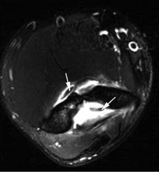 B
B
Fig. 5.2 Axial (A) and sagittal (B) T2-weighted images showing loose bodies in the coronoid and olecranon fossae and in the trochlear ulnar joint (arrows on each). Adapted with permission from Brunton LM, Anderson MW, Pannunzio ME, Khanna AJ,
Chhabra AB: Magnetic resonance imaging of the elbow: update on current techniques and indications. J Hand Surg Am 2006;31: 1001–1011.
A  B
B
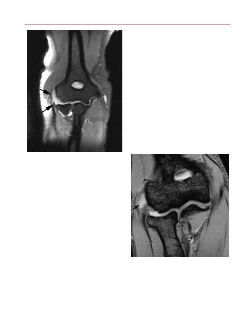
Fig. 5.3 MCL injury of the right elbow. (A) A coronal fat-suppressed T2-weighted image showing discontinuity (complete tear) of the MCL with surrounding edema and hemorrhage (arrow). (B) A coronal fat-suppressed T1-weighted arthrogram showing a partial tear of the MCL as a “T-sign” with leakage of intraarticular contrast at the un-
dersurface of the MCL at its distal insertion on the sublime tubercle (arrow). Part A is adapted with permission from Brunton LM, Anderson MW, Pannunzio ME, Khanna AJ, Chhabra AB: Magnetic resonance imaging of the elbow: update on current techniques and indications. J Hand Surg Am 2006;31:1001–1011.
5 The Elbow 121
Full-thickness tears of the anterior bundle of the MCL can be detected accurately by MRI. The MCL is seen on MRI as a vertically oriented, uniformly low signal intensity structure coursing between the medial epicondyle and the sublime tubercle on coronal images. The abnormalities detectable by MRI include ligament attenuation, redundancy, or discontinuity. Increased signal intensity often is found within and adjacent to the ligament on fat-suppressed T2-weighted images, indicating edema or hemorrhage (Fig. 5.3A).16 Concomitant findings, such as adjacent muscle edema and ulnar neuritis, are not unusual. MRI is less reliable for detecting partial-thickness tears, but the addition of intraarticular contrast may improve the sensitivity in this regard (Fig. 5.3B).17,18
LCL Complex Injury
The LCL complex of the elbow consists of four separate components:
•RCL
•LUCL
•Annular ligament
•Variable accessory LCL
Injury to this complex from an acute elbow dislocation may lead to chronic mechanical symptoms or recurrent instability when early treatment is inadequate. Tears of the LUCL and RCL are common in acute elbow dislocations. Chronic insu ciency of the LUCL is believed to lead to posterolateral rotatory instability, which is often di cult to diagnose clinically.19
The ability of MRI to visualize tears of the LCL complex historically has been less dependable, especially in patients with chronic instability.20 One study of asymptomatic individuals revealed inconsistent MRI signal characteristics within the LUCL.21 Other investigators, however, have found MRI to be e ective in detecting abnormalities of the LUCL in the setting of posterolateral rotatory instability when 3D gradient-echo and FSE sequences were used.22 Findings are similar to those seen in MCL injuries, with combinations of ligament attenuation, redundancy, and/or discontinuity evident on coronal or axial images (Fig. 5.4).12,16 MR arthrography in this setting has not been studied su ciently to date.
Biceps Tendon Injury
Injury to the distal biceps tendon is relatively common. Complete rupture occurs almost exclusively in men after forced extension of the flexed elbow against an eccentric load. However, symptoms arising from a partial tear may have an insidious onset, and such tears usually occur from repetitive microtrauma in an already degenerative tendon. Although MRI is not always necessary to diagnose a complete distal biceps tendon rupture, it can distinguish between tendino-
Fig. 5.4 An axial T2-weighted image of the right elbow showing thickening and abnormal signal (sprain vs. partial tear) of the LUCL with adjacent high signal intensity edema posterior to the radial head (arrow). Adapted with permission from Brunton LM, Anderson MW, Pannunzio ME, Khanna AJ, Chhabra AB: Magnetic resonance imaging of the elbow: update on current techniques and indications. J Hand Surg Am 2006;31:1001–1011.
sis, partial tears, and complete tears. MRI may also allow for the diagnosis of concomitant injuries. A complete rupture is characterized by the absence of the low signal intensity tendon tissue at the radial tuberosity insertion on axial images. The less experienced clinician may have di culty localizing the biceps tendon insertion on the axial images and may find it easier to make or confirm the diagnosis on the sagittal T2-weighted images. A large amount of high signal intensity fluid and/or hemorrhage in the antecubital fossa usually accompanies an acute distal biceps tendon injury. The extent of tendon retraction is assessed on sagittal images, although the course of the tendon often is oriented obliquely to this plane (Fig. 5.5). An abnormally thickened or attenuated distal biceps tendon with increased signal intensity may represent tendinopathy or a partial tear. Associated bone marrow edema within the radial tuberosity is likely related to microavulsive injury. Increased fluid within the bicipitoradial bursa may accompany a partial tear in some patients.12,23
Triceps Tendon Injury
Ruptures of the triceps tendon are far less common than are injuries to the distal biceps tendon. Complete tears result from a traumatic event and are manifested clinically by the inability to extend the elbow against gravity. Extensive

122 II Upper Extremity
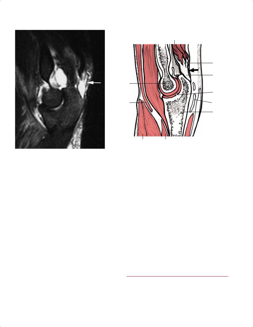
Fig. 5.5 Distal biceps tendon injury. (A) An axial T2weighted image of the right elbow showing rupture of the distal biceps tendon (short arrow) from its insertion (long arrow) at the radial tuberosity, accompanied by surrounding edema. (B) A sagittal T2-weighted image showing a complete tear of the biceps tendon with prominent edema and hemorrhage surrounding the thickened, retracted tendon (arrow). (C) Artist’s sketch. Part B is adapted with permission from Brunton LM, Anderson MW, Pannunzio ME, Khanna AJ, Chhabra AB: Magnetic resonance imaging of the elbow: update on current techniques and indications. J Hand Surg Am 2006;31:1001–1011.
B
posterior elbow swelling, ecchymosis, and a palpable tendon defect are also common findings. The tendon typically is detached from its insertion on the olecranon process of the proximal ulna, and a small osseous avulsion may be seen on conventional radiographs.
MRI has a role in diagnosing triceps tendon injury in patients with equivocal clinical findings. A complete rupture is accompanied by extensive adjacent soft-tissue edema, and the retracted tendon margin is seen on sagittal images (Fig. 5.6).12,16 Partial injury or tendinopathy may result only in thickening or thinning of the tendon, usually without related
A
C
abnormal signal intensity. Findings of olecranon bursitis may accompany triceps tendon injury in some patients.
■Degenerative Conditions: Lateral and Medial Epicondylitis
The term epicondylitis may be a misnomer because mucoid degeneration and tearing of the extensor carpi radialis brevis tendon (in the case of lateral epicondylitis), rather than inflammation, are the key histologic features of the disorder.24
|
|
|
|
|
|
|
|
5 The Elbow |
123 |
|||
|
Biceps m. Brachialis m. |
Triceps m. |
|
|
|
|
|
|
||||
|
|
|
|
|
|
|
|
|
|
Triceps |
|
|
|
|
|
|
|
|
|
|
|
|
|
||
|
|
|
|
|
|
|
|
|
|
|
||
|
|
|
|
|
|
|
|
|
|
|
||
|
|
|
|
|
|
|
|
|
|
tendon |
|
|
|
|
|
|
|
|
|
|
|
|
avulsion |
|
|
|
|
|
|
|
|
|
|
|
|
Edema |
|
|
|
Trochlea |
|
|
|
|
Olecranon |
|
|||||
|
|
|
|
|
|
|
|
|
|
|
||
|
|
|
|
|
|
|
|
|
|
Ulnotrochlear |
|
|
|
|
|
|
|
|
|
|
|
|
ridge |
|
|
|
Biceps |
|
|
|
|
Olecranon |
|
|||||
|
|
|
|
|
bursa |
|
||||||
|
tendon |
|
|
|
|
|
||||||
|
|
|
|
|
|
|
|
|||||
|
|
|
|
|
|
|
|
|
|
Ulna |
|
|
|
Pronator teres m. |
|
|
Flexor digitorum |
|
|
|
|
|
|
||
|
|
|
|
|
|
|
|
|||||
|
Brachioradialis m. |
|
|
|
|
|
|
|||||
|
|
|
|
|
|
superficialis |
|
|
|
|
|
|
A |
|
B |
|
|
|
|
||||||
|
|
|
|
|||||||||
|
|
|
|
|||||||||
Fig. 5.6 A sagittal T2-weighted image (A) and artist’s sketch (B) revealing a torn, retracted triceps tendon with a large amount of high signal intensity edema between the olecranon process and the tendon end (arrows on each). Part A is adapted with permission from
Nevertheless, in the absence of a more accurate term, lateral epicondylitis (also known as tennis elbow) is the most common overuse disorder of the elbow, occurring most frequently in individuals who participate in racquet sports and in mid- dle-aged persons who perform repetitive wrist extension in their daily activities. Patients complain of lateral elbow pain that increases with the o ending activity. Examination reveals tenderness over the insertion of the common extensor tendon at the lateral epicondyle, and the pain is exacerbated by resisted wrist extension with the elbow fully extended. Although the diagnosis usually is obvious by history and physical examination, concurrent underlying pathology cannot be ruled out easily. The di erential diagnosis for lateral elbow pain includes the following:
•Degenerative joint disease
•OCD
•Radial tunnel syndrome
•Posterolateral rotatory instability
•Occult fracture
•Loose bodies
It is important to consider these other etiologies when nonoperative treatment of lateral epicondylitis for more than 6 to 12 months has failed.
Brunton LM, Anderson MW, Pannunzio ME, Khanna AJ, Chhabra AB: Magnetic resonance imaging of the elbow: update on current techniques and indications. J Hand Surg Am 2006;31:1001–1011.
Usually, MRI is used to rule out concomitant causes of refractory elbow pain rather than to diagnose lateral epicondylitis.25 Abnormalities of the LCL complex are not unusual in patients with severe lateral epicondylitis (Fig. 5.7).26 The typical finding of isolated lateral epicondylitis is increased signal intensity in a thickened common extensor tendon. If the common extensor tendon appears normal, adjacent softtissue edema usually predominates (Fig. 5.8).12,16 Although asymptomatic individuals may have abnormal MRI findings, these findings are usually limited to tendon thickening and intermediate signal intensity on T1-weighted or T2-weighted images without adjacent soft-tissue edema.27
Despite the relatively low prevalence of medial epicondylitis, MRI has an additional role in excluding other causes of medial elbow pain, such as MCL injury or ulnar neuritis, in patients for whom nonoperative treatment fails. A thickened common flexor tendon and local soft-tissue edema are the typical MRI features.
■ Infectious Conditions
Depending on the acuity of presentation, various imaging modalities are used in the evaluation of patients with signs
124 II Upper Extremity
Fig. 5.7 A coronal T2-weighted image of the right elbow showing concurrent severe lateral epicondylitis (upper arrow) with high signal intensity within a thickened common extensor tendon and discontinuity of the underlying RCL (lower arrow).
and symptoms of musculoskeletal infection. Conventional radiographs, CT, and radioisotope studies all have roles in the diagnosis of an infectious process, although MRI is the single most useful study for determining the precise anatomic location and extent of infection.
Acute osteomyelitis causes inflammation of medullary bone, which can be detected by MRI well before findings are evident on conventional radiographs. On T1-weighted images, areas of marrow involvement show a low signal intensity that contrasts with the surrounding bright normal marrow. STIR images, or other fat-suppressed T2-weighted images, are the most sensitive for infection, showing a ected areas as high signal intensity. Sometimes these regions of high T2-weighted signal are accompanied by a well-defined low-intensity margin. Because these findings may be similar in patients with tumor or early osteonecrosis, correlation with the history and physical examination is critical.28
The role of MRI in patients with presumed septic arthritis is less clear. Although the presence of an elbow joint e u- sion can be well depicted by MRI, characterizing the synovial
fluid as normal, bloody, or purulent is less reliable. MRI often is used to determine the extent of involvement in patients with severe cellulitis, septic bursitis, or other superficial infections that are unresponsive to nonoperative measures. MRI also is an excellent method for diagnosing a suspected deep abscess about the elbow that may require surgical incision and drainage.29
The MRI findings in septic and nonseptic olecranon bursitis may be markedly similar. Abnormalities may include the following:
•Marginal lobulation
•Bursal septation
•Heterogeneous internal signal
•Poorly defined margins
•Adjacent soft-tissue edema
•Thickening or edema of the triceps tendon
•Localized bone marrow edema
The most consistent finding in septic olecranon bursitis is rim and soft-tissue enhancement on postgadolinium T1weighted images.30
Fig. 5.8 A coronal STIR image of the right elbow showing isolated lateral epicondylitis, indicated by the thickened common extensor tendon with amorphous features and high signal intensity (arrow). Adapted with permission from Brunton LM, Anderson MW, Pannunzio ME, Khanna AJ, Chhabra AB: Magnetic resonance imaging of the elbow: update on current techniques and indications. J Hand Surg Am 2006;31:1001–1011.
5 The Elbow 125
Fig. 5.9 A coronal oblique STIR image of the right elbow showing an unstable OCD fragment of the capitellum with joint fluid tracking behind the fragment (arrow). Adapted with permission from Brunton LM, Anderson MW, Pannunzio ME, Khanna AJ, Chhabra AB: Magnetic resonance imaging of the elbow: update on current techniques and indications. J Hand Surg Am 2006;31:1001–1011.
fragment. Fluid surrounding the fragment on fat-suppressed T2-weighted images or surrounding contrast material on MR arthrography are signs of an unstable fragment (Fig. 5.9). Enhancement of a fragment after intravenous gadolinium contrast has been administered suggests adequate blood supply and fragment viability.31
Compression Neuropathies
The most commonly occurring compression neuropathies about the elbow include the following:
•Cubital tunnel syndrome (ulnar nerve)
•Radial tunnel syndrome (posterior interosseous nerve— pain)
•Posterior interosseous nerve syndrome (posterior interosseous nerve—motor deficits)
•Pronator syndrome (median nerve)
Each syndrome has key historical and physical examination findings that have been addressed elsewhere.32 Electromyography and nerve conduction studies are the most frequently used adjunctive diagnostic tests. Advanced imaging may have a role in evaluating patients for whom nonoperative treatment fails or those with recurrence after surgical intervention.
MRI is most useful for assessing cubital tunnel syndrome, especially when electrodiagnostic testing is equivocal, or for excluding accompanying pathology in patients in whom this syndrome recurs. MRI may be more sensitive than nerve conduction studies in the diagnosis of ulnar neurop-
■ Other Pathologic Conditions
OCD
OCD of the humeral capitellum is an idiopathic disorder affecting adolescents with a history of repetitive overuse of the elbow. A ected individuals commonly present with pain and swelling of the lateral aspect of the elbow. Mechanical symptoms and/or limitations of motion may be indicative of an associated loose body. Conventional radiographs taken in the early stages of OCD may show only subtle changes within the capitellum.
In contrast, MRI can be helpful in the early detection of OCD. Focal areas of subchondral low signal intensity on T1-weighted images are characteristic. Fat-suppressed T2weighted images provide the most sensitive evaluation for a potential OCD lesion at the capitellar articular surface; however, these images are the least specific. With a patient in whom the clinical suspicion is relatively high, the fat-suppressed T2-weighted image can be used as a “screening” examination and then correlated with the T1-weighted image where anatomic detail may be better seen. MRI can also help to evaluate the stability and viability of the OCD
Fig. 5.10 An axial STIR image of the right elbow showing a thickened ulnar nerve with a mottled appearance and surrounding increased signal intensity consistent with edema from inflammation (ulnar neuritis) (arrow). Adapted with permission from Brunton LM, Anderson MW, Pannunzio ME, Khanna AJ, Chhabra AB: Magnetic resonance imaging of the elbow: update on current techniques and indications. J Hand Surg Am 2006;31:1001–1011.
126 II Upper Extremity
athy at the elbow.33 Prolonged compression of peripheral nerves leads to a spectrum of MRI findings, including the following:
•Thickening of the nerve
•Increased signal intensity within the nerve
•A mottled appearance of the nerve
•A surrounding fluid collection, which may represent varying degrees of inflammatory changes (Fig. 5.10)
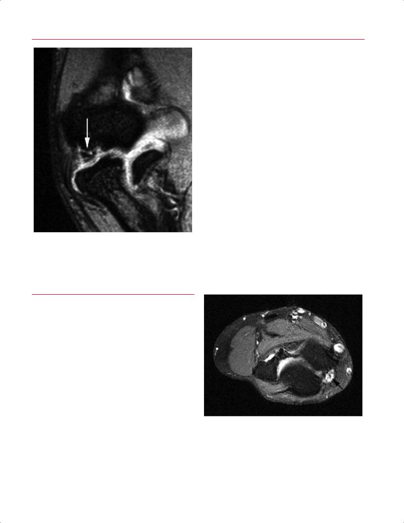
The median and radial nerves are more di cult to evaluate; however, MRI can detect adjacent space-occupying lesions that may produce compression. Patients with chronic neuropathy also may show changes within adjacent denervated muscles, such as fatty atrophy on T1-weighted images (Fig. 5.11) or muscle edema on T2-weighted images.5
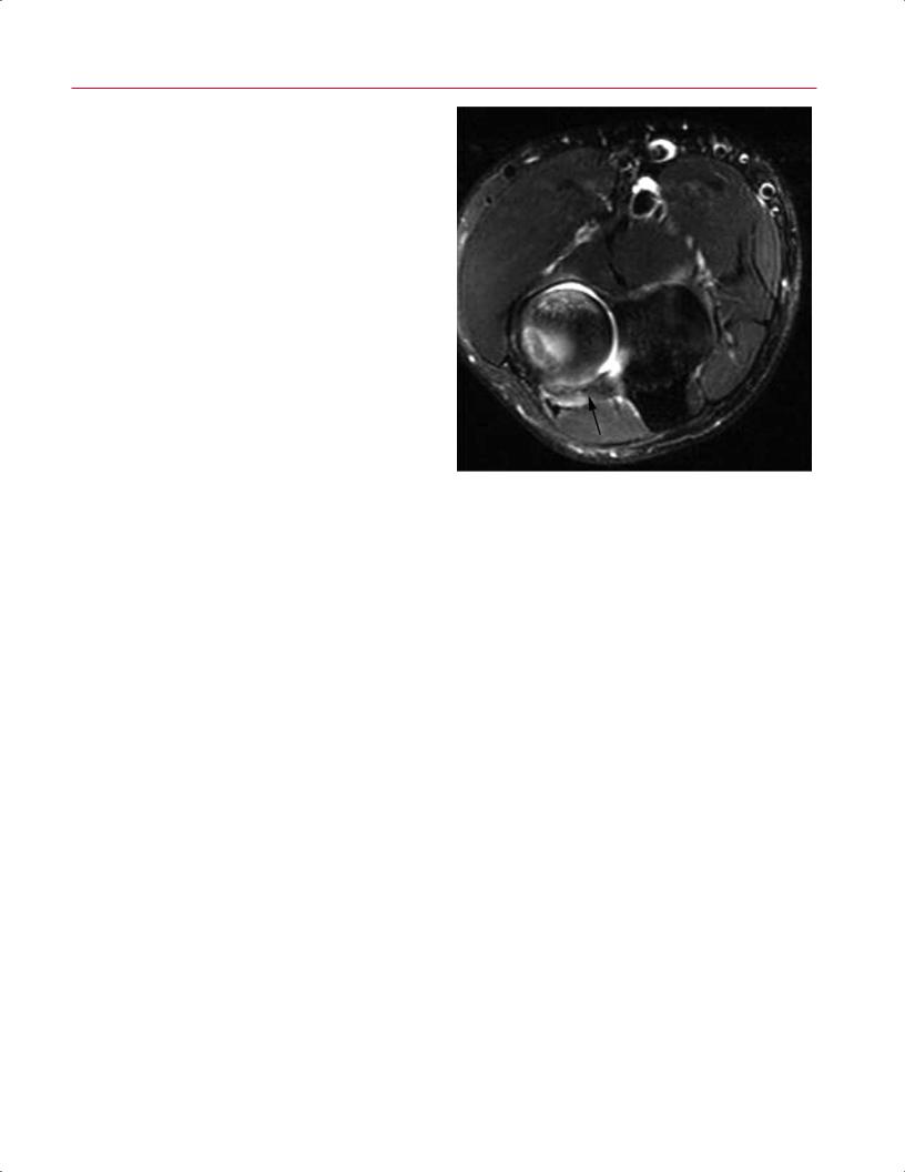
Fig. 5.11 A sagittal T1-weighted image revealing a lipoma (L) within the supinator muscle (Sup). The supinator shows di use high signal intensity, which matches that of the lipoma and subcutaneous fat, indicating fatty atrophy of the denervated muscle from long-standing compression of the posterior interosseous nerve by the lipoma. Adapted with permission from Brunton LM, Anderson MW, Pannunzio ME, Khanna AJ, Chhabra AB: Magnetic resonance imaging of the elbow: update on current techniques and indications. J Hand Surg Am 2006;31:1001–1011.
Soft-Tissue Masses
Although soft-tissue masses are less common around the elbow than in the lower extremity, MRI is essential for evaluating such lesions. The primary role of MRI is di erentiating determinate from indeterminate lesions (see Chapter 15). Determinate lesions are those for which a well-trained
Synovial Disorders
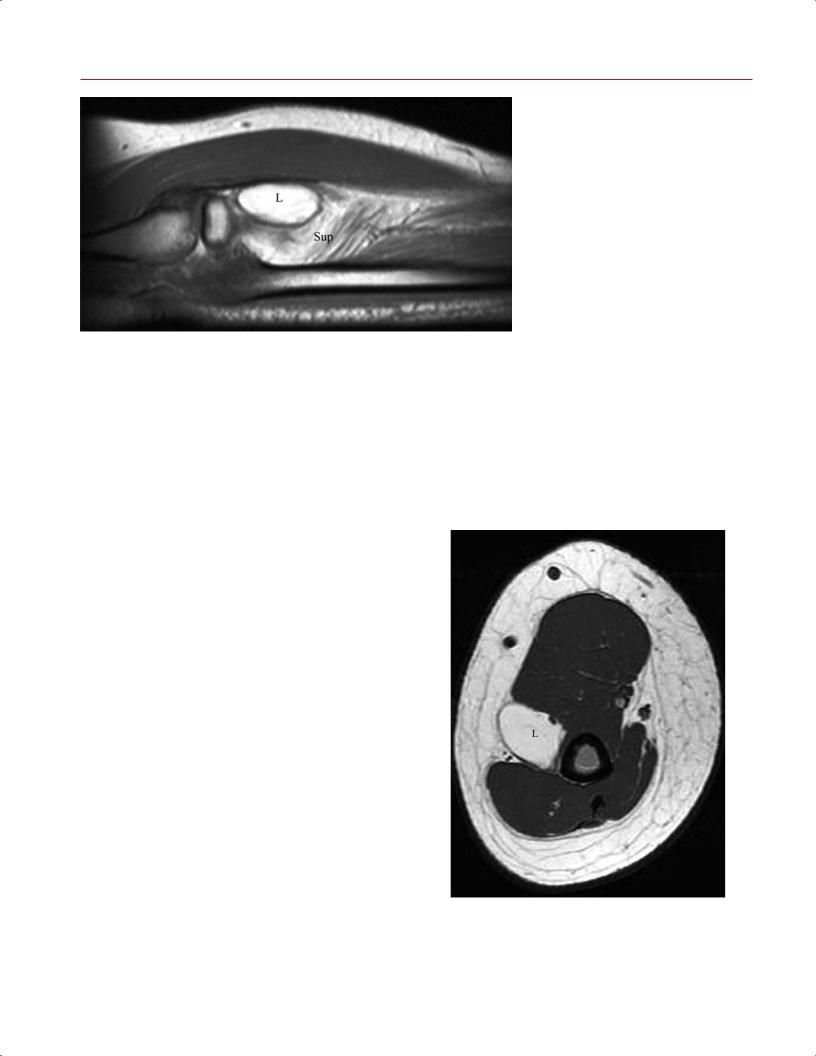
MRI can depict numerous abnormal synovial-based processes, such as RA, crystal deposition disorders, PVNS, and idiopathic synovial osteochondromatosis.13 When synovial fluid analysis is equivocal, MRI may aid in di erentiating conditions that produce a joint e usion. With the use of intravenous gadolinium, MRI has become a powerful tool for detecting the early stages of inflammatory arthritides, such as RA, because of the striking enhancement of the proliferative synovium (see Chapter 6 for specific MRI findings in patients with RA). Although PVNS and synovial chondromatosis are commonly seen in the hip and knee and are quite rare in the elbow, these syndromes should be considered in the differential diagnosis of the patient with multiple loose bodies in the elbow. Gradient-echo imaging shows accentuated “signal dropout” in areas of PVNS, relating to the presence of hemosiderin in this disease process. Other nonspecific MRI findings of PVNS include variable synovial proliferation, bone erosion, and joint e usion.34,35
Fig. 5.12 An axial T1-weighted image showing the similarity in signal intensity between the well-circumscribed lipoma (L) and the adjacent subcutaneous fat.

5 The Elbow 127
musculoskeletal radiologist or clinician can provide a single |
• Myositis ossificans (chronic stage) |
|
diagnosis after MRI evaluation with a high degree of cer- |
• PVNS |
|
tainty based on the imaging appearance. Such lesions in- |
A lipoma matches the signal intensity of subcutaneous fat |
|
clude the following12,36,37: |
||
on all pulse sequences, including fat-suppressed sequences |
||
|
||
• Lipomas |
(Fig. 5.12). Indeterminate lesions are those lesions in which |
|
• Ganglions |
the radiologist or clinician is unable to provide a definitive |
|
• Hemangiomas |
diagnosis and therefore warrant a needle or open biopsy (see |
|
• Neurofibromas |
Chapter 15 for additional information on MRI of soft-tissue |
|
• Muscle tears |
masses).36 |
References
1.Fritz RC, Breidahl WH. Radiographic and special studies: recent advances in imaging of the elbow. Clin Sports Med 2004;23:567–580
2.Kijowski R, Tuite M, Sanford M. Magnetic resonance imaging of the elbow. Part I: normal anatomy, imaging technique, and osseous abnormalities. Skeletal Radiol 2004;33:685–697
3.Potter HG. Imaging of posttraumatic and soft tissue dysfunction of the elbow. Clin Orthop Relat Res 2000;370:9–18
4.Burstein D, Gray M. New MRI techniques for imaging cartilage. J Bone Joint Surg Am 2003;85(suppl 2):70–77
5.Kijowski R, Tuite M, Sanford M. Magnetic resonance imaging of the elbow. Part II: abnormalities of the ligaments, tendons, and nerves. Skeletal Radiol 2005;34:1–18
6.Chung CB, Lektrakul N, Gigena L, Resnick D. Magnetic resonance imaging of the upper extremity: advances in technique and application. Clin Orthop Relat Res 2001;383:162–174
7.O’Dwyer H, O’Sullivan P, Fitzgerald D, Lee MJ, McGrath F, Logan PM. The fat pad sign following elbow trauma in adults: its usefulness and reliability in suspecting occult fracture. J Comput Assist Tomogr 2004;28:562–565
8.Skaggs DL, Mirzayan R. The posterior fat pad sign in association with occult fracture of the elbow in children. J Bone Joint Surg Am 1999;81:1429–1433
9.Itamura J, Roidis N, Mirzayan R, Vaishnav S, Learch T, Shean C. Radial head fractures: MRI evaluation of associated injuries. J Shoulder Elbow Surg 2005;14:421–424
10.Pudas T, Hurme T, Mattila K, Svedström E. Magnetic resonance imaging in pediatric elbow fractures. Acta Radiol 2005;46:636–644
11.Gri th JF, Roebuck DJ, Cheng JCY, et al. Acute elbow trauma in children: spectrum of injury revealed by MR imaging not apparent on radiographs. AJR Am J Roentgenol 2001;176:53–60
12.Melloni P, Valls R. The use of MRI scanning for investigating soft-tis- sue abnormalities in the elbow. Eur J Radiol 2005;54:303–313
13.Chen FS, Diaz VA, Loebenberg M, Rosen JE. Shoulder and elbow injuries in the skeletally immature athlete. J Am Acad Orthop Surg 2005;13:172–185
14.Dubberley JH, Faber KJ, Patterson SD, et al. The detection of loose bodies in the elbow: the value of MRI and CT arthrography. J Bone Joint Surg Br 2005;87:684–686
15.Cain EL Jr, Dugas JR, Wolf RS, Andrews JR. Elbow injuries in throwing athletes: a current concepts review. Am J Sports Med 2003;31: 621–635
16.Fritz RC, Steinbach LS. Magnetic resonance imaging of the musculoskeletal system: Part 3. The elbow. Clin Orthop Relat Res 1996;324:321–339
17.Hill NB Jr, Bucchieri JS, Shon F, Miller TT, Rosenwasser MP. Magnetic resonance imaging of injury to the medial collateral ligament of the elbow: a cadaver model. J Shoulder Elbow Surg 2000;9:418–422
18.Schwartz ML, Al-Zahrani S, Morwessel RM, Andrews JR. Ulnar collateral ligament injury in the throwing athlete: evaluation with sa- line-enhanced MR arthrography. Radiology 1995;197:297–299
19.O’Driscoll SW, Bell DF, Morrey BF. Posterolateral rotatory instability of the elbow. J Bone Joint Surg Am 1991;73:440–446
20.Grafe MW, McAdams TR, Beaulieu CF, Ladd AL. Magnetic resonance imaging in diagnosis of chronic posterolateral rotatory instability of the elbow. Am J Orthop 2003;32:501–503, discussion 504
21.Terada N, Yamada H, Toyama Y. The appearance of the lateral ulnar collateral ligament on magnetic resonance imaging. J Shoulder Elbow Surg 2004;13:214–216
22.Potter HG, Weiland AJ, Schatz JA, Paletta GA, Hotchkiss RN. Posterolateral rotatory instability of the elbow: usefulness of MR imaging in diagnosis. Radiology 1997;204:185–189
23.Williams BD, Schweitzer ME, Weishaupt D, et al. Partial tears of the distal biceps tendon: MR appearance and associated clinical findings. Skeletal Radiol 2001;30:560–564
24.Kraushaar BS, Nirschl RP. Tendinosis of the elbow (tennis elbow). Clinical features and findings of histological, immunohistochemical, and electron microscopy studies. J Bone Joint Surg Am 1999;81:259–278
25.Aoki M, Wada T, Isogai S, Kanaya K, Aiki H, Yamashita T. Magnetic resonance imaging findings of refractory tennis elbows and their relationship to surgical treatment. J Shoulder Elbow Surg 2005;14:172–177
26.Bredella MA, Tirman PFJ, Fritz RC, Feller JF, Wischer TK, Genant HK. MR imaging findings of lateral ulnar collateral ligament abnormalities in patients with lateral epicondylitis. AJR Am J Roentgenol 1999;173:1379–1382
27.Kijowski R, De Smet AA. Magnetic resonance imaging findings in patients with medial epicondylitis. Skeletal Radiol 2005;34:196–202
28.Boutin RD, Brossmann J, Sartoris DJ, Reilly D, Resnick D. Update on imaging of orthopedic infections. Orthop Clin North Am 1998;29:41– 66
29.Berquist TH, Broderick DF. Musculoskeletal infection. In: Berquist TH, ed. MRI of the Musculoskeletal System. 5th ed. Philadelphia: Lippincott Williams & Wilkins; 2006:916–947
30.Floemer F, Morrison WB, Bongartz G, Ledermann HP. MRI characteristics of olecranon bursitis. AJR Am J Roentgenol 2004;183:29–34
31.Kijowski R, De Smet AA. MRI findings of osteochondritis dissecans of the capitellum with surgical correlation. AJR Am J Roentgenol 2005;185:1453–1459

128 II Upper Extremity
32.Lubahn JD, Cermak MB. Uncommon nerve compression syndromes of the upper extremity. J Am Acad Orthop Surg 1998;6:378–386
33.Vucic S, Cordato DJ, Yiannikas C, Schwartz RS, Shnier RC. Utility of magnetic resonance imaging in diagnosing ulnar neuropathy at the elbow. Clin Neurophysiol 2006;117:590–595
34.Khanna AJ, Cosgarea AJ, Mont MA, et al. Magnetic resonance imaging of the knee. Current techniques and spectrum of disease. J Bone Joint Surg Am 2001;83(suppl 2 Pt 2):128–141
35.Cheng XG, You YH, Liu W, Zhao T, Qu H. MRI features of pigmented villonodular synovitis (PVNS). Clin Rheumatol 2004;23:31–34
36.Frassica FJ, Khanna JA, McCarthy EF. The role of MR imaging in soft tissue tumor evaluation: perspective of the orthopedic oncologist and musculoskeletal pathologist. Magn Reson Imaging Clin N Am 2000;8:915–927
37.Chen AL, Youm T, Ong BC, Rafii M, Rokito AS. Imaging of the elbow in the overhead throwing athlete. Am J Sports Med 2003;31:466–473

6 The Wrist and Hand
Lance M. Brunton, Mark W. Anderson, and A. Bobby Chhabra
■Six Specialized Pulse Sequences and Imaging Protocols
The routine use of MRI for the distal upper extremity, compared with its use for larger joints, has been hindered by the technical challenges of imaging small anatomic structures. MRI of the wrist and hand typically is performed at a magnetic field strength of 0.2 to 3.0 T, using systems with dedicated surface coils. Recent advances in high field strength magnets and extremity surface coil design, however, have resulted in additional improvement in image quality. One prospective study revealed significantly higher contrast-to- noise ratios between muscle and bone and between bone and cartilage for all sequences with a 3-T system than with a 1.5-T system.1 3-T systems are now becoming available for clinical use, but most hand and wrist examinations are still performed with 1.5-T magnets.
The hand or wrist is best imaged with the patient prone and the a ected extremity placed above the head with the forearm pronated. Typically, the closer the body part being imaged is to the isocenter of the magnet, the better the image quality that is obtained. Because this position is uncomfortable for many patients, scanning may also be performed with the patient supine, the arm positioned at the patient’s side, and the forearm in neutral, with the attendant risk of some degradation of image quality. A small field of view and 1- to 3-mm-thick sections are preferable.
Scanning is routinely performed in the axial, coronal, and sagittal planes. Most centers use a standard screening examination, consisting of an array of specified imaging planes and their corresponding pulse sequences. Based on the indication, additional pulse sequences may be added. Standard pulse sequences include the following:
•T1-weighted
•T2-weighted FSE with fat suppression
•STIR
•Gradient-echo
T1-weighted images provide good anatomic detail and are especially useful for identifying fat (given its high signal intensity on these images) in lipomatous masses or atrophied muscles. Fatsuppressed, T2-weighted sequences are the most sensitive for detecting many pathologic conditions
by accentuating the presence of abnormal fluid, edema, or hemorrhage related to traumatic injury or an inflammatory process. Gradient-echo sequences, especially with a 3D, thin-section technique, are useful for evaluating the carpal ligaments and articular cartilage (see Chapter 14).
MR arthrography of the wrist can be performed with sin- gle-, double-, or triple-compartment (radiocarpal, intercarpal, or distal radioulnar compartments) injection of contrast material. Approximately 2 to 5 mL of contrast are administered under sterile technique and fluoroscopic guidance. A single-compartment radiocarpal technique is used most often, with normal dissemination of contrast material between the proximal carpal row and the distal radius. Fat-suppressed, T1-weighted images are typically obtained in all three planes, in addition to other sequences. MR arthrography of the wrist has proven useful in the evaluation of the scapholunate ligament, the lunotriquetral ligament, and the TFCC.2
Intravenous contrast provides valuable information for the evaluation of a soft-tissue mass or possible synovial process involving the wrist and hand, especially when combined with fat-suppressed T1-weighted images. It is useful for di erentiating solid and cystic lesions, showing rim-enhancing soft-tissue abscesses, and determining bone vascularity in cases of suspected osteonecrosis. Intravenous contrast may also be used to perform indirect arthrography, in which the contrast may migrate into the joint over time. This e ect is most notable in an inflamed joint. The potential information gained from these procedures must always be weighed against the additional cost and risk associated with an invasive procedure.
■ Traumatic Conditions
Traumatic conditions a ecting the wrist and hand that are commonly evaluated by MRI include occult fractures, tears of the TFCC, and injuries to the interosseous carpal ligaments and small joint ligaments and tendons.
Occult Fractures of the Scaphoid and
Other Bones
At initial presentation, a patient with wrist pain after a traumatic injury to the outstretched hand may not have radio-
129