

Книги по МРТ КТ на английском языке / MRI for Orthopaedic Surgeons Khanna ed 2010
.pdf30 I Initial Concepts
extensor tendon originating from the lateral epicondyle, and the medial images show the components of the common flexor tendon originating from the medial epicondyle. In the sagittal plane, the ulnar nerve is often well visualized running posterior to the medial epicondyle. The common flexor tendon lies in close association with the ulnar nerve, and both structures can often be visualized on the same sagittal image.
■ Wrist/Hand
Axial Images
The best use of axial images is for evaluating the median and ulnar nerves (Fig. 2.18) and the contents of the carpal tunnel and Guyon canal. Additionally, the flexor and extensor tendons crossing the wrist joint are best visualized in this plane (Fig. 2.19). Many of the muscles in the forearm exert
|
Hypothenar m. |
Flexor pollicis |
Flexor |
|
Flexor digitorum |
|
|
longus tendon |
|
superficialis tendon |
|||
|
|
Trapezium |
retinaculum |
|
|
|
|
|
|
Flexor carpi |
|
Ulnar a. |
Flexor digitorum |
|
|
|
Ulnar n. |
profundus tendon |
||
|
|
|
radialis tendon |
|||
|
|
|
|
|
|
Thenar m. |
|
First metatarsal |
|
Median n. |
|
|
|
|
|
|
|
|
|
|
|
Extensor pollicis |
|
|
|
|
|
|
brevis tendon |
|
|
|
|
Hamate |
|
|
|
|
|
|
|
|
Radial a. |
|
|
|
|
|
|
Extensor pollicis |
|
|
|
|
ECU tendon |
|
|
|
|
|
|
|
|
longus tendon |
|
|
|
|
|
|
Extensor carpi radialis |
|
|
|
|
Extensor digiti |
|
longus tendon |
Trapezius |
Extensor carpi |
|
|
minimi tendon |
|
|
|
radialis brevis |
|
|
Extensor indicis tendon |
|
|
Second metacarpal |
tendon |
|
|
|
|
|
|
|
|
||
A |
|
|
Third metacarpal |
Capitate |
Extensor digitorum tendons |
|
|
|
|
Extensor pollicis |
|
|
|
|
|
|
|
|
|
|
|
Extensor carpi radialis |
longus tendon |
|
Extensor digiti |
||
|
longus tendon |
Extensor digitorum |
||||
|
minimi tendon |
|||||
|
|
|
tendons |
|
||
|
|
|
|
|
|
|
Extensor carpi radialis brevis tendon
ECU tendon (compartment VI)
Extensor pollicis brevis tendon
|
Abductor pollicis |
|
|
|
longus tendon |
Radius |
Ulna |
B |
|
||
|
|
|
|
|
|
Extensor indicis tendon |
|
Fig. 2.18 Axial illustrations of the musculotendinous units and neurovascular structures of the wrist with retraction of the skin and subcutaneous tissues along the volar (A) and dorsal (B) aspect of the wrist.

2 Normal MRI Anatomy of the Musculoskeletal System 31
A
|
Extensor digiti |
Extensor digitorum |
Extensor carpi |
||
|
minimi tendon |
tendons |
Extensor pollicis |
radialis brevis tendon |
|
|
|
|
|
||
|
ECU tendon |
|
longus tendon |
|
|
|
|
|
|
||
|
|
|
|
Extensor carpi |
|
|
Ulna |
|
|
radialis longus |
|
|
|
|
|
tendon |
|
|
Flexor digitorum |
|
|
Extensor pollicis |
|
|
profundus tendons |
|
|
||
|
|
|
brevis tendon |
||
|
|
|
|
||
|
|
|
|
Radius |
|
|
|
|
|
Abductor pollicis |
|
|
|
|
|
longus tendon |
|
|
Ulnar |
|
|
Radial a. |
|
|
n. |
|
|
||
|
Ulnar |
|
|
Flexor pollicis |
|
|
a. |
|
|
||
|
|
|
|
longus tendon |
|
|
Flexor carpi |
|
|
Flexor carpi |
|
|
ulnaris tendon |
|
|
||
|
|
|
|
radialis tendon |
|
|
Flexor digitorum |
Palmaris longus |
|
||
B |
tendon |
Median n. |
|||
superficialis tendons |
|||||
Fig. 2.19 An axial proton-density image (A) and artist’s sketch (B) of the wrist, proximal to the carpal tunnel, showing the large, low signal intensity musculotendinous units along the volar and dorsal aspects of the left distal forearm.
32 I Initial Concepts
force not only on the elbow but also on the wrist and hand. Muscles in the distal forearm can be classified by location (superficial or deep).
There are five superficial muscles in the dorsal aspect of the distal forearm:
•Extensor carpi radialis brevis
•Extensor carpi radialis longus
•Extensor digitorum
•Extensor digiti minimi
•ECU
Four superficial muscles are found in the distal forearm:
•Flexor carpi radialis
•Flexor carpi ulnaris
•Flexor digitorum superficialis
•Palmaris longus (not always present)
There are four deep muscles in the dorsal aspect of the distal forearm:
•Abductor pollicis longus
•Extensor pollicis brevis
Superficial palmar arch
Palmar aponeurosis cut
Hook of hamate
Volar carpal lig.
(roof of Guyon’s tunnel)
Pisiform bone
Flexor carpi ulnaris tendon
Ulnar a.
Ulnar n.
Palmaris longus tendon (cut)
Flexor pollicis brevis m.
Abductor pollicis m.
Superficial radial a.
Common digital branches of median n.
Transverse carpal lig.
Superficial radial a.
Median n.
Antebrachial fascia
Radial a.
Flexor carpi radialis tendon
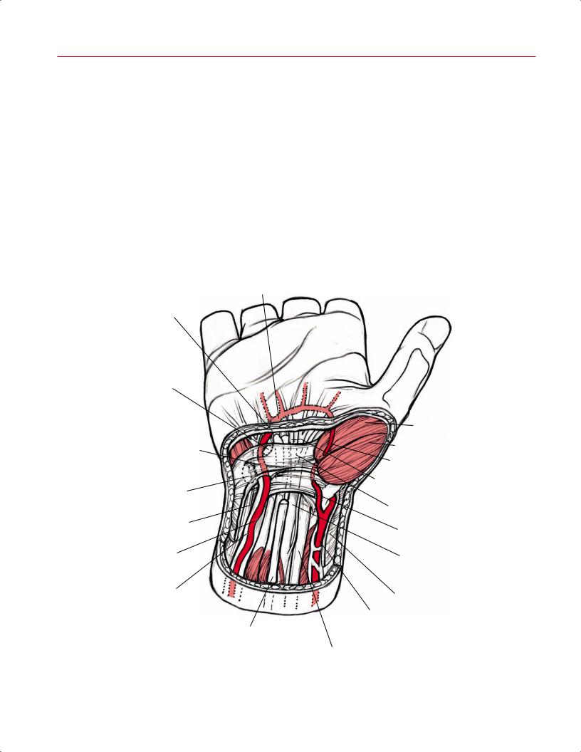
Fig. 2.20 A 3D illustration of the volar aspect of the dissected right wrist and hand, revealing the neurovascular structures, including the ulnar and median nerves and ulnar and radial arteries.

|
2 Normal MRI Anatomy of the Musculoskeletal System 33 |
||
|
|
|
|
• Extensor pollicis longus |
the coronoid process of the ulna, the mid-radius, and the |
||
• Extensor indicis |
interosseous membrane. It inserts along the volar aspect of |
||
Three deep muscles are found in the volar aspect of the |
the base of the distal phalanx of the thumb. The pronator |
||
quadratus originates from the volar aspect of the distal ulna |
|||
distal forearm: |
|||
and inserts along the volar aspect of the distal radius. |
|||
|
|||
• Flexor digitorum profundus |
The extensor carpi radialis longus originates along the |
||
• Flexor pollicis longus |
supracondylar ridge of the humerus and the lateral inter- |
||
• Pronator quadratus |
muscular septum. It inserts along the dorsal aspect of the |
||
The flexor carpi radialis originates from the common |
second (index finger) metacarpal base. The extensor carpi |
||
radialis brevis originates from the common extensor tendon |
|||
flexor tendon at the medial epicondyle and inserts on the |
|||
at the lateral epicondyle and inserts at the dorsal aspect of |
|||
base of the second and third metacarpals. The muscle belly |
|||
the third metacarpal base. The extensor digitorum (Fig. 2.18) |
|||
of the flexor carpi radialis is intermediate in signal intensity, |
|||
originates from the common extensor tendon at the lateral |
|||
and its tendon should be low in signal intensity. The pal- |
|||
epicondyle and inserts along the dorsal aspect of each finger |
|||
maris longus lies medial to the flexor carpi radialis, originat- |
|||
(digits 2 through 5) (Fig. 2.21). The extensor digiti minimi |
|||
ing from the common flexor tendon at the medial epicondyle |
|||
(Fig. 2.23) originates from the common extensor tendon at |
|||
and inserting along the flexor retinaculum and palmar apo- |
|||
the lateral epicondyle and inserts along the dorsal aspect of |
|||
neurosis. Both the flexor retinaculum and palmar aponeu- |
|||
the fifth digit. The extensor digiti minimi runs immediately |
|||
rosis are seen as dark bands within the superficial palmar |
|||
superficial to the radioulnar joint. The ECU originates from |
|||
aspect of the wrist. The flexor carpi ulnaris originates from |
|||
the dorsal aspect of the mid-ulna and from the lateral epi- |
|||
the common flexor tendon and the olecranon; inserts on the |
|||
condyle. It inserts at the base of the fifth metacarpal. Proxi- |
|||
fifth metacarpal, hamate, and pisiform; and lies in close as- |
|||
mal to its insertion, it can be seen coursing along the groove |
|||
sociation with the ulnar nerve, which is seen as a thin, in- |
|||
of the distal ulna. The pisiform is actually a sesamoid bone |
|||
termediate signal intensity structure running just deep and |
|||
within the ECU. The TFCC is composed of the following: |
|||
lateral to the flexor carpi ulnaris (Fig. 2.20). |
|||
|
|
||
The flexor digitorum superficialis originates at the medial |
• Fibrocartilaginous articular disc |
||
epicondyle, coronoid process, and radial head. It inserts along |
|||
• Dorsal and volar radioulnar ligaments |
|||
the volar aspect of the middle phalanx of each finger (Fig. |
|||
• Ulnolunate ligament |
|||
2.21). Proximal to its insertions, the flexor digitorum super- |
|||
• Ulnotriquetral ligament |
|||
ficialis divides into four musculotendinous units within the |
|||
• UCL |
|||
distal forearm, before entering the carpal tunnel. Within the |
|||
• ECU subsheath |
|||
carpal tunnel, the median nerve most often lies immediately |
|||
• A variable meniscus homologue |
|||
superficial to the flexor digitorum superficialis tendons (Figs. |
|||
|
|
||
2.18, 2.19, and 2.20), although it may have an intertendinous |
The abductor pollicis longus originates from the dorsal |
||
position. However, within the forearm, the median nerve |
aspect of the proximal third of the radius and ulna and from |
||
lies immediately deep to the flexor digitorum superficialis |
the interosseous membrane. It inserts at the dorsal aspect |
||
muscle. The large, dark musculotendinous units of the flexor |
of the first metacarpal base. The extensor pollicis brevis |
||
digitorum superficialis are easily identified as they converge |
originates at the posterior aspect of the distal third of the ra- |
||
within the forearm to enter the carpal tunnel. In contrast to |
dius and the interosseous membrane. It inserts at the dorsal |
||
the large, low signal intensity musculotendinous units of the |
aspect of the base of the proximal phalanx of the thumb, |
||
flexor digitorum superficialis, the median nerve is a thinner |
forms the lateral margin of the anatomic snu box, and runs |
||
structure with intermediate signal intensity, which can be |
in close association with the abductor pollicis longus. These |
||
more challenging to identify. Thus, the musculotendinous |
tendons run deep to the extensor retinaculum at the level of |
||
units of the flexor digitorum superficialis may be used as a |
the distal radial groove (Fig. 2.23). The extensor retinaculum |
||
landmark for the adjacent median nerve. The flexor digito- |
is seen in the dorsal aspect of the wrist as a thin, dark band |
||
rum profundus (Fig. 2.22) originates from the anteromedial |
immediately deep to the subcutaneous tissues, which are |
||
aspect of the ulnar and interosseous membrane and inserts |
very bright on non–fat-suppressed images. The extensor pol- |
||
along the distal phalanges of each finger (Fig. 2.21). As does |
licis longus originates from the posterior aspect of the mid- |
||
the flexor digitorum superficialis, the flexor digitorum pro- |
ulna and the interosseous membrane, and it inserts at the |
||
fundus divides into four musculotendinous units within the |
dorsal aspect of the base of the distal phalanx of the thumb. |
||
forearm before entering the carpal tunnel. Within the car- |
The extensor indicis originates from the posterior aspect of |
||
pal tunnel, the tendons of the flexor digitorum profundus |
the distal ulna and the interosseous membrane, and it inserts |
||
lie immediately deep to the tendons of the flexor digitorum |
at the dorsal aspect of the second digit (index finger). The ab- |
||
superficialis and are easy to identify based on this predict- |
ductor pollicis brevis originates at the scaphoid, trapezius, |
||
able relationship. The flexor pollicis longus originates from |
and flexor retinaculum. It inserts at the base of the proximal |
||
34 I Initial Concepts
|
Insertion of extensor tendon |
|
Interosseous m. |
Long extensor |
Phalanx |
|
|
|
tendon |
||
|
at base of distal phalanx |
Dorsal digital |
|
|
Extensor expansion |
|
|
Metacarpal |
|
||
|
Insertion of central band |
expansion |
|
|
|
|
|
|
|
||
A |
extensor tendon |
|
|
1 |
|
|
Flexor digitorum |
|
|
|
|
|
|
|
|
|
|
|
profundus muscle |
|
|
|
|
|
insertion |
|
|
|
|
Flexor digitorum |
IP joint Flexor digitorum |
|
|
|
superficialis muscle |
profundus tendon |
|
|
|
insertion |
|
Flexor digitorum |
Lumbrical m. Digital n. |
Digital a. |
|
Flexor digitorum |
superficialis tendon |
Interosseous m. |
Flexor tendons |
|
|
|||
Metacarpal phalanx |
profundus muscle |
Distal wing |
2 |
|
|
insertion |
|
|
|
|
|
|
|
|
B
Insertion of extensor tendon at base of distal phalanx
C
|
Long |
|
extensor |
Insertion of central band |
tendon |
extensor tendon |
Proximal wing |
Lumbrical m. |
|
|
|
Collateral ligs. |
Long extensor tendon |
Single tendon |
|
Distal IP joint
|
|
|
|
|
|
|
|
|
|
|
Interosseous m. |
|
|
|
|
|
|
|
|
|
|
|
Lumbrical m. |
|
|
|
|
|
|
|
Flexor digitorum superficialis tendon |
||||
|
|
Flexor digitorum profundus tendon |
|
|
|
||||||
|
|
Proximal phalanx |
|
|
|
|
|
||||
|
|
Insertion of central |
|
|
|
|
Metacarpal |
||||
D |
|
|
|
|
|||||||
|
band extensor |
|
|
|
|
|
|||||
|
|
|
|
|
|
||||||
|
|
tendon |
|
|
|
|
|
||||
|
|
Distal insertion of |
|
|
|
|
Interosseous m. |
||||
|
|
|
|
|
|
|
|||||
|
|
lumbrical m. |
|
|
|
|
Lumbrical m. |
||||
|
|
|
|
|
|
|
|
|
|
|
|
|
|
|
|
|
|
Insertion of interosseous m. |
|
||||
|
|
|
|
|
|
|
|
|
MCP |
|
|
|
|
Proximal IP joint |
|
|
|
|
Flexor digitorum |
||||
E |
|
Distal IP joint |
|
|
|
|
|
|
profundus tendon |
||
|
|
|
|
|
|
|
|
||||
|
|
|
|
|
|
|
|
|
|
|
Flexor digitorum |
|
|
|
|
|
|
|
|
|
|
|
|
|
|
|
|
|
|
|
|
|
|
|
|
|
|
|
|
|
|
|
|
|
|
|
superficialis tendon |
|
|
|
|
|
|
|
|
|
|
|
|
|
|
|
|
|
|
|
|
|
|
|
|
|
|
|
|
|
|
C1 |
A2 |
A1 |
Palmar aponeurosis |
||
|
|
|
|
|
|
||||||
A5 C3 A4 C2 A3
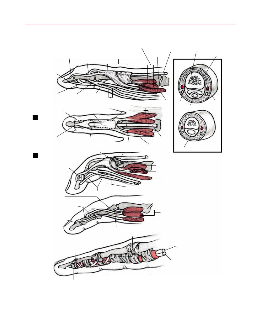
Fig. 2.21 Multiview illustrations of the muscles and ligaments in the fingers: (A) lateral superficial, (B) dorsal superficial, (C) lateral superficial in flexion, (D) lateral deep, and (E) pulley system (numbers and letters designate specific pulleys). Inset: axial cross-section through the middle (1) and distal (2) phalanxes.
2 Normal MRI Anatomy of the Musculoskeletal System 35
Fig. 2.22 A sagittal T1-weighted image of the wrist showing the normal appearance of the flexor digitorum profundus and flexor digitorum superficialis tendons, as well as the triangular fibrocartilage.
phalanx of the thumb. Unlike the other muscles described in this section, the abductor pollicis brevis has no function at the wrist joint, and thus is considered a hand muscle.
In the axial plane, the distal radioulnar joint is examined at the sigmoid notch of the radius. Particular osseous structures, such as the hook of the hamate, and the articulations between carpal bones can be assessed on axial images. The extrinsic carpal ligaments (Fig. 2.24), seen as uniformly low intensity structures in close relationship with the wrist capsule, may be accentuated with intraarticular contrast. Axial images of the digits provide cross-sectional visualization of small anatomic structures such as the flexor tendons, extensor mechanism, volar plates, and neurovascular bundles (Figs. 2.21 and 2.25).
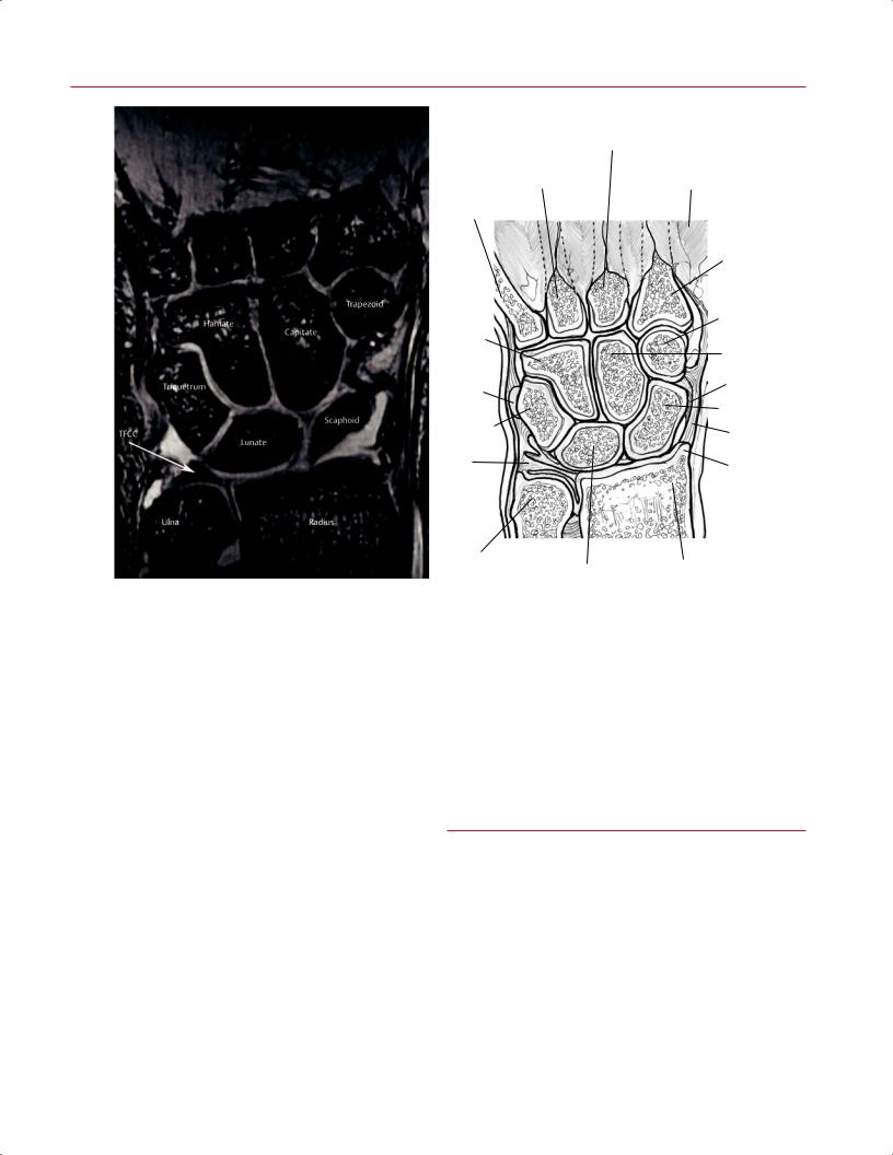
Coronal Images
Coronal plane images provide useful anatomic information about the wrist and hand, and show well the osseous structures and their respective relationships (Fig. 2.26). Small
structures such as the scapholunate and lunotriquetral ligaments are usually best seen on coronal images, and with thin sections; these structures are routinely well delineated. The radiocarpal compartment is separated from the radioulnar joint by the triangular fibrocartilage. Most information about the triangular fibrocartilage is also gained from coronal images. The triangular fibrocartilage looks like a curvilinear bowtie, and it extends horizontally from the ulnar surface of the distal radius to the base of the ulnar styloid process. It is low in signal intensity. In the coronal plane, the triangular fibrocartilage and interosseous ligaments are often seen on the same sections as the ECU tendon. Within the hand, the ligaments of the IP and MCP joints (in particular, the UCL of the thumb) are most readily assessed in this plane. However, thumb imaging should be performed separately with
Proximal |
Distal |
IP joint |
IP joint |
Terminal extensor
Central slip insertion
Sagittal band
Extensor indicis tendon
Abductor |
|
pollicis |
|
brevis m. |
|
First dorsal |
|
interosseous m. |
|
Extensor pollicis |
|
longus tendon |
|
Abductor pollicis |
|
longus tendon |
|
Extensor carpi radialis |
|
longus tendon |
|
Extensor carpi radialis |
|
brevis tendon |
Abductor pollicis |
|
|
|
longus m. |
Intertendinous connections
Extensor digiti quinti (minimi) tendon
Extensor digitorum communis tendon
Extensor indicis tendon
ECU tendon
Synovial sheaths
Extensor retinaculum
Extensor indicis tendon
Extensor pollicis |
|
brevis m. |
Extensor digitorum communis m. |
|
Fig. 2.23 Illustration of the dorsal aspect of the right hand showing the muscles and tendons.
|
Triquetrotrapezial lig. |
Scaphoid |
Capitate |
|
|
Pisometacarpal lig. |
|
|
|
|
|
|
|
|
|
|
|
||
|
Trapezium |
|
|
|
|
Pisohamate lig. |
|
|
|
|
|
|
|
|
|
|
Hook of hamate |
|
|
|
|
|
|
|
|
|
Palmar metacarpal lig. |
|
|
|
|
|
|
|
|
|
Ulnar arcuate lig. |
|
|
|
Thumb |
|
|
|
|
|
|
|
|
|
metacarpal |
|
|
|
|
|
|
Capitate |
|
|
|
|
Hamate |
|
|
|
|
|
|
|
|
|
|
|
|
|
|
|
|
|
RCL |
|
Triquetoscaphoid lig. |
|
Radial arcuate |
|
|||
|
|
|
|
|
|||||
|
Extensor indicis |
|
Triquetrum |
|
|
|
|
lig. (deltoid lig.) |
|
|
tendon |
|
|
|
Triquetrol |
|
|
|
|
|
|
|
|
|
|
|
|
||
|
Extensor carpi |
|
Radiotriquetral lig. |
|
lunate lig. |
|
Trapezium |
|
|
|
radialis longus tendon |
|
ECU tendon |
|
|
|
|
|
|
|
Extensor carpi |
|
|
|
Flexor carpi |
|
|
|
|
|
|
|
|
|
ulnaris tendon |
|
|
|
|
|
radialis brevis tendon |
|
Extensor digiti |
|
|
|
|
|
|
|
Abductor pollicis |
|
minimi tendon |
|
|
|
RCL |
|
|
|
|
|
|
|
UCL |
|
|
|
|
|
longus tendon |
|
Extensor digitorum |
|
Scaphoid |
|
|||
|
|
|
|
|
|||||
|
|
|
communis |
|
|
|
|
|
|
|
Extensor pollicis |
|
|
|
|
|
|
|
|
|
|
tendons |
|
|
|
|
Radioscaphocapitate |
|
|
|
brevis tendon |
|
Pisoulnocarpal |
|
|
||||
|
|
Extensor |
|
lig. |
|
||||
|
|
|
|
|
|||||
|
Extensor pollicis |
|
lig. |
|
|
|
|
|
|
|
|
retinaculum |
|
|
|
Lunate |
|
||
|
longus tendon |
|
|
|
Ulnar |
|
|
||
|
|
|
|
|
|
|
|||
|
|
ECU m. |
|
|
|
|
|
||
|
|
|
|
|
styloid |
|
Radiolunate lig. |
|
|
|
Abductor pollicis longus m. |
|
|
|
|
|
|
||
|
|
|
|
|
|
|
Radioscapholunate lig. |
|
|
|
|
|
Extensor indicis m. |
|
|
|
|
||
|
|
|
|
|
|
|
|
||
|
Extensor pollicis brevis m. |
|
Extensor digiti |
|
Ulnotriquetral |
|
Short radiolunate lig. |
|
|
|
|
|
|
lig. |
|
Ulnolunate lig. |
|
||
|
|
|
minimi tendon |
|
|
|
|||
A |
Extensor pollicis longus m. |
|
|
|
Ulna |
Radius |
B |
||
|
Extensor digitorum m. |
|
|
Volar radioulnar lig. |
|||||
|
|
|
|
|
|
|
|
||
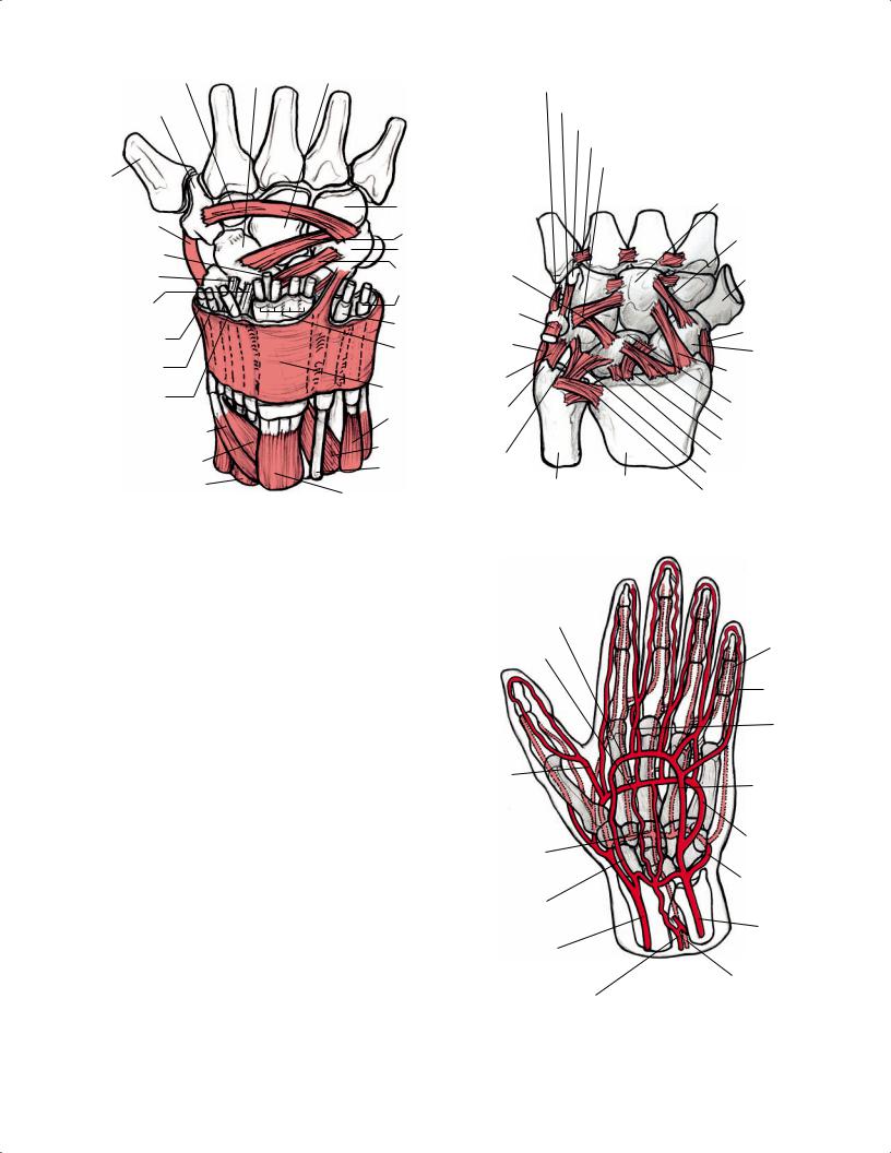
Fig. 2.24 Illustrations of the wrist showing the dorsal (A) and volar (B) aspects of the left distal radius and ulna, carpal bones, musculotendinous units, and intercarpal ligaments.
specialized planes. The abductor pollicis longus and extensor pollicis brevis tendons are seen within the medial/volar aspect of the wrist at the level of the volar aspect of the scaphoid and lunate. Also within the volar aspect of the wrist are the radioscaphocapitate ligament and the radiolunotriquetral ligament, which extend in the ulnar direction from the radial styloid. Located slightly ulnar to these ligaments is the radioscapholunate ligament (Fig. 2.24). These three carpal ligaments are low in signal intensity and are often seen on the same image in the coronal plane.
Despite the advantages of coronal images for evaluation of the TFCC and intercarpal ligaments, the variable courses of the tendons and neurovascular structures crossing the wrist and hand joints make evaluation of these structures di cult in the coronal plane, and therefore these structures are primarily assessed on axial images. On the dorsal coronal images, the extensor digiti minimi tendon is seen as it courses obliquely along the ulnar side of the triquetrum. The extensor pollicis longus and the extensor carpi radialis brevis are separated by Lister’s tubercle, which shows fatty marrow signal intensity. On the volar coronal images, the flexor tendons are seen coursing deep to the flexor retinaculum, which appears as a dark, transverse band. The flexor tendons appear as hypointense bands and can be followed as they pass through the carpal tunnel between the trapezium and the hook of the hamate. The median nerve is intermediate in signal, slightly brighter than that of the adjacent tendons.
Superficial palmar arch
Deep palmar arch |
Dorsal |
|
digital a. |
||
|
||
|
Proper |
|
|
digital a. |
|
|
Common |
|
|
palmar a. |
|
Princeps |
|
|
pollicis a. |
Superficial bra |
|
|
||
|
of ulnar a. |
|
|
Deep palmar |
|
Dorsal carpal arch |
branch |
|
|
||
|
Dorsal carpal |
|
Superficial palmar |
branch of ulnar a |
|
|
||
branch of radial a. |
|
|
|
Ulnar a. |
|
Radial a. |
|
|
|
Posterior interosseous a. |
|
Anterior interosseous a. |
|
Fig. 2.25 A 3D illustration of the volar (red) and dorsal (pink) vasculature in the left wrist and hand.
2 Normal MRI Anatomy of the Musculoskeletal System 37
Third metacarpal
Fourth metacarpal |
Interosseous m. |
|
Fifth metacarpal
Second metacarpal
|
|
|
Trapezoid |
|
|
Hamate |
|
|
|
|
|
|
Capitate |
|
|
Pisiform |
|
RCL |
|
|
|
|
|
|
|
|
|
Scaphoid |
|
|
Triquetrum |
|
RCL |
|
|
TFCC |
|
Radial |
|
|
|
|
styloid |
|
|
Ulnar |
|
|
|
|
head |
Radius |
|
|
A |
Lunate |
|
B |
|
|
|
|||
|
|
|
|
|
Fig. 2.26 A coronal T2*-weighted image (A) and artist’s sketch (B) of the wrist at the level of the TFCC. The carpal bone architecture is well delineated on this image, which was acquired on a high field strength magnet with an eight-channel surface coil.
The radial and ulnar styloid may be evaluated for fractures on coronal images. The joints, including the distal radioulnar, radiocarpal, intercarpal, and carpometacarpal joints, can also be evaluated for degenerative or traumatic changes. The articular surfaces of the carpal bones show intermediate signal intensity.
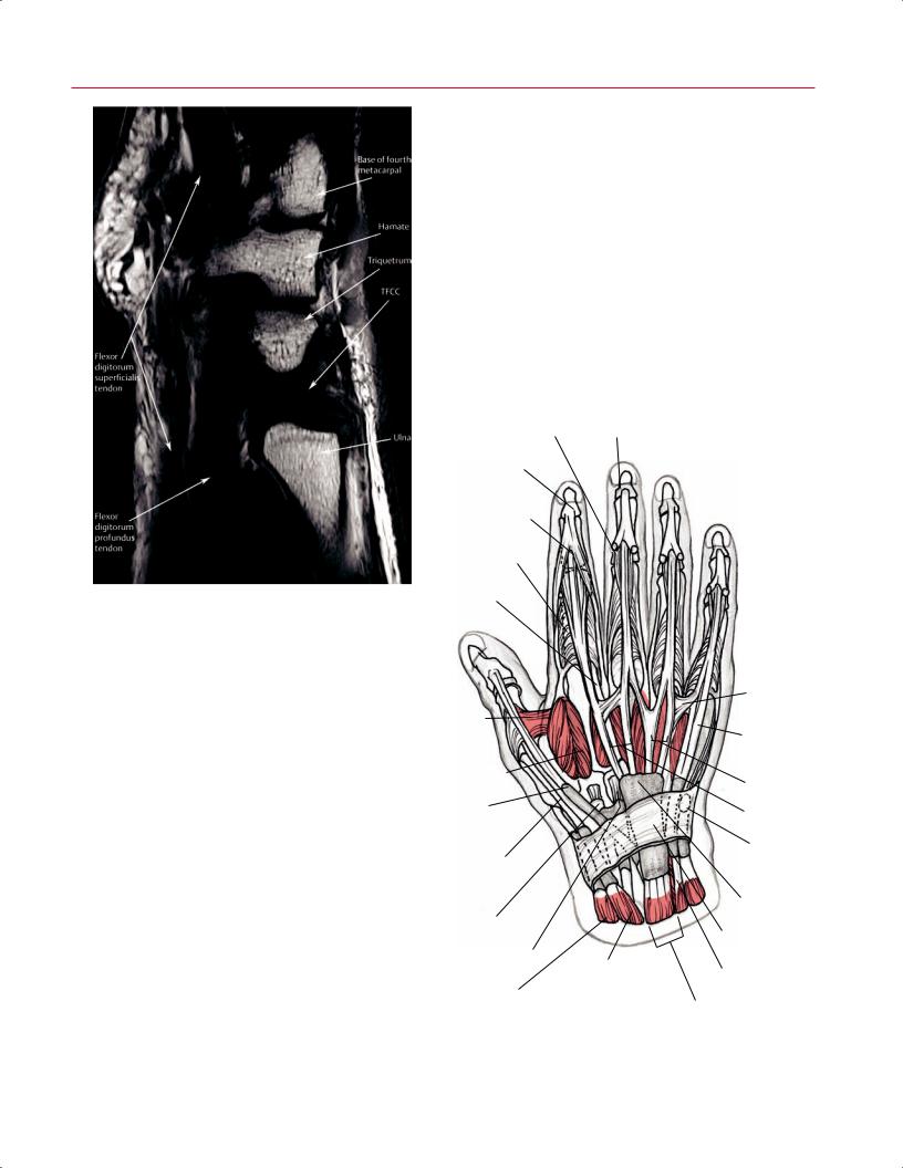
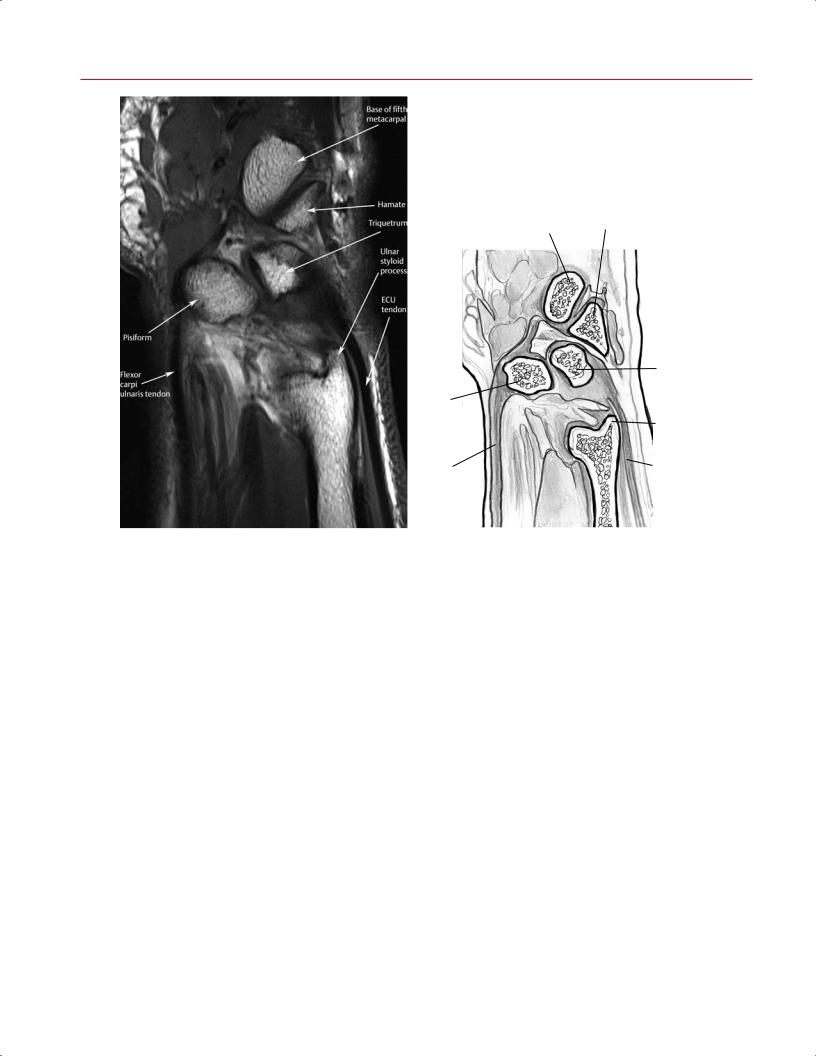
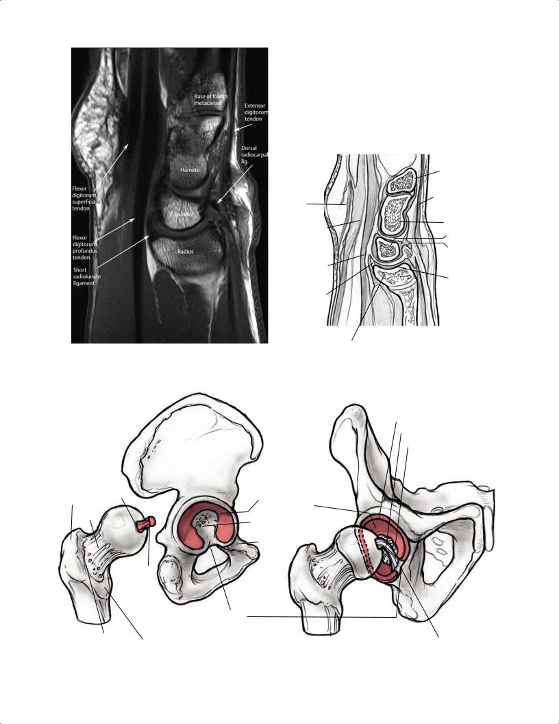
Sagittal Images
The sagittal plane is useful for depicting the longitudinal course of structures and providing an orthogonal section for bones in the wrist and hand. Information from sagittal images can be used to evaluate the TFCC (Fig. 2.22) and overall carpal alignment, especially with regard to the scapholunate and capitolunate articulations (Figs. 2.27 and 2.28). Deformity of the scaphoid and abnormalities of the pisotriquetral joint are also examined in this plane. Fractures of the other carpal bones, including the hook of the hamate and the lu-
nate, are also well visualized. The course of the flexor and extensor tendons is well shown on sagittal images of the digits, as is the course of the median nerve in the wrist.
■ Hip
Axial Images
The hip (femoroacetabulum), a diarthrodial joint, is an articulation between the acetabulum and the head of the femur (Fig. 2.29). The acetabulum is formed by the union of the ilium, ischium, and pubis; is normally oriented approximately 45 degrees caudally; and has between 14 and 26.5 degrees of anteversion (averages: men, 18.5 ± 4.5 degrees; women, 21.5 ± 5.0 degrees).8 The normal femoral neck–shaft angle ranges between 125 and 140 degrees.8,9 Given the complex 3D geometry of the hip joint, MRI of the hip should use all
38 I Initial Concepts
Base of fifth
Hamate
metacarpal
H
Triquetrum
Pisiform
Ulnar styloid process
ECU tendon
Flexor carpi ulnaris tendon
A |
B |
Fig. 2.27 A sagittal T1-weighted image (A) and artist’s sketch (B) of the wrist at the level of the pisiform, showing the flexor and ECU tendons within the forearm and wrist.
three standard planes of imaging and a specialized plane along (parallel to) the femoral neck. The muscles of the hip can be grouped according to function or anatomic location (medial, anterior, lateral, and posterior). There is no single accepted grouping technique, but some form of a systematic review should be performed. Functional muscle grouping is a logical technique:
•Abductors: gluteus medius, gluteus minimus, and tensor fascia lata
•Adductors: pectineus, gracilis, adductor longus, adductor brevis, and adductor magnus
•Flexors: iliopsoas, rectus femoris, sartorius
•Extensors: gluteus maximus, hamstrings
•External rotators: piriformis, obturator externus, obturature internus, superior gemelli, inferior gemelli, and quadratus femoris
Although most of the musculature in the region of the hip is best evaluated on axial images (Figs. 2.30 and 2.31), the
hamstrings and gluteus medius and minimus are more easily evaluated in the coronal plane (Fig. 2.32).
Generally, in addition to the conventional axial images, oblique axial images oriented along the axis of the femoral neck are also obtained. These oblique images allow visualization of the head–neck junction and can aid in evaluating anatomy associated with femoroacetabular impingement. Axial images allow for good visualization of the anterior and posterior portions of the labrum (Fig. 2.31), which are seen as low signal intensity structures. Axial images are also useful for identifying the regional neurovascular bundles, specifically the sciatic nerve, obturator, and femoral neurovascular bundles; in these bundles, the nerves are seen in cross-section as intermediate signal structures, and often discrete fascicles can be discerned.
The gluteus maximus, gluteus medius, and gluteus minimus can be identified and di erentiated by fascial divisions. The tensor fascia latae is located anterior to the gluteus medius (Fig. 2.31). At the level of the inferior pubic ramus, the
|
Base of fourth |
|
metacarpal |
|
Extensor |
Flexor |
digitorum |
retinaculum |
tendon |
|
Hamate |
Flexor digitorum
superficialis Lunate
tendon
Dorsal RCL
Flexor digitorum
profundus tendon
Lunate fossa
Short radiolunate ligament
A |
Radius |
B |
Fig. 2.28 A sagittal T1-weighted image (A) and artist’s sketch (B) of the wrist at the radiolunate articulation showing the normal relationship of the radius, lunate, hamate, and fourth (ring finger)
metacarpal. Long segments of the flexor digitorum superficialis and profundus tendons are seen as they pass through the carpal tunnel.
Labrum
Ligamentum teres
Fat pad
|
Greater |
Fovea |
|
Articular |
|
|
|
|
surface |
|
|||
|
trochanter |
|
|
|||
|
|
|
of acetabulum |
|
||
|
|
|
|
|
||
|
Neck |
|
|
Acetabular fossa |
|
|
|
|
|
|
|
||
|
|
|
|
Superior ramus |
|
|
|
|
|
|
pubis |
|
|
|
|
|
Round |
|
|
|
|
|
|
lig. |
|
|
|
|
|
|
|
Acetabular |
|
|
|
|
|
|
notch |
|
|
|
|
|
Lesser |
|
|
|
A |
Intertrochanteric |
Acetabular lig. |
B |
|||
trochanter |
||||||
|
crest |
|
|
|||
Fig. 2.29 Disarticulated lateral (A) and partially disarticulated anterior oblique (B) 3D illustrations of the femoroacetabular articular surfaces.