

Книги по МРТ КТ на английском языке / MRI for Orthopaedic Surgeons Khanna ed 2010
.pdf100 II Upper Extremity
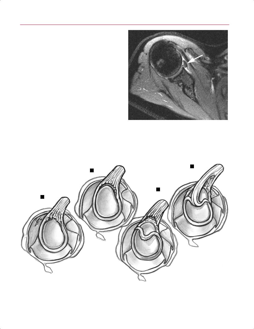
Fig. 4.6 An axial fat-suppressed proton-density image of a reverse soft-tissue Bankart lesion in the left shoulder showing an avulsion of the posterior-inferior labrum from the glenoid rim (small arrow) and a reverse Hill-Sachs impaction fracture on the anteromedial aspect of the humeral head (large arrow).
Other MRI findings associated with episodes of shoulder instability include injuries to the rotator cu , especially in patients more than 40 years old. Anterior dislocations are associated with tears of the supraspinatus and subscapularis, whereas posterior dislocations are associated with tears of the subscapularis or fractures of the lesser tuberosity.
Labral Pathology and SLAP Lesions
MRI has proved to be a sensitive, specific, and accurate modality for evaluating the glenoid labrum,7,17,18 which routinely is assessed in all three imaging planes.19 Coronal oblique and axial plane images, however, provide the most diagnostic information.20 The axial plane best shows the anterior and posterior labrum, whereas the coronal oblique plane visualizes the superior and inferior portions.21 The normal labrum is approximately 3 mm in craniocaudal dimension from base to apex and is 4 mm in width at its base of insertion into the glenoid cartilage.22 The classic morphology is described as smooth, with triangular anterior and posterior wedges, as seen on axial images.21 The intact fibrous labrum shows low signal intensity on all pulse sequences. The peripheral attachment of the labrum joins the capsule and glenohumeral ligaments, creating the capsulolabral complex. This capsulolabral or labral ligamentous complex is best appreciated on sagittal oblique MR images. The sagittal oblique plane also is useful for evaluating displaced bucket-handle tears of the labrum.22 Labral tears and SLAP lesions (superior labrum from anterior to posterior, relative to the bi-
Fig. 4.7 An axial intermediate weighted MR arthrographic image of the left shoulder showing a medium-sized reverse Hill-Sachs defect (arrowheads) involving 25% to 50% of the articular surface of the humeral head. Reprinted by permission from Saupe N, White LM, Bleakney R, et al.: Acute traumatic posterior shoulder dislocation: MR findings. Radiology 2008;248:185–193.
ceps tendon anchor) are best appreciated on fluid-sensitive sequences.17
There is considerable variation in the attachment and morphology of the glenoid labrum. Labral anatomy anteriorly and posteriorly is best evaluated with axial images. Above the equator of the glenoid, the labrum may not be attached to the glenoid, but inferior to the middle of the glenoid, the labrum often is confluent with the articular cartilage. The greatest variation in labrum anatomy occurs in the anterosuperior glenoid.23 To detect true abnormalities, knowledge of these normal variants of labral anatomy as visualized on MRI is very important. Specifically, the absence of a labral attachment in the anterosuperior quadrant is a normal variant and should be di erentiated from pathologic labral tears. Therefore, one must be cautious about interpreting an isolated labral tear in the anterosuperior quadrant.
A normal sublabral foramen, or hole, between the labrum and the glenoid rim is often misinterpreted as an anterior labral disruption or tear. When present, the sublabral foramen is located in the 1 o’clock to 3 o’clock positions, anterior to the biceps-labral complex, and represents space between the anterosuperior labrum and the adjacent glenoid cartilage.22 A sublabral foramen can vary in size from a few millimeters to the entire anterosuperior quadrant above the level of the subscapularis tendon.24
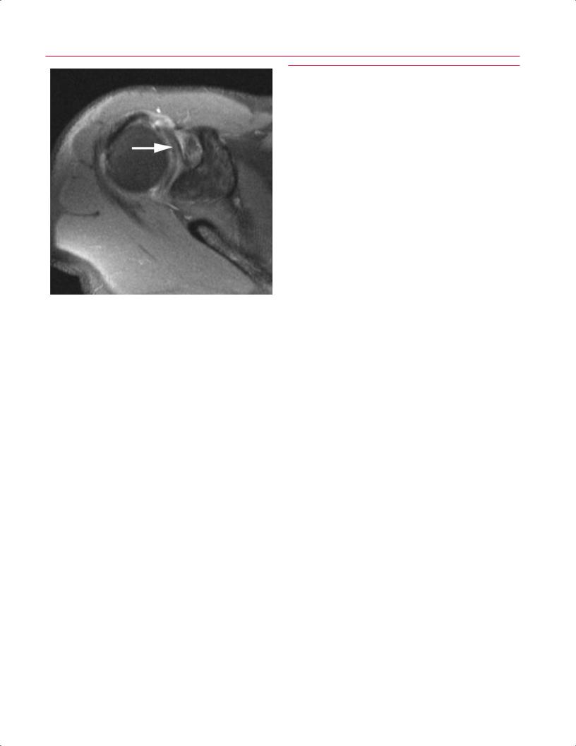
Another normal anatomic variant, known as the Buford complex (Fig. 4.8), consists of three elements25:
4 The Shoulder 101
• A cord-like MGHL
• An MGHL that attaches directly to the superior labrum anterior to the biceps (at the base of the biceps anchor)
• The absence of anterosuperior labral tissue
The sagittal oblique plane shows the course of the cord-like MGHL attaching directly to the superior labrum at the anterior base of the biceps tendon.
The sublabral recess, or sublabral sulcus, is an additional |
|
anatomic variant that should be recognized. In this variant, |
|
the superior labrum (from the 11 o’clock to 1 o’clock posi- |
|
tions) is meniscoid in morphology.22,26 On coronal oblique |
|
views, a recess appears as an area of high signal intensity on |
|
T2-weighted images between the labrum and the superior |
|
glenoid. The recess curves medially and follows the contour |
|
between the superior labrum and adjacent hyaline cartilage |
|
of the superior glenoid.22,26 The caudal meniscoid portion of |
|
the labrum is not attached to the superior portion of the gle- |
|
noid, but the base of the superior labrum remains attached |
|
near the insertion of the long head of the biceps tendon.22,26 |
Fig. 4.8 An axial fat-suppressed proton-density image of the right |
In 1990, Snyder and associates27 were the first to describe |
shoulder showing the Buford complex, which consists of three ele- |
SLAP lesions. Patients with SLAP lesions usually present with |
ments: a cord-like MGHL (arrow), an MGHL that attaches directly to |
|
the superior labrum anterior to the biceps (at the base of the biceps |
|
anchor), and an absence of anterosuperior labral tissue. |
SLAP LESIONS
D
B
C
A
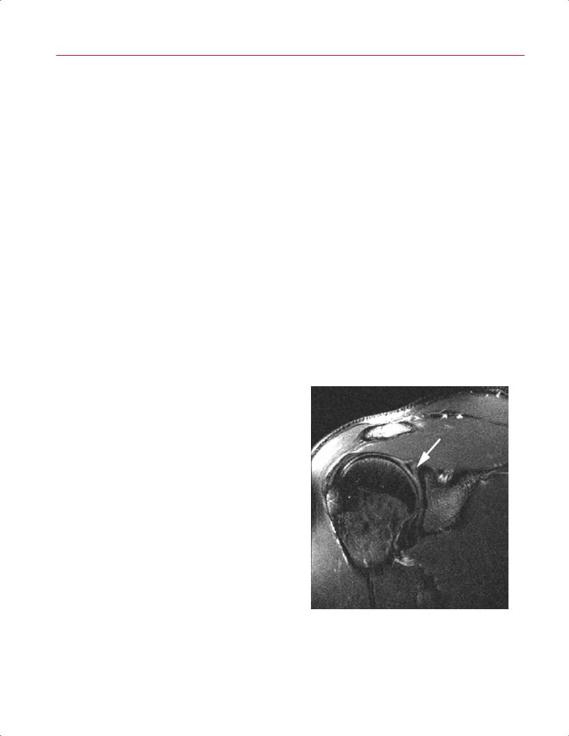
Fig. 4.9 Types of SLAP lesions. (A) Type I, a frayed and degenerative superior labrum with a normal (stable) biceps tendon anchor. (B) Type II, detachment of the superior labrum and biceps anchor. (C) Type III, a bucket-handle tear of a meniscoid superior labrum without extension into the biceps tendon. The biceps anchor is stable
and the remaining labrum is intact. (D) Type IV, a bucket-handle tear of a meniscoid superior labrum with extension into the biceps tendon. (Based on Powell SE, Nord KD, Ryu RKN: The diagnosis, classification, and treatment of SLAP lesions. Oper Tech Sports Med 2004;12:99–110.)
102 II Upper Extremity
A 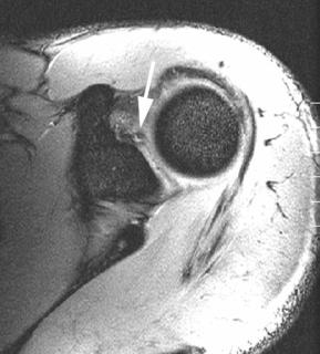
 B
B
Fig. 4.10 Type II SLAP lesion. (A,B) Axial fat-suppressed gradient-echo images of the left shoulder showing increased signal intensity within the superior glenoid labrum (arrow) in a patient with a type II SLAP lesion verified by arthroscopy.
complaints of deep pain (typically with overhead activities) and/or mechanical symptoms of catching, locking, popping, or grinding.28 Frequently, there is a history of overhead activities or sports. The mechanism of injury often is traction or compression.28
The most common classification of SLAP tears includes four distinct lesions, although recently new lesions and subclassifications have been added (Fig. 4.9).27,29
In type I lesions, there is a frayed and degenerative superior labrum with a normal (stable) biceps tendon anchor. On MRI, the labral contour appears blunted or irregular with a slight increase in signal intensity on T2-weighted images.30,31
Type II lesions have similar labral fraying but have detachment of the superior labrum and biceps anchor, making them unstable. MRI findings of type II SLAP lesions include a line of high-intensity signal coursing across the base of the hyperintense labrum to the periphery (Figs. 4.10 and 4.11). The long head of the biceps tendon has normal signal and shape and is attached to the avulsed labrum.31 The sublabral recess can be di erentiated from a type II SLAP lesion by two MRI signs on T2-weighted images present in these SLAP lesions, but not in sublabral recesses26:
•High signal intensity in the labrum posterior to the biceps anchor, which extends to the articular surface
•An area of high signal intensity irregular or curved laterally and distinct from the medial curvilinear area of signal intensity at the labral–glenoid junction seen in a sublabral recess
In type III lesions, there is a bucket-handle tear of a meniscoid superior labrum without extension into the biceps tendon. The biceps anchor is stable and the remaining
Fig. 4.11 A coronal oblique fat-suppressed T2-weighted image of the right shoulder showing increased signal intensity within the superior glenoid labrum (arrow) posterior to the long head of the biceps tendon origin in a patient with a type II SLAP lesion verified by arthroscopy.
4 The Shoulder 103
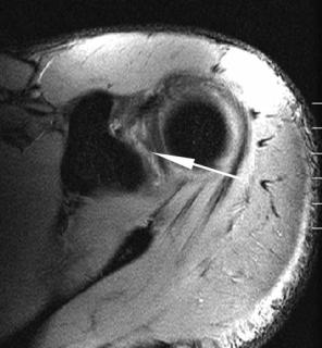
Fig. 4.12 An axial fat-suppressed proton-density image of a type IV SLAP lesion in the right shoulder showing increased signal intensity within the superior glenoid labrum with extension into the biceps anchor (arrow).
labrum is intact. On MRI, type III SLAP lesions are characterized by a line of high signal intensity coursing across the base of the hyperintense labrum but extending beyond the equator in the case of a nondisplaced bucket-handle tear. In the case of a displaced bucket-handle tear, a discrete piece of fibrocartilage is identified within the joint capsule. On MRI, the displaced piece of fibrocartilage appears as a low signal intensity intraarticular fragment surrounded by high signal intensity joint fluid.32 In type III SLAP lesions, the superior labrum also is deficient, yet the biceps tendon can be followed to the supraglenoid tubercle.31
In type IV SLAP lesions, there is a bucket-handle tear of a meniscoid superior labrum with extension into the biceps tendon. On MRI, a type IV SLAP lesion is characterized by a line of high signal intensity coursing across the base of the normally hypointense labrum to the periphery and extending beyond the equator with a deficient labrum (Fig. 4.12).30 In addition, there is hyperintensity and splitting of fibers of the biceps tendon.31
SLAP lesions frequently are associated with other shoulder pathology that can be visualized on MRI. The presence of paralabral cysts, almost invariably associated with labral tears,21,33,34 should incite the search for a labral tear. The imaging features include a cystic-appearing mass adjacent to the labrum or capsule with increased signal on T2-weighted sequences.21 Rotator cu tears (both partial thickness and full thickness) and Bankart lesions are commonly associated with SLAP lesions.35,36
■ Degenerative Conditions
The shoulder is a ected by numerous degenerative conditions, including the following:
•Subacromial impingement syndrome
•Rotator cu tears
•Glenohumeral osteoarthritis
•AC osteoarthritis
•Pathologic conditions a ecting the biceps tendon
Subacromial Impingement Syndrome
Subacromial impingement syndrome is a continuum of abnormalities and clinical symptoms; in its final stages, it may be associated with full-thickness rotator cu tears. Impingement syndrome is secondary to compression of the rotator cu by abnormal morphology of structures within the supraspinatus outlet. Patients with chronic impingement syndrome typically present with the insidious onset of di use shoulder pain in the area of the deltoid muscle; the pain is activity related, worse with overhead activities, and often worse at night.37 However, it must be stated that there is often no correlation between clinical symptoms and rotator cu pathology found on MRI or during autopsy.
Osseous factors that contribute to subacromial impingement syndrome include the following38,39:
•Anterior acromial spurs
•The shape of the acromion
•The slope of the acromion
•The morphology of the AC joint (e.g., hypertrophic bone, calcification of the coracoacromial ligament, callus formation)
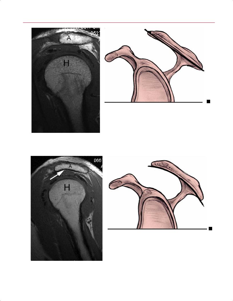
The shape of the acromion (acromial morphology) is best evaluated with sagittal oblique MR images. Bigliani and associates38 classified acromial morphology into three types:
•Type I, a flat undersurface (Fig. 4.13)
•Type II, a smooth curved inferior surface (Fig. 4.14)
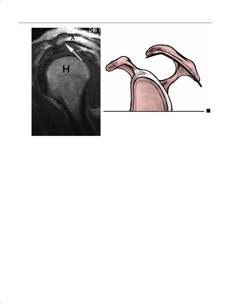
•Type III, an anteroinferior hook or beak (Fig. 4.15)
The type II and type III acromions that create narrowing of the supraspinatus outlet are associated with a predisposition to rotator cu disease.40,41
In addition to acromial morphology in the sagittal plane, the slope of the acromion in the coronal plane may predispose patients to impingement syndrome and subsequent rotator cu tears. Normally, the anterior acromion should be nearly horizontal when viewed in the oblique coronal plane. Lateral downsloping of the acromion narrows the supraspinatus outlet and may predispose to impingement syndrome.42 Sagittal oblique MR images can be used to assess the morphology of the acromion from posterior to anterior,
104 II Upper Extremity
A
Fig. 4.13 Type I acromion. (A) A sagittal oblique T1-weighted image. A, acromion; H, humeral head. (B) Artist’s sketch. Straight line indicates flat inferior surface.
A
Fig. 4.14 Type II acromion. (A) A sagittal oblique T1-weighted image (arrow, curved inferior surface). A, acromion; H, humeral head. (B) Artist’s sketch. Curved line indicates curved inferior surface.
4 The Shoulder 105
A
Fig. 4.15 Type III acromion. (A) A sagittal oblique T1-weighted image (arrow, anterolateral hook or beak). A, acromion; H, humeral head. (B) Artist’s sketch. Bent line indicates an anteroinferior hook or beak.
whereas coronal oblique MR images can be used to evaluate the lateral acromial slope.
The spectrum of MRI changes in subacromial impingement syndrome have been characterized and documented.43,44 Rotator cu disease is evaluated on the basis of tendon morphology and changes in the observed signal intensity within the specific cu tendons. In addition, pathologic processes within the supraspinatus outlet, including abnormalities of the acromion, AC joint, and the subacromial-subdeltoid bursa, may be identified in the spectrum of findings in patients with impingement symptoms.
Changes in the subacromial bursa often are seen on MRI in association with impingement syndrome.45,46 Bursal inflammation is seen as decreased signal intensity, or loss of peribursal fat, on T1-weighted images and as increased signal intensity (from associated fluid, inflammation, or bursal proliferative disease) on T2-weighted or fat-suppressed T2weighted sequences.42,47 Low signal intensity within a thickened subacromial bursa on T1-weighted and T2-weighted images indicates a proliferative process in chronic bursitis, which also is associated with rotator cu disease.48
Capsular hypertrophy of the AC joint has also been identified on coronal oblique MR images in patients with impingement syndrome.44 Other MRI findings associated with impingement syndrome include inferior acromial and AC spurs, which are visualized as alterations in the nor-
mal smooth contour of the inferior acromion and AC joint. However, recent studies have suggested that small acromial spurs may be a normal finding associated with advancing age, whereas spurs larger than 5 mm are strongly associated with the presence of rotator cu pathology.41,49
Normal rotator cu tendons display uniformly low signal intensity on all pulse sequences. In cu tendon degeneration (tendinosis), there is intermediate signal intensity on T1weighted or proton-density images, with no evidence of fluid signal intensity on T2-weighted images (Fig. 4.16). Fat-sup- pressed T2-weighted sequences are sensitive to changes of degeneration, and, in the absence of a partial or complete rotator cu tear, they display areas or regions of hyperintensity.50 These areas of increased signal intensity often occur adjacent to acromial or AC spurs (as described above) or a low-lying acromion.44 More severe changes or degeneration may be characterized by intermediate to increased signal intensity on T1-weighted and proton-density images, which persist without additional increase in signal intensity on T2-weighted images. Other findings of rotator cu tendinosis include areas of thickening and irregularity within the tendon.42
Caution must be used to avoid confusing rotator cu abnormalities with the magic angle phenomenon. This phenomenon, which results in spuriously increased signal intensity on sequences with a short TE (including T1-weighted and pro- ton-density sequences), occurs in tissues containing highly
106 II Upper Extremity
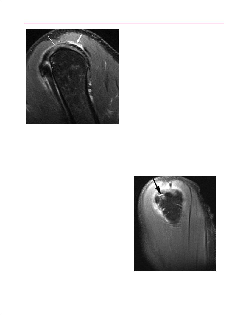
Fig. 4.16 A sagittal oblique T2-weighted image showing hyperintense signal in the tendon (thin arrow) without a discrete defect, consistent with supraspinatus tendinosis. Compare this finding with the fluid signal intensity in the subacromial bursa (thick arrow), consistent with subacromial bursitis.
structured collagen fibers that lie at an angle of 55 degrees to the main magnetic field.51 In the shoulder, this phenomenon is common just proximal to the insertion of the supraspinatus tendon on the greater tuberosity. The increased signal intensity associated with the magic angle phenomenon disappears on T2-weighted images, making it possible to di erentiate this phenomenon from true pathology.51
Intratendinous lesions are defined as tears that do not communicate with the bursal or articular surface.
Coronal oblique and sagittal oblique images are ideal for the evaluation of partial-thickness tears of the supraspinatus and infraspinatus tendons. Similarly, axial and sagittal oblique images are best for evaluating the subscapularis and teres minor tendons. The diagnosis of this type of tear is suggested by increased signal in the rotator cu that only partially traverses the rotator cu substance.55 Typically, there is abnormal morphology of the tendon insertion onto bone with evidence of partial tendon discontinuity on T1-weighted images. Partial-thickness tears show low to intermediate signal intensity on T1-weighted images, intermediate to high signal intensity on proton-density images, and fluid signal intensity on T2-weighted sequences (Figs. 4.17 and 4.18). Increased signal intensity is the result of tracking of fluid within the bursal or articular surface of the cu .
The diagnosis of a partial-thickness rotator cu tear is most di cult at either end of the spectrum: low-grade par- tial-thickness tears tend to be mistaken for tendinosis and high-grade partial-thickness tears may be mistaken for full-thickness rotator cu tears (Fig. 4.19).42 MRI findings in degeneration and partial-thickness rotator cu tears may overlap, and tendon abnormalities must be evaluated on the basis of bursal, intrasubstance, and articular surface morphology and on signal intensity changes on T1-weighted, proton-density, and T2-weighted sequences.8
Full-thickness tears of the supraspinatus and infraspinatus are best visualized with the coronal oblique and sagittal
Rotator Cu Tears
Rotator cu tears represent the end of the spectrum of abnormalities associated with chronic subacromial impingement syndrome. Therefore, the clinical presentation often is similar to that described for impingement syndrome. Some patients may present with acute pain and weakness after traumatic rotator cu rupture, whereas other patients with rotator cu tears may have minimal to no symptoms. Patients also may report weakness and limited active range of motion despite normal passive motion.37 The supraspinatus and infraspinatus tendons are the most common tendons torn.52 MR images, particularly in the coronal oblique and sagittal oblique planes, are most useful in showing tears of these two tendons.40 These tears are best shown with fatsuppressed T2-weighted images.53 Rotator cu tears can be characterized as partial thickness or full thickness.54 Partialthickness tears may involve the articular or bursal surface or, less commonly, they may be completely intratendinous.
Fig. 4.17 A sagittal oblique T2-weighted image showing a partialthickness rotator cu tear (arrow) on a background of tendinosis.

4 The Shoulder 107
Fig. 4.18 A coronal oblique T2-weighted image of the left shoulder showing a high-grade partial-thickness rotator cu tear (arrow).
oblique images. Full-thickness tears of the subscapularis and teres minor are best visualized using the axial and sagittal oblique images. The direct signs of a full-thickness rotator cu tear consist of a complete tendon defect or complete discontinuity of the tendon with retraction, and abnormal increased signal intensity within the tendon defect (Table 4.1).53,56–60 A complete tear cannot be diagnosed unequivo-
Fig. 4.19 A sagittal oblique T2-weighted image showing a full-thick- ness rotator cu tear (arrow).
cally without visualization of a complete tendon defect or a direct communication between the glenohumeral joint and the subacromial-subdeltoid bursa. The defect, or tendinous gap, is seen as an interruption or loss of continuity of the normally low signal intensity tendon. This gap appears as a continuous band of increased signal intensity that traverses the full thickness of the rotator cu , extending from the glenohumeral joint to the subacromial bursa.56 The signal intensity is increased on T1-weighted and proton-density images and increased additionally on T2-weighted and fatsuppressed T2-weighted sequences.42 The increased signal intensity is secondary to interposed joint fluid or granulation tissue at the cu tear site. Large cu tears may fill in with fibrous or granulation tissue that may have low-signal intensity on T2-weighted images.47
Indirect signs of rotator cu tears can be used in conjunction with the primary assessment of changes in tendon signal intensity and morphology to help in the diagnosis of full-thickness rotator cu tears. Indirect signs of rotator cu abnormality include the following (Table 4.1)61,62:
•Fluid within the subacromial-subdeltoid bursa
•Obliteration of the peribursal fat stripe
•The presence of a glenohumeral e usion
•Atrophy of the involved muscle
Subacromial-subdeltoid bursal fluid is associated with a fullthickness rotator cu tear, but it is not specific for this condition. This fluid is manifested on MRI as high signal intensity changes in the subacromial-subdeltoid bursa that equal the intensity of fluid on T2-weighted images. Changes in subacromial and subdeltoid peribursal fat also may be considered indirect signs of cu abnormality. Because peribursal fat may be replaced by low signal intensity granulation tissue or scar or high signal intensity fluid, which is often limited to the site of the cu tear, this abnormality is considered a secondary sign when a cu tear is not clearly visualized.
Most rotator cu tears can be identified by a good history and physical examination. MRI is most useful in planning and predicting the prognosis for successful rotator cu repair. Identification of tendon retraction and cu muscle atrophy on MRI is imperative in the evaluation of a fullthickness tear. Large, retracted cu tears with atrophy of the
Table 4.1 Characteristics of Rotator Cu Tears on MRI
|
Direct Signs |
Indirect Signs |
|
|
Visualization of tendon defect |
Subacromial-subdeltoid fluid |
|
|
Indication of direct |
Retraction of supraspinatus |
|
|
communication between |
musculotendinous junction |
|
|
glenohumeral joint and |
|
|
|
subacromial bursa |
|
|
|
Retraction of supraspinatus or |
Changes in subacromial and |
|
|
infraspinatus tendon |
subdeltoid peribursal fat |
|
|
|
Fatty atrophy of rotator cu |
|
|
|
muscles |
|
|
|
|
|

108 II Upper Extremity
Fig. 4.20 A coronal oblique proton-density MR image showing a fullthickness supraspinatus tear with tendon retraction (arrow). Note the slightly increased signal intensity of the muscle, consistent with early atrophy.
muscle may not be repairable. Retraction of the torn rotator cu tendon is assessed by medial displacement of the musculotendinous junction from its normal position, which lies in a 45-degree arc lateral to the highest point of the humeral head.47 Other authors consider the supraspinatus tendon retracted if the musculotendinous junction is located more medial than the middle third of the humeral head (Fig. 4.20).56 Retraction of the supraspinatus or infraspinatus tendons is best seen on coronal oblique images that show the medial and lateral extension of the cu tear. Similarly, axial images are best for identifying subscapularis and teres minor retraction. T2-weighted sagittal oblique images provide additional information, allowing identification of articular and bursal surface extension and the quantification of the size of the tear in the AP direction.42
Large or chronic rotator cu tears frequently are accompanied by atrophy of the rotator cu muscle. Muscle atrophy can be identified in two forms, which often are present simultaneously: fatty replacement and decreased muscle bulk. T1-weighted or proton-density images are best suited for this assessment. Fatty replacement is best shown on T1weighted images, which display high signal intensity (equal to fat) horizontal streaks parallel to the long axis of the involved muscle (Fig. 4.20).44 In addition, MRI can visualize the diminution in cross-sectional size of the involved muscle, which is best seen on the sagittal oblique images.56
Glenohumeral Osteoarthritis
injuries and glenohumeral instability, present with complaints of shoulder pain and sti ness. In osteoarthritis of the shoulder, the glenoid articular cartilage and subchondral bone typically are worn posteriorly, the humeral head may be subluxed posteriorly, and the articular cartilage of the humeral head is eroded centrally, with a surrounding rim of remaining articular cartilage and osteophytes.63 Although glenohumeral osteoarthritis usually is diagnosed with conventional radiographs, early arthritis frequently is missed with this modality because conventional radiographs do not directly image the cartilage and therefore cannot detect early chondral damage. Rather, conventional radiographs rely on signs such as joint space narrowing and secondary changes such as the presence of subchondral cysts and osteophyte formation, but these findings often are not seen until the disease process is more advanced. MRI, the optimal imaging modality for assessing articular cartilage, can provide morphologic information, such as fissuring and the presence of partialor full-thickness cartilage defects.64,65
Articular cartilage of the glenohumeral joint is best evaluated on gradient-echo or fat-suppressed T2-weighted sequences with images in the coronal oblique and axial planes.64,66,67 Normal articular cartilage has a uniform thickness and homogeneous intermediate signal intensity on T1weighted, fat-suppressed T2-weighted, and gradient-echo sequences.66,67 Osteoarthritis is associated with multiple defects and erosions in the articular cartilage; in glenohumeral osteoarthritis, these changes typically occur centrally
Osteoarthritis in the glenohumeral joint is similar to that in other joints, such as the hip and knee. Patients with this condition, who tend to be older than those with rotator cu
Fig. 4.21 A coronal T1-weighted image of the left shoulder showing subchondral cyst formation (arrow) in a patient with glenohumeral osteoarthrosis.

4 The Shoulder 109
in the humeral head and posteriorly on the glenoid.63 This altered morphology of the articular cartilage can be seen on MRI as surface irregularities, partial-thickness focal defects, and full-thickness focal defects because the normally intermediate signal intensity cartilage is occupied by fluid signal intensity joint fluid.66 With osteoarthritis, there usually are multiple defects of variable size and depth or di use cartilage thinning.64 Other MRI findings of osteoarthritis include bone marrow edema, osteophytes, subchondral cysts, and subchondral sclerosis (Fig. 4.21).64 Less commonly, signal intensity abnormalities, such as foci of high signal intensity in morphologically normal cartilage, may suggest early osteoarthritis.66
AC Osteoarthritis
AC joint osteoarthritis is a common condition associated with the normal aging process.68 Patients with AC joint osteoarthritis who manifest symptoms typically present with anterior shoulder pain that is exacerbated by activities such as bench presses and shoulder elevation or adduction. Physical examination reveals focal tenderness over the AC joint, pain with passive cross-body adduction, and pain with resisted AC extension. Although degenerative changes of the AC joint are extremely common, most patients with this condition are asymptomatic.
MRI findings of AC joint arthritis include the following (Fig.
4.22)68–70:
•Subacromial fat e acement
•Joint space narrowing
•Osseous irregularity of the joint
•Capsular distention
•Osteophyte formation
These AC joint arthritic changes are seen on MRI in more than 80% of asymptomatic patients and in approximately 95% of asymptomatic patients more than 30 years old.68,70 A study has suggested that the presence of reactive bone marrow edema on fat-suppressed T2-weighted MR images in the distal clavicle or acromion, or both sides of the AC joint, is a reliable predictor of symptomatic AC joint pathology, and is not seen in asymptomatic patients.70 AC joint degenerative changes commonly are associated with other shoulder pathology, such as impingement syndrome, rotator cu tears, and glenohumeral osteoarthritis.68
Abnormalities of the Long Head of the
Biceps Tendon
The biceps tendon may be followed on multiple sequences from its origin at the supraglenoid tubercle through the bicipital groove.21 The long head of the biceps tendon should be inspected on coronal oblique, axial, and sagittal oblique images. The origin of the biceps tendon and its proximal
Fig. 4.22 A coronal oblique fat-suppressed T2-weighted image of the right shoulder shows hypertrophy of the AC joint, joint effusion, and subchondral bone marrow edema related to AC joint osteoarthrosis.
intraarticular portion are best visualized on sagittal oblique and coronal oblique images, but more distally, as it courses within the bicipital groove, it is best visualized on axial images. The normal MRI appearance of the long head of the biceps tendon includes low signal intensity on all pulse sequences and identification in its normal anatomic location (Figs. 4.23 and 4.24).21 On axial images at the level of the bicipital groove, the tendon appears as an oval-shaped low signal intensity structure surrounded by a thin moderate signal intensity synovial sheath.47 Pathologic conditions involving the long head of the biceps tendon include tendinosis, partialthickness tears, complete tears (rupture), subluxation, and dislocation.
The clinical presentation of patients with disorders of the long head of the biceps tendon varies with the underlying abnormality; such patients often have concomitant chronic impingement syndrome or rotator cu tears. Patients with biceps tendinosis, partial tears, and subluxation may have pain and tenderness in the bicipital groove.71 This pain may radiate down the arm anteriorly to the biceps muscle belly, but it is often di cult to distinguish from that of impingement syndrome.71 When long-standing, the pain often will resolve spontaneously after rupture, and complete tears are often asymptomatic.71
The MRI findings in tendinosis include high signal intensity within the tendon on all pulse sequences, and thickening and inhomogeneity of the tendon (Fig. 4.25).21,72 This finding