

Книги по МРТ КТ на английском языке / Functional Neuroimaging in Child Psychiatry Ernst 1 ed 2000
.pdf
Preface
ªUnprecedentedº, ªrevolutionaryº, ªextraordinaryº are terms commonly used to describe the promise that the emergence of functional neuroimaging techniques presents for neuroscience. Indeed, until recently, the possibilities that such techniques now provide for the advancement of knowledge fell within the realm of ªneuroscience Wctionº.
Many recent books have been dedicated to various facets of neuroimaging. This volume is unique in its focus on children, as well as in its eVort to encompass the Welds of genetics and neuropsychology the interface of which with neuroimaging will soon become the state of the art in neuroimaging research. This book is addressed to clinicians and investigators. Clinicians will learn about the basic principles, advantages, and disadvantages of neuroimaging techniques and the advances made in our understanding of the mechanisms underlying childhood psychiatric disorders through neuroimaging research. Investigators may rely on this volume as a reference book to aid them in (i) deciding which technique is most appropriate for a given research question, (ii) evaluating the ethical issues raised by the involvement of children in research in order to weigh them in reference to the progress that has been made in understanding certain childhood psychiatric disorders through the use of neuroimaging, and (iii) considering how the Welds of molecular genetics and neuropsychology can be exploited in planning for a brain imaging study.
The book is divided into Wve sections. Part 1 is dedicated to the description and future potential of the various functional imaging techniques currently used to study the human brain. Readers should come away from this section with an understanding of the relative advantages and limitations of each of these techniques and become Xuent in the language of nuclear medicine and neuromagnetic imaging. To this purpose, a glossary of the terms commonly
xi

xiiPreface
used in functional neuroimaging is provided at the end of the book. In addition, the issues raised by the application of these techniques to pediatric studies are clearly and uniquely addressed. Part 2 comprises the ethical issues raised by the inclusion of children in functional neuroimaging protocols. Part 3 gives an overview of neural development and cognitive development and addresses the issues of controlling for, as well as exploiting, these maturational changes in neuroimaging research, particularly in functional magnetic resonance imaging studies involving cognitive activation. Part 4 reviews the progress made in research on child psychiatric disorders. Because of the paucity of research in children, the review of imaging Wndings in adults often predominates and provides hypotheses to be tested in children. Each group of psychiatric disorders is described separately, and the speciWc problems (scientiWc, technical, and ethical) of studying children with speciWc disorders using neuroimaging are covered. Finally, Part 5 addresses future
directions. It introduces the important tools of genetics and neuropsychology that are expected to act synergistically to potentiate the yield of neuroimaging research and discusses how neuroimaging Wndings can be interpreted within the context of neurodevelopmental disorders.
In our concluding remarks, we mention the development of new brain imaging techniques and strategies that were not covered previously. We also express our opinions regarding the most important advances and future applications of this exciting Weld of research in child psychiatry.
The authors wish to thank Stephen Foote, PhD, Director of the Division of Neuroscience and Basic Behavioral Science, NIMH for his support of this work and to thank Barry Horwitz, PhD, Language Section, Voice, Speech, and Language Branch, National Institute on Deafness and Other Communication Disorders for his helpful review of the Wnal chapter.
Monique Ernst and Judy Rumsey

Foreword
Research is limited by the instruments that it uses: from the telescope came the solar system: from the microscope came the germ theory. A subtext of these discoveries is ªseeing is believingº. For all too long, psychiatric illness has been deWned by what could not be seen: a lesion on autopy transformed a psychiatric disorder into a neurologic disorder. The ken of clinical responsibility of psychiatry has, therefore, been delimited to conditions with behavioral and intrapsychic symptoms without visible neuropathology. This situation, of course, has been fertile ground for the persistence of stigma, as psychiatric disorder was equated with moral weakness among the lay public and derided as ªfunctionalº by nonpsychiatric clinicians.
Fortunately, the solution for this seeming oxymoron, mental illness, came from technology. Since the mid-1980s increasingly powerful devices have been developed to image the structure, function, neurophysiology, and chemical composition of the living human brain. The impact of these technologies has been informative for brain science but transformative for psychiatry. For example, no longer can schizophrenia be considered a political act or psychologic deviance when one can see the atrophic cortex, hypofunctional frontal lobe, enhanced release of striatal dopamine and reduced levels of N-acetyl- aspartate.
Functional Neuroimaging in Child Psychiatry, edited by Monique Ernst and Judy Rumsey, takes current imaging opportunities and discusses their applicability to the realm of the child and adolescent subject. The power of this important textbook is that it addresses the critical fact that children are not simply miniature adults. Children present unique opportunities but also formidable challenges. The textbook provides superb reviews of the major methods ± the use of radioactive tracers in PET and SPECT, fMRI and spectroscopy, and magnetoencephalography ± with an eye toward pediatric applications.
xiii
xiv |
J. Coyle |
|
|
Perhaps one of the most diYcult barriers confronting pediatric brain imaging is the misperception of the ethical constraints on such studies. Arnold and his colleagues provide a very thoughtful commentary on risks, risk±beneWt assessment, informed consent, and patient selection. In particular, they cover in some detail radiation biology in considering the risk of exposure to radioactive ligands in pediatric SPECT and PET studies, making comparisons to other sources of radiation that can be readily understood. This chapter should be required reading for investigators as well as members of Institutional Review Boards (IRBs) involved in pediatric imaging.
The most compelling aspect of brain imaging is the ability to monitor neuronal function with increasingly higher resolution. These technical advances have transformed imaging research from a passive diagnostic method to a strategy for mapping the dynamics of neural systems involved in cognitive function, emotional states, and motor±sensory processing. Cognitive neuroscientists have now become major innovators in developing tasks that illuminate dysfunctional circuits. In this regard, those involved in pediatric functional brain imaging will face the
enormously complex but informative unfolding of circuit maturation, plasticity, and cognitive functions during brain development. Consequently, the textbook takes particular care in critically reviewing selected psychological instruments that can be brought to bear on neurocognitive development.
At the time of writing this Foreword, those sequencing the human genome in the Human Genome Project are whirling along with an estimated completion date of 2003 for mapping the entire human genome. Then begins the really exciting biological research aimed at determining how a mere 100000 genes interact to create the human body and its most complex organ, the brain. Brain imaging during human development will play a major role in this endeavor. For this reason, I believe that Functional Neuroimaging in Child Psychiatry is a critically important book: it provides the roadmap to this extraordinary research frontier.
Joseph T. Coyle
Chairman, Department of Psychiatry, Harvard Medical School, Boston, MA,
USA

Fig. 3.2. Feasibility of fMRI studies in pediatric populations. Task-related signal change in a child and in an adult during a phonologic awareness task. The graphs show the amount of head motion generated in both individuals and correction after processing of the data.

(a)
(b)
Fig. 5.2. Magnetic Weld distribution to a current dipole in a homogeneous conductor. Vector plots of the magnetic Welds to tangential (a) and radial (b) orientations of a current dipole to the detector are shown. The length of the arrows is proportional to the
Weld amplitude (high Weld amplitudes near the source have been left out for overall Wgure clarity). Contour plots of the sampled Weld distributions in a single plane are also shown (measurements made normal to the surface and outside of conductor). Red indicates outgoing Weld and blue the in-going Weld. Note that the magnetic
Welds of the radially oriented dipole close inside the conductor; this produces no measurable Weld at the detectors (shown to illustrate general relationship between dipole and detector).
(a)
(b)
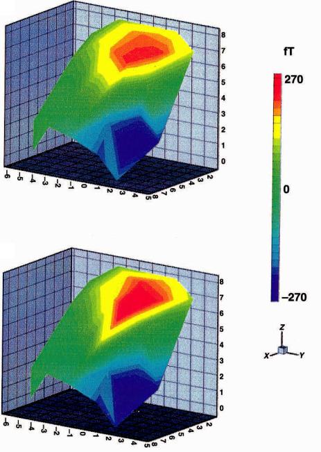
Fig. 5.13. Contour plots of the sensory evoked Weld data in Fig. 5.12. The three-dimensional topography of the M20 response (the Wrst large deXection seen in Fig. 5.12b) is illustrated. Positive values indicate outgoing magnetic Xux (red) and negative numbers indicate ingoing Xux (blue). (a) The theoretical Weld produced by the Wnal iteration of the forward solution in a single equivalent current dipole (ECD) model; (b) the actual measured data. The scaling is the same for both (a) and (b) and is shown to the right of the Wgure. The x, y, z coordinate system (scaled in centimeters) is also illustrated to the right of the Wgure and shows the orientation of the two plots in the
Wgure. The x axis is deWned as the line connecting the two preauricular points (positive x exits near the right ear); the y axis is the line normal to the x axis through the nasion (positive y exits the bridge of the nose), and the z axis is formed normal to x and y at their intersection (positive z exits the top of the head). The point 0, 0, 0, is the center of the head. Note the close correspondence between the theoretical Weld and measured Weld, which is predicted from the low residual error for this component in Fig. 5.12a.

(a)
(b)
Fig. 5.15. Current density in a homogeneous volume conductor. Two orientations of current dipole are shown as red arrows where the arrows show the direction of intracellular current Xow: (a) tangential to the surface and (b) radial to surface. Vector plots of the current density (blue arrows) are illustrated inside the rectangular volume conductor: this is the ªvolume currentº and is responsible for the scalp-measured EEG. The length of the arrows is proportional to the log10 of the current density magnitude. Contour plots of the surface distribution of the normal current density are shown at the same slice plane through the top of the volume conductor. Compare these Wgures with those in Fig. 5.2. Note that the radially oriented MEG source produces no externally measurable Weld, but the same orientation is seen as a single electric potential extremum in this Wgure. Note also that while the EEG potential distribution appears to have the same spatial resolution as the MEG Weld distribution in Fig. 5.2, the blurring eVects of the skull are not illustrated here. Consequently, this Wgure would be a more accurate depiction of the potential distribution measured directly from the pial surface of the cortex.

Fig. 9.4. Dorsolateral prefrontal cortical activity for children (n 5 6) and adults (n 5 8) during performance of a spatial working memory task. AC, anterior commissure.
Fig. 11.3. Metabolite signal intensity maps of N-acetyl-containing compounds (NAA), choline-containing compounds (Cho) and creatine/phosphocreatine (Cre) with coaxial MRI for a patient with childhood-onset schizophrenia. The upper slabs are taken at the level of the hippocampal region and the lower slabs at the level of the dorsolateral prefrontal cortex. The left side of the brain is on the right of the Wgure, and vice versa. Color images are scaled to the highest value of each metabolite signal intensity for each 1H MRS imaging slice, so that the pattern of regional distribution of metabolite signal intensities within the same slice can be compared between subjects, although the color intensity from the same anatomic location cannot be compared between subjects. The spectra shown are representative of the hippocampus and dorsolateral prefrontal cortex.