

Книги по МРТ КТ на английском языке / Functional Neuroimaging in Child Psychiatry Ernst 1 ed 2000
.pdf194 L. K. Jacobsen and A. Bertolino
scanning. Childhood-onset patients were medication free for 9±32 days (mean 19.5"5.9) prior to PET scanning. Patients were compared with 26 healthy control adolescents matched for age, gender, Tanner stage, and handedness. However, 19 of the normal control subjects had a sibling with attention-deWcit hyperactivity disorder (ADHD), who had participated in a separate PET study (Ernst et al., 1994). PET scanning was performed using a method designed to minimize radiation exposure to subjects (Zametkin et al., 1993). As in the adult study, all subjects performed a computerized auditory CPT with eyes patched during FDG uptake (approximately 30 min).
Image analysis included both a region of interest (ROI) approach used previously to analyze data from the adult study and a newer pixel-based approach. For the ROI analysis, Wve slices most closely resembling anatomically deWned templates were selected. Raw pixel values were converted into regional glucose metabolic rates (rCMRGlu, in 100mg/minper g tissue) (Brooks, 1982) and these were then measured by an independent rater, blind to diagnosis, for 60 standard ROIs distributed within the Wve planes. Global CMRGlu was calculated by averaging values for glucose metabolism across all areas of the cortex sampled that were rich in gray matter. Regional CMRGlu values were normalized by dividing each subject's absolute CMRGlu for each ROI by his/her global CMRGlu.
For the pixel-based analysis, Statistical Parametric Mapping (SPM), version 1995 (Friston et al., 1991) was used. After transforming each image into a standard stereotactic space (Talairach and Tournoux, 1988), images were smoothed using a 10mm!10mm!6mm Gaussian Wlter. Proportional scaling was used to control for intersubject diVerences in global CMRGlu. Between group comparisons were performed on a pixel-by-pixel basis, with the resulting value of t for each pixel being transformed to a Z score. Pixels corresponding to Z scores greater than or equal to 2.58 (or p#0.005) were then subjected to a test of signiWcance based on spatial extent (Friston et al., 1994). Spatially contiguous pixels (clusters) of a size corresponding to p#0.005 were considered signiWcant.
As in the adult study, results of the ROI-based analysis indicated that global CMRGlu in childhood-onset schizophrenia patients (14.13"2.11mg/min per 100g) was comparable to that found in controls (14.01"2.39 mg/min per 100g). Also, similar to Wndings in the adult study, adolescents with childhood-onset schizophrenia had signiWcantly lower metabolic rates than controls in their middle frontal gyrus (anterior frontal region of plane C) and superior frontal gyrus (anterior medial frontal region of plane E) (Fig. 11.1). The middle frontal gyral
region was identical to that identiWed as hypometabolic in the adult sample, while the other frontal region was one plane lower than that identiWed in the adult sample. Comparison of the eVect sizes observed in the childhoodand the adult-onset samples failed to indicate more severe hypofrontality in early-onset schizophrenia. Within the early-onset schizophrenic group, metabolic rates of the brain regions showing hypofrontality was not signiWcantly correlated with negative symptoms, as measured by the Scale for the Assessment of Negative Symptoms (SANS; Andreasen, 1983) total score, duration of illness, or total number of months that patients had received antipsychotic medication prior to study entry. (Comparable correlations were not reported for the adult study of Cohen et al. (1987).)
In addition to hypofrontality, adolescents with earlyonset illness showed signiWcantly higher metabolic rates in the region of the supramarginal gyrus (parietal region of plane B) and in the inferior frontal gyrus/insula (posterior frontal region of plane E) than did the controls. The hypermetabolism in the region of the inferior frontal gyrus/insula was similar to that seen in a subgroup of schizophrenic adults with poor CPT performance, whose ªhitsº (correct responses) were comparable to those of the childhood-onset sample, although they made fewer false alarm errors.
Because of technical failures during the childhood-onset study, performance data were available for only 13 schizophrenic and 20 control subjects, although all subjects performed the task. For the subgroups for whom CPT data were available, schizophrenic patients had signiWcantly fewer CPT hits (correct button presses) and signiWcantly more CPT false alarms (incorrect button presses) than did healthy controls. When the metabolic data were analyzed for the subgroups for whom performance data were available, all diagnostic diVerences were lost except for the posterior frontal region of plane E, where the patients had elevated metabolic rates.
Within the childhood-onset schizophrenic group, CPT false alarm rate was negatively correlated with CMRGlu for the right parietal (supramarginal) region on plane B (r5 0.62, p#0.05) and the region of the right inferior frontal gyrus/insula (r5 0.57, p#0.05). In controls, these same correlations were positive and signiWcant for the right supramarginal region (r5 0.73, p#0.01) and failed to reach signiWcance for the inferior frontal gyrus/insula. Therefore, the hypermetabolic Wndings may reXect greater cognitive eVort being expended by the patients in order to attend and perform, albeit poorly.
In contrast to the ROI-based analysis, the SPM analysis failed to reveal any regions of hypometabolism in schizo-
Functional imaging in childhood-onset schizophrenia |
195 |
|
|
|
|
(a)
(b)
Fig. 11.1. Comparison of region-of-interest results of glucose metabolic studies of (a) 16 patients with childhood-onset schizophrenia studied by Jacobsen et al. (1997) and (b) 16 adult-onset schizophrenics studied by Cohen et al. (1987). Both childhoodand adult-onset patients demonstrated reductions in frontal metabolism, relative to age-matched controls, while performing a Continuous Performance Task. Only the childhood-onset group demonstrated biparietal hypermetabolism. Plane A is the most superior slice and lies 94 mm above the canthomeatal (CM) line; plane B is 84 mm and plane C is 67 mm above the CM line. Plane D contains the basal ganglia and thalamus and is 53 mm above the CM line. Plane E contains the lower frontal and temporal lobes and is 40 mm above the CM line. L, left, R, right.
phrenia but rather identiWed several hypermetabolic foci. The SPM method revealed increased metabolic rates in a region of the right pre/postcentral gyrus (Brodmann area 4/6), the left insula, the left inferior frontal gyrus (Brodmann area 47), and bilaterally in the cerebellum, a region not examined in the Cohen et al. (1987) study. The increase in bilateral cerebellar metabolic rate remained signiWcant when the analysis was repeated after excluding the three schizophrenic subjects who had the most movement artifact, identiWed by visual inspection of PET scans prior to transforming them into standard stereotactic space. Subsequent ROI sampling of the left and right cerebellum from one slice (corresponding to level 0°12, atlas of Matsui and Hirano (1978) conWrmed the bilateral hypermetabolism observed for this region.
Hypofrontality was demonstrated only when using the ROI-based approach, while cerebellar hypermetabolism was demonstrated with both data analytic approaches. While the pixel-based approach theoretically permits more reWned localization, particularly when comparing
diVerent conditions within subjects, its accuracy may be compromised by deviations in brain anatomy associated with psychiatric disorder (schizophrenia, attention-deWcit disorder) and/or with normal brain development, as well as by the need to interpolate to Wll datapoints lying between the acquired slices. Because only the ROI-based analysis was used in the adult schizophrenia study, agerelated comparisons are, of necessity, limited to this approach.
While the similarities in results for the childhoodand the adult-onset samples generated using the ROI-based analysis suggest a Wnal common pathophysiology, the available data failed to suggest a straightforward relationship between the earlier onset of symptoms and the severity of neurodevelopmental lesion eVects. An important limitation of this study, which may bear upon the relatively weak hypofrontality observed in the schizophrenic patients, was the inclusion of 19 siblings of subjects with ADHD as pediatric controls. Decreased frontal metabolism has been observed in both adults and adolescents with

196L. K. Jacobsen and A. Bertolino
ADHD (Zametkin et al., 1990; Ernst et al., 1994), albeit inconsistently (Ernst et al., 1997). While none of the controls in the present study met DSM criteria (American Psychiatric Association, 1994) for current or previous ADHD and all were older than the maximum age before which symptoms must be present in order to be diagnosed with ADHD, any decrease in frontal metabolism in these normal subjects would have obscured the hypofrontality observed in schizophrenic patients. Nonetheless, the failure of the pixel-based analysis to provide evidence of hypofrontality further suggests that this severe form of schizophrenia is not associated with more severe reductions in frontal metabolism than have been seen with PET in adult-onset schizophrenia.
The Wnding of cerebellar hypermetabolism in childhoodonset schizophrenia, seen with both data analytic approaches, is notable in light of recent evidence implicating the cerebellum in higher cortical processes (Kim et al., 1994). In healthy subjects, cerebellar activation has been demonstrated during verb generation and motor sequence learning, with activation declining as task performance became more automatic (Jenkins et al., 1994; Raichle et al., 1994), possibly reXecting a shift in mental strategy (Fiez, 1996). Previous reports implicating cerebellar dysfunction in schizophrenia include one report of decreased cerebellar metabolism in medicated adult patients studied at rest (Volkow et al., 1992) and one report of greater increases in cerebellar blood Xow during cognitive task performance in schizophrenic adults (Steinberg at al., 1995). More recently, dysfunction of a prefrontal±thalamic±cerebellar network in schizophrenia was suggested by Wndings of reduced blood Xow in these regions during practiced and novel memory tasks (Andreasen et al., 1996). These reports, together with observations from the present study, suggest that abnormal neural circuitry in schizophrenia involves the cerebellum, with the increase in cerebellar metabolism possibly resulting from failure of patients to shift to less eVortful, more automatic strategies for performing the CPT.
Functional brain imaging: proton MRS
As with other functional imaging modalities, few previous 1H MRS studies have been conducted in childhood-onset schizophrenia. Thomas and colleagues (1996) conducted a 1H MRS study of 10 children with schizophrenia and 12 healthy controls in which 1H spectra were measured in frontal gray matter bilaterally and in occipital gray matter. As has been observed in adult-onset schizophrenia (Bertolino et al., 1996, 1998a,b), the ratio of NAA/Cre was signiWcantly lower in the frontal lobes of the schizophrenic children. There were no group diVerences in metabolite
ratios in the occipital gray matter, suggesting some regional speciWcity for the frontal Wnding. Brooks and colleagues (1998) conducted a 1H MRS study in children with schizophrenia spectrum symptoms (mean age 11 years) in which 1H spectra were measured in the left frontal lobe. A decreased ratio of NAA/Cre was found for patients with schizophrenia spectrum disorder in this location. Although this Wnding appears to be consistent with Wndings from 1H MRS studies of adult schizophrenia, the data from this study suVer from the lack of diagnostic clarity inherent when studying spectrum disorders and from the absence of information about metabolite ratios in other brain regions in these patients.
The above 1H MRS studies of pediatric schizophrenia measured 1H spectra from a small number of large voxels, precluding an adequate assessment of regional metabolite ratios. In order to provide a more thorough assessment of metabolite concentrations, 1H MRS imaging in which 1H spectra were measured from a large number of small single-volume elements was applied to a subgroup of the NIMH childhood-onset schizophrenia sample (Bertolino et al., 1998c) distinct from the subsample participating in the cerebral glucose metabolism study described above. Prior use of this technique to study patients with adultonset schizophrenia demonstrated low levels of NAA in the hippocampal area and the dorsolateral prefrontal cortex, Wndings that were independent of treatment and consistent with developmental neuronal pathology (Bertolino et al., 1998a,b). Consequently, it was of major interest whether there were similarities in neurochemical patterns, as well as potential diVerences in the magnitude of any abnormalities, seen in childhoodversus adult-onset schizophrenia.
In this 1H MRS imaging study, 14 children and adolescents (mean age"SD 16.4"1.7; 11 males) with childhoodonset schizophrenia and 14 healthy children and adolescents matched for sex and age (mean age 16.1"2.1 years) were examined. All patients were on antipsychotic medication at the time of scanning. Exclusion criteria for both groups included a history of signiWcant head injury, alcohol or drug abuse, and serious medical/neurologic illness. Multiple slice 1H MRS imaging was performed on a conventional 1.5 T MR imaging system equipped with selfshielded gradients, using the method of Duyn et al. (1993) as modiWed by Bertolino and colleagues (1996, 1998a,b). The volume of brain to be included in the slices was chosen from a set of T1-weighted 3mm thick oblique axial images acquired in a plane parallel to the angle of the sylvian Wssure, which maximized the cross-sectional proWle of the hippocampus. Phase-encoding procedures were used to obtain a 32!32 array of spectra from volume elements in
Functional imaging in childhood-onset schizophrenia |
197 |
|
|
|
|
4.0
Normal controls
/Cre |
3.5 |
Patients |
|
|
|
* |
|
|
|
|
|
|
|
|
|
|
|
|
|
||
NAA |
|
|
|
|
|
|
|
|
|
|
3.0 |
|
|
|
|
|
|
|
|
|
|
|
|
|
|
|
|
|
|
|
|
|
ratios |
2.5 |
|
* |
|
|
|
|
|
|
|
|
|
|
|
|
|
|
|
|
|
|
regional |
2.0 |
|
|
|
|
|
|
|
|
|
1.5 |
|
|
|
|
|
|
|
|
|
|
|
|
|
|
|
|
|
|
|
|
|
Mean |
1.0 |
|
|
|
|
|
|
|
|
|
0.5 |
|
|
|
|
|
|
|
|
|
|
|
|
|
|
|
|
|
|
|
|
|
|
0 |
PUT |
HIP |
STG |
ORFC |
DLPFC |
A CING P CING |
OCC |
FWM |
CSO |
|
THAL |
|||||||||
|
|
|
|
|
|
ROI |
|
|
|
|
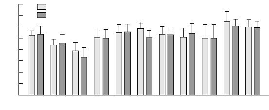
Fig. 11.2. Mean regional ratios of N-acetyl-containing compounds (NAA) to creatine plus phosphocreatine (Cre) for 14 patients with childhood-onset schizophrenia and 14 ageand sex-matched normal controls. Schizophrenic patients demonstrated regionally speciWc reductions in NAA/Cre ratios in the hippocampal regions and dorsolateral prefrontal cortex (*p#0.05). THAL, thalamus; PUT, putamen; HIP, hippocampal area; STG, superior temporal gyrus; ORFC, orbitofrontal cortex; DLPFC, dorsolateral prefrontal cortex; A CING and P CING, anterior and posterior cingulate, respectively; OCC, occipital cortex; FWM, prefrontal white matter; CSO, centrum semiovale.
each selected slice. Each volume element (voxel) had nominal dimensions of 7.5mm!7.5mm!15mm (0.84ml). The 1H MRS imaging sequence involved a spinecho slice selection with repetition time (TR) of 2200ms and echo time (TE) of 272ms and included suppression of water and most of the signal arising from lipids in skull marrow and in surface tissues. The total length of the procedure was approximately 1h. Foam padding was used to stabilize subjects' heads.
The raw 1H MRS imaging data were processed by Wrst locating the NAA, Cho, and Cre peaks for all voxels. Voxels in which these metabolite signals could not be identiWed (e.g., voxels located outside the head and on or near the skull's surface) were manually nulled. The signal strength in a range of 0.2 parts per million around the NAA, Cho, and Cre signal positions was integrated to produce four 32!32 arrays of metabolite signals corresponding to the four brain sections from which spectra were acquired. Obtaining absolute metabolite concentrations necessitates controlling for a number of additional variables, including Weld inhomogeneities, eddy current artifacts, and changes in metabolite T1 and T2 relaxation times owing to pathology, which would require signiWcant additional scan time. Because total scan time had to be limited to what could be tolerated by this clinical population, metabolite signals were reported as ratios of the area under each peak: NAA/Cre, NAA/Cho and Cho/Cre.
A rater blind to diagnosis manually drew ROIs on the T1- weighted coaxial MR images and then transferred them to the same location on the metabolite maps. These ROIs
were drawn with reference to standard anatomic atlases (Talaraich and Tournoux 1988; Duvernoy and Cabanis, 1991) bilaterally in the hippocampal area, dorsolateral prefrontal cortex, superior temporal gyrus, orbitofrontal cortex, occipital cortex, anterior and posterior cingulate, centrum semiovale, prefrontal white matter, thalamus and putamen as described in Bertolino et al. (1996). Voxels that were contained within the anatomic ROI, but not present on the metabolite maps, were removed. Consequently, the Wnal calculations were performed only on voxels containing 1H spectra (approximately 700 voxels per study). A computer program calculated the average value of the area under each peak in all voxels within the ROIs on the metabolite maps.
A three-dimensional MRI dataset was also acquired to permit manual measurement of hippocampal and prefrontal lobe volumes to provide anatomic data to correlate with the 1H MRS imaging data (Bertolino et al., 1996, 1998a). A physician also rated each patient at the time of scanning for positive and negative symptoms using the SAPS (Scale for the Assessment of Positive Symptoms), SANS, and BPRS (Brief Psychiatric Rating Scale).
As illustrated in Figs. 11.2 and 11.3 (p. 242), patients showed a signiWcant bilateral reduction of NAA/Cre in the hippocampus (p#0.05) and dorsolateral prefrontal cortex (p#0.04) relative to their controls. There were no group diVerences for Cho/Cre for these regions. There was a trend for NAA/Cho to be reduced in the dorsolateral prefrontal cortex (p#0.06). There was no main eVect of hemisphere or interaction of diagnosis by hemisphere for any

198L. K. Jacobsen and A. Bertolino
metabolite ratio, thus indicating bilateral, rather than lateralized, abnormalities. The eVect sizes of the NAA/Cre reductions (0.6 in hippocampus and 1.3 in dorsolateral prefrontal cortex) were comparable to values previously found in patients with adult-onset schizophrenia (0.5 and 1.3, respectively; Bertolino et al., 1996).
To evaluate whether the ratio diVerences seen in the patients were attributable to changes in the numerator or denominator terms, the mean integrated areas of NAA, Cho and Cre signals were normalized to the corresponding mean integrated areas of the centrum semiovale (i.e., NAA hippocampus/NAA centrum semiovale, Cho hippocampus/Cho centrum semiovale, etc.) The centrum semiovale was used as a reference region because metabolite ratios from this region have a low coeYcient of variation and because this region has not been implicated in the pathophysiology of schizophrenia (Bertolino et al., 1998b). Analysis of these normalized metabolite ratios showed a trend for NAA to be reduced bilaterally in the hippocampus (p#0.06) and a reduction of NAA in the dorsolateral prefrontal cortex (p#0.1). As in previous studies of patients with adult-onset schizophrenia (Bertolino et al., 1996, 1998a,b), no diVerences were found for Cho, which can be inXuenced by several physiologic, pharmacologic (Satlin et al., 1997), and pathologic processes, including neurodegenerative disease (Brenner et al., 1993; Gadian et al., 1994; Hetherington et al., 1995). Neither were diVerences found for Cre. The metabolite ratio diVerences observed between patients and healthy subjects in the childhoodonset schizophrenia study primarily reXected diVerences in NAA, a marker of neuronal integrity. The observation of reduced NAA in conjunction with normal Cho may imply the presence of neuronal damage unaccompanied by gliosis, such as might occur with a neurodevelopmental defect.
The NAA reductions were not found to be related to anatomic deviations nor to a number of clinical variables. Correlations between the volumes of the hippocampus and of the prefrontal lobe and the metabolite ratios in these regions were not signiWcant (all r values were between 0.14 and 0.32). Correlations with symptoms, as measured by rating scales, and other clinical variables (age, age at onset of disease, current dose and years of treatment with neuroleptic drugs, and length of illness) failed to reveal signiWcant relationships (all r values between 0.33 and 0.40). These Wndings suggest that the NAA reductions seen in schizophrenia are not related to progressive pathology, further reinforcing the likelihood that they reXect developmental neuropathology.
These MRS imaging Wndings are consistent with earlier Wndings in patients with adult-onset schizophrenia of
reductions of NAA in mesial temporal limbic and prefrontal cortices (Nasrallah, 1993; Maier et al., 1995; Renshaw et al., 1995; Bertolino et al., 1996; Yurgelun-Todd et al., 1996; Deicken et al., 1997), although some controversy about these reductions persists (Fukuzako et al., 1995; Stanley et al., 1996; Bartha et al., 1997). Furthermore, emerging evidence suggests that NAA is reXecting more than neuronal density (Arnold et al., 1990; Brenner et al., 1993; de Stefano et al., 1995; Rango et al., 1995; Vion-Dury et al., 1995; Falconer et al. 1996; Hugg et al., 1996; Cheng et al., 1997) and is sensitive to neuronal function.
Conclusions and future directions
The functional imaging studies described in this chapter are among the Wrst to be conducted in childhood-onset schizophrenia and provide valuable evidence that the underlying pathophysiology in this disorder is similar to that in adult-onset schizophrenia. Studies of regional CBF and glucose metabolism have provided evidence of taskrelated hypofrontality in early-onset disease, consistent with Wndings in adult schizophrenia. A comparison of glucose metabolism in childhoodand adult-onset schizophrenia using similar methods has revealed generally equivalent degrees of hypofrontality during the performance of an attentional task. MRS imaging Wndings in childhood-onset schizophrenia are consistent both for the extent and the localizaton of diVerences with earlier studies of patients with adult-onset schizophrenia that showed regionally speciWc reductions of NAA in mesial temporolimbic and prefrontal cortices suggestive of neuronal involvement in these areas. Overall, emerging neuroimaging Wndings support the hypothesis of a common pathophysiologic process speciWcally aVecting mesial temporolimbic and prefrontal circuitries in patients with childhoodand adult-onset schizophrenia.
While these data suggest a similar pathophysiology for childhoodand adult-onset schizophrenia, the limited data available fail to suggest a straightforward relationship between the earlier onset of symptoms and the severity of eVects of any neurodevelopmental lesion. Comparable eVect sizes seen in both the PET study of CMRGlu and the MRS study fail to indicate greater brainrelated abnormalities in early-onset schizophrenia. Limitations in statistical power, age diVerences in the childhoodand adult-onset samples, and/or diVerences in the control groups may have contributed to the failure to Wnd such eVects.
The failure to Wnd more pronounced abnormalities in functional imaging studies of childhood-onset schizo-
Functional imaging in childhood-onset schizophrenia |
199 |
|
|
|
|
phrenia may seem inconsistent with the longitudinal structural MRI studies of the NIMH childhood-onset schizophrenia study described earlier in this chapter, which reported progressive reductions in the volume of temporal and mesial-temporal structures (including the hippocampus), the midsagittal thalamic area, and the whole brain, together with a progressive increase in the size of the ventricles (Rapoport et al., 1997; Jacobsen et al., 1998). However, patients in the MRS study were, on average, 2 years older than those participating in the PET study and those entering the longitudinal MRI study. The longitudinal MRI study of the NIMH sample has shown that the above-mentioned brain structures are changing more rapidly than is normal during the period from 14 to 16 years of age. If structural changes are associated with changes in function, the MRS study completed when adolescents with schizophrenia were, on average, 16.4 years old may have reXected such changes, leading to the observation of abnormalities similar to those seen in adult-onset schizophrenia. In contrast, the PET study, performed when patients were, on average, 14.2 years old, may have been conducted prior to the occurrence of these brain functional changes, thus resulting in a failure to observe more robust hypofrontality. Other factors possibly contributing to the minor inconsistency in Wndings from these two studies include the study of medicated patients in the MRS imaging study and unmedicated patients in the PET study and the use of siblings of patients with ADHD as normal controls in the FDG study. Finally, the two methods may be sensitive to diVerent dimensions of pathology.
While the timing of brain-related changes in schizophrenia require further study, the Wndings discussed in this chapter suggest a window of progressive changes in brain morphology, and possibly physiology, that occur before the age of 16. Future functional brain imaging studies of patients with childhood-onset schizophrenia should focus on studying patients prior to and within this window. Ideally, future studies should employ longitudinal designs and include subjects between the ages of 7 and 18 years. Such data would provide invaluable information about the precise role of neurodevelopment in the symptoms and accompanying abnormalities of brain function observed in schizophrenia. The study of younger patients may elucidate the causes of the unusually early onset of illness.
Special challenges inherent in performing research with patients with childhood-onset schizophrenia include obtaining valid consent and assent, reducing movement during scanning, and signiWcantly poorer performance of activation tasks than that achieved by healthy controls.
Minors participating in any research and their parents need to understand potential risks and discomforts associated with the research before providing assent and consent, respectively, for participation. Procedures, risks and discomforts should be described in simple language.
Patients with childhood-onset schizophrenia tend to move during scanning, which compromises image quality. The use of simulated scanning prior to data collection may improve the ability of these patients to remain still and, thus, enhance the quality of acquired data. To the best of our knowledge, there are no reports to date on the eVectiveness of exposing psychotic children to a simulated scanning experience prior to their participating in an actual functional imaging experiment. However, Rosenberg and colleagues (1997) have demonstrated that simulated scanning prior to actual scanning reduces anxiety during actual scanning in children with obsessivecompulsive disorder.
The same factors that lead to increased movement during scanning on the part of patients with childhoodonset schizophrenia can also interfere with the performance of activation tasks, leading to signiWcant group diVerences in task performance. As with studies of adults with schizophrenia, children and adolescents with schizophrenia may incorporate the experiment into their delusional system and/or be actively hallucinating during the experimental procedure, particularly if the study is performed during medication washout. While the presence of a parent or trusted adult during the procedure often helps, some patients may be unable to complete the study. The tradeoVs between maintaining patients on medication to optimize task performance and facilitate cooperation must be balanced against the introduction of potential artifacts resulting from psychotropic medications, with decisions depending, in part, on the speciWc hypotheses to be tested.
Despite the diYculties encountered in applying functional neuroimaging techniques to the study of childhoodonset schizophrenia, there is much to be gained from such an endeavor. Improvements in technology that decrease scanning time and increase the child friendliness of the scanning environment may help to overcome some of the limitations associated with severe psychotic illness. Further study, particularly employing longitudinal designs, may elucidate the developmental course of brainrelated changes that contribute to early symptoms, both typical and atypical, and their evolution into adult-like schizophrenic illness. Such mapping of developmental brain changes may provide a window through which to view precursors to more typical adult-onset psychosis as well.

200 L. K. Jacobsen and A. Bertolino
iReferencesi
Abi-Dargham, A., Laruelle, M., Aghajanian, G. K., Charney, D. and Krystal, J. (1997). The role of serotonin in the pathophysiology and treatment of schizophrenia. J. Neuropsychiatry Clin. Neurosci., 9, 1±17.
Abi-Dargham, A., Gil, R., Krystal, J. et al. (1998). Increased striatal dopamine transmission in schizophrenia: conWrmation in a second cohort. Am. J. Psychiatry, 155, 761±7.
Alaghband-Rad, J., McKenna, K., Gordon, C. T. et al. (1995). Childhood-onset schizophrenia: the severity of premorbid course. J. Am. Acad. Child Adolesc. Psychiatry, 34, 1273±83.
Altshuler, L. L., Casanova, M. F., Goldberg, T. E. and Kleinman, J. E. (1990). The hippocampus and parahippocampus in schizophrenic, suicide and control brains. Arch. Gen. Psychiatry, 47, 1029±34.
American Psychiatric Association (1994). Diagnostic and Statistical Manual of Mental Disorders, 4th edn (DSM-IV).Washington, DC: American Psychiatric Association.
Andreasen, N. C. (1983). Scale for the Assessment of Negative Symptoms (SANS). Iowa City, IA: The University of Iowa.
Andreasen N. C., Nasrallah H. A., Dunn, V. et al. (1986). Structural abnormalities in the frontal system in schizophrenia: a magnetic resonance imaging study. Arch. Gen. Psychiatry, 43, 136±44.
Andreasen, N. C., Rezai, K., Alliger, R. et al. (1992). Hypofrontality in neuroleptic-naive patients and in patients with chronic schizophrenia: assessment with xenon 133 single-photon emission computed tomography and the Tower of London. Arch. Gen. Psychiatry, 49, 943±58.
Andreasen N. C., Arndt, S., Swayze, V. et al. (1994a). Thalamic abnormalities in schizophrenia visualized through magnetic resonance image averaging. Science, 266, 294±7.
Andreasen, N. C., Flashman, L., Flaum, M. et al. (1994b). Regional brain abnormalities in schizophrenia measured with magnetic resonance imaging. J. Am. Med. Assoc., 272, 1763±9.
Andreasen, N. C., O'Leary, D. S., Cizadlo, T., Arndt, S., Rezai, K., Ponto, L. L., Watkins, G. L. and Hichwa, R. D. (1996). Schizophrenia and cognitive dysmetria: a positron-emission tomography study of dysfunctional prefrontal-thalamic-cere- bellar circuitry. Proc. Natl. Acad. Sci. USA, 93, 9985±90.
Arnold D. L., Matthews, P. M., Francis, G. and Antel, J. (1990). Proton magnetic resonance spectroscopy of human brain in vivo in the evaluation of multiple sclerosis: assessment of the load of disease. Magn. Reson. Med., 14, 154±9.
Arnold, S. E., Hyman, B. T., van Hoesen, G. W. and Damasio, A. R. (1991). Some cytoarchitectural abnormalities of the entorhinal cortex in schizophrenia. Arch. Gen. Psychiatry, 48, 625±32.
Asarnow, R. F., Brown, W. and Strandburg, R. (1995). Children with a schizophrenic disorder: neurobehavioral studies. Eur. Arch. Psychiatry Clin. Neurosci., 245, 70±9.
Barker, P. B., Breiter, S. N., Soher, B. J. et al. (1994). Quantitative proton spectroscopy of canine brain: in vivo and in vitro correlations. Magn. Reson. Med., 32, 157±63.
Bartha, R., Williamson, P. C., Drost, D. J. et al. (1997). Measurement of glutamate and glutamine in the medial prefrontal cortex of
never treated schizophrenic patients and healthy controls by proton magnetic resonance spectroscopy. Arch. Gen. Psychiatry,
54, 959±65.
Benes, F. M., Wickramasinghe, R., Vincent, S. L., Khan, Y. and Todtenkopf, M. (1997). Uncoupling of GABA(A) and benzodiazepine receptor binding activity in the hippocampal formation of schizophrenic brain. Brain Res., 755, 121±9.
Berman, K. F., Torrey, E. F., Daniel, D. G. and Weinberger, D. R. (1992). Regional cerebral blood Xow in monozygotic twins discordant and concordant for schizophrenia. Arch. Gen. Psychiatry, 49, 927±34.
Bertolino, A., Nawroz, S., MattayV. S. et al. (1996). A speci®c pattern of neurochemical pathology in schizophrenia as assessed by multislice proton magnetic resonance spectroscopic imaging.
Am. J. Psychiatry, 153, 1554±63.
Bertolino, A., Saunders, R. C., Mattay, V. S., Bachevalier, J., Frank, J. A. and Weinberger, D. R. (1997). Proton magnetic resonance spectroscopic imaging in monkeys with mesial temporolimbic lesions. Cereb. Cortex, 7, 740±8.
Bertolino, A., Callicott, J. H., Elman, et al. (1998a). Regionally speciWc neuronal pathology in untreated patients with schizophrenia: a proton magnetic resonance spectroscopic imaging study. Biol. Psychiatry, 43, 641±8.
Bertolino, A., Callicott, J. H., Nawroz, S. et al. (1998b). Reproducibility of proton magnetic resonance spectroscopic imaging in patients with schizophrenia. Neuropsychopharmacology, 18, 11±19.
Bogerts, B., Ashtari, M., Degreef, G., Alvir, J. M. J., Bilder, R. M. and Lieberman, J. A. (1990a). Reduced temporal limbic structure volumes on magnetic resonance images in Wrst episode schizophrenia. Psychiatry Res. 35, 1±13.
Bogerts, B., Falkai, P., Haupts, M. et al. (1990b). Post-mortem volume measurements of limbic system and basal ganglia structures in chronic schizophrenics: initial results from a new brain collection. Schizophr. Res., 3, 295±301.
Bogerts, B., Lieberman, J. A., Ashtari, M. et al. (1993). Hippocampus-amygdala volumes and psychopathology in chronic schizophrenia. Biol. Psychiatry, 33, 236±46.
Breier, A., Buchanan, R.W., Elkashef, A., Munson, R. C., Kirkpatrick, B., Cellad, F. (1992). Brain morphology and schizophrenia. A magnetic resonance imaging study of limbic prefrontal cortex and caudate structures. Arch. Gen. Psychiatry, 49, 921±6.
Brenner, R. E., Munro, P. M.G., Williams, S.C.R. et al. (1993). The proton NMR spectrum in acute EAE: the signiWcance of the change in the Cho:Cre ratio, Magn. Reson. Med., 29, 737±45.
Brooks, R. A. (1982). Alternative formula for glucose utilization using labeled deoxyglucose. J. Nucl. Med., 23, 538±9.
Brooks, W. M., Hodde Vargas, J., Vargas, L. A., Yeo, R. A., Ford, C. C. and Hendren, R. L. (1998). Frontal lobe of children with schizophrenia spectrum disorders: a proton magnetic resonance spectroscopic study. Biol. Psychiatry, 43, 263±9.
Brozowski, T. J., Brown, R. M., Rosvold, H. E. and Goldman, P. S. (1979). Cognitive deWcit caused by regional depletion of dopamine in prefrontal cortex of rhesus monkey. Science, 205, 929±32.
Buchsbaum, M. S., DeLisi, L. E., Holcomb, H. H. et al. (1984).
Functional imaging in childhood-onset schizophrenia |
201 |
|
|
|
|
Anteroposterior gradients in cerebral glucose use in schizophrenia and aVective disorders. Arch. Gen. Psychiatry. 41, 1159±66.
Buckley, P. F., Moore, C., Long, H. et al. (1994). 1H Magnetic resonance spectroscopy of the left temporal and frontal lobes in schizophrenia: clinical neurodevelopmental and cognitive correlates. Biol. Psychiatry, 36, 792±800.
Chabrol, H., Guell, A., Bes, A. and Moron, P. (1986). Cerebral blood
Xow in schizophrenic adolescents. [Letter to the Editor] Am. J. Psychiatry, 143, 130.
Chakos, M. H., Lieberman, J. A., Alvir, J., Bilder, R. and Ashtari, M. (1995). Caudate nuclei volumes in schizophrenic patients treated with typical antipsychotics or clozapine. Lancet, 345, 456±7.
Cheng, L. L., Ma, M. J., Becerra, L. et al. (1997). Quantitative neuropathology by high resolution magic angle spinning proton magnetic resonance spectroscopy. Proc. Natl. Acad. Sci. USA, 94, 6408±13.
Chua, S. E. and McKenna, P. J. (1995). Schizophrenia ± a brain disease? A critical review of structural and functional cerebral abnormality in the disorder. Br. J. Psychiatry, 166, 563±82.
Cohen, R. M., Semple, W. E., Gross, M. et al. (1987). Dysfunction in a prefrontal substrate of sustained attention in schizophrenia.
Life Sci. 40, 2031±9.
Creese, I., Burt, D. R. and Snyder, S. H. (1976). Dopamine receptor binding predicts clinical and pharmacological potencies of antischizophrenic drugs. Science, 192, 481±3.
Deicken, R. F., Zhou, L., Corwin, F., Vinogradov, S. and Weiner, M. W. (1997). Decreased left frontal lobe N-Acetylaspartate in schizophrenia. Am. J. Psychiatry, 154, 688±90.
DeLisi, L. E., Buchsbaum, M. S., Holcomb, H. H. et al. (1985). Clinical correlates of decreased anteroposterior metabolic gradients in positron emission tomography (PET) of schizophrenic patients. Am. J. Psychiatry. 142, 78±81.
de Stefano, N., Matthews, P. M. and Arnold, D. L. (1995). Reversible decreases in N-acetylaspartate after acute brain injury. Magn. Reson. Med., 34, 721±7.
Done, D. J., Crow, T. L., Johnstone, E. C. and Sacker, A. (1994). Childhood antecedents of schizophrenia and aVective illness: social adjustment at ages 7 and 11. BMJ, 309, 599±703.
Duvernoy, H. M. and Cabanis, E. A. (1991). The Human Brain Surface, Three-dimensional Sectional Anatomy, and MRI. New York: Springer-Verlag.
Duyn, J. H., Gillen, J., Sobering, G., van Zijl, P. C. and Moonen C.T.W. (1993). Multisection proton MR spectroscopic imaging of the brain. Radiology, 188, 277±82.
Ebmeier, K. P., Lawrie, S. M., Blackwood, D. H. R., Johnstone, E. C. and Goodwin, G. M. (1995). Hypofrontality revisited: a high resolution single photon emission computed tomography study in schizophrenia. J. Neurol. Neurosurg. Psychiatry, 58, 452±6.
Ernst, M., Liebenauer, L. L., King, A. C., Fitzgerald, G. A., Cohen, R. M. and Zametkin, A. J. (1994). Reduced brain metabolism in hyperactive girls. J. Am. Acad. Child Adolesc. Psychiatry, 33, 858±68.
Ernst, M., Cohen, R. M., Liebenauer, L. L., Jons, P. H. and Zametkin,
A. J. (1997). Cerebral glucose metabolism in adolescent girls with attention deWcit/hyperactivity disorder. J. Am. Acad. Child Adolesc. Psychiatry, 36, 1399±1406.
Falconer, J. C., Liu, S. J., Abbe, R. A. and Narayana, P. A. (1996). Time dependence of N-acetyl-aspartate, lactate, and pyruvate concentrations following spinal cord injury. J. Neurochem., 66, 717±22.
Falkai, P. and Bogerts, B. (1986). Cell loss in the hippocampus of schizophrenics. Eur. Arch. Psychiatry Neurol. Sci., 236, 154±61.
Feinberg, I. (1982). Schizophrenia: caused by a fault in programmed synaptic elimination during adolescence? J. Psychiatr. Res., 17, 319±34.
Fiez, J. A. (1996). Cerebellar contributions to cognition. Neuron, 16, 13±15.
Findling, R. L., Friedman, L., Buck, J. et al. (1996). Hippocampal volume in adolescent schizophrenia. Schizophr. Res., 18, 185.
Flaum, M., Swayze, V. W., O'Leary, D. S. et al. (1995). Brain morphology in schizophrenia: eVects of diagnosis, laterality and gender. Am. J. Psychiatry, 152, 704±14.
Frazier, J. A., Giedd, J. N., Kaysen, D. et al. (1996). Brain magnetic resonance imaging rescan after two years of clozapine maintenance. Am. J. Psychiatry, 153, 564±6.
Friedman, L., Findling, R. L., Buch, J. et al. (1996). Structural MRI and neuropsychological assessments in adolescent patients with either schizophrenia or aVective disorders. Schizophr. Res.,
18, 189±90.
Friston, K. J., Frith, C. D., Liddle, P. F. and Frackowiak, R. S. J. (1991). Comparing functional (PET) images: the assessment of signiWcant change. J. Cereb. Blood Flow Metab., 11, 690±9.
Friston, K. J., Worsley, K. J., Frackowiak, R. S. J., Mazziotta, J. C. and Evans, A. C. (1994). Assessing the signiWcance of focal activations using their spatial extent. Hum. Brain Map., 1, 210±20.
Fukuzako, H., Takeuchi, K., Hokazono, Y. et al. (1995). Proton magnetic resonance spectroscopy of the left medial temporal and frontal lobes in chronic schizophrenia: preliminary report.
Psychiatry Res. Neuroimaging, 61, 193±200.
Fuster, J. M. and Alexander, G. E. (1971). Neuron activity related to short term memory. Science, 173, 652±4.
Gadian, D. G., Connelly, A., Duncan, J. S. et al. (1994). 1H magnetic resonance spectroscopy in the investigation of intractable epilepsy. Acta Neurol. Scand. Suppl. 152, 116±21.
Giedd, J. N., Castellanos, F. X., Rajapakse, J. C. et al. (1996). Quantitative analysis of gray matter volumes in childhood-onset schizophrenia and attention deWcit/hyperactivity disorder. Soc. Neurosci. Abst., 22, 1166.
Goldman, P. S. (1974). An alternative to developmental plasticity: heterology of CNS structure in infants and adults. In Plasticity and Recovery of Function in the Central Nervous System, eds. D. G. Stein, J. J. Rosen and N. Butters, pp. 149±74. New York: Academic Press.
Goldman, P. S. and Alexander, G. E. (1977). Maturation of prefrontal cortex in the monkey revealed by local reversible cryogenic depression, Nature, 267, 613±15.
Goldman-Rakic, P. S. (1987). Circuitry of primate prefrontal cortex and regulation of behavior by representational memory. In
Handbook of Physiology, vol. 5, eds. V. B. Mountcastle, F. Plum

202 L. K. Jacobsen and A. Bertolino
and S. R. Geiger, pp. 373±417. Bethesda, MD: American Physiological Society.
Goldman-Rakic, P. S. (1991). Prefrontal cortical dysfunction in schizophrenia: the relevance of working memory. In
Psychopathology and the Brain, eds. B. J. Carroll and J. E. Barrett, pp. 1±23. Hillsdale, NJ: Laurence Erlbaum.
Goldman-Rakic, P. S. (1994). SpeciWcation of higher cortical functions. In Atypical Cognitive DeWcits in Developmental Disorders: Implications for Brain Functions, eds. S. H. and J. Grafman, pp. 3±17. Hillsdale NJ: Laurence Erlbaum.
Gordon, C. T., Frazier, J. A., McKenna, K. et al. (1994). Childhoodonset schizophrenia: an NIMH study in progress. Schizophr. Bull., 20, 697±712.
Harvey, I., Ron, M. A., Du Boulay, G., Wicks, D., Lewis, S. W. and Murray, R. M. (1993). Reduction of cortical volume in schizophrenia on magnetic resonance imaging. Psychol. Med., 23, 591±604.
Hetherington, H., Kuzniecky, R., Pan, J. et al. (1995). Proton nuclear magnetic resonance spectroscopic imaging of human temporal lobe epilepsy at 4.1 T. Ann. Neurol., 38, 396±404.
Hollis, C. (1995). Child and adolescent (juvenile onset) schizophrenia: a case control study of premorbid developmental impairments. Br. J. Psychiatry, 166, 489±95.
Hugg, J. W., Kuzniecky, R. I., Gilliam, F. G., Morawetz, R. B. and Hetherington, H. P. (1996). Normalization of contralateral metabolic function following temporal lobectomy demonstrated by
1H magnetic resonance spectroscopic imaging. Ann. Neurol., 40, 236±9.
Jacobsen, L. K. and Rapoport, J. L. (1998). Childhood onset schizophrenia; implications of clinical and neurobiological research. J. Child Psychol. Psychiatr. Allied Disciplines, 39, 101±13.
Jacobsen, L. K., Giedd, J. N., Vaituzis, A. C. et al. (1996a). Temporal lobe morphology in childhood onset schizophrenia. Am. J. Psychiatry, 153, 355±61.
Jacobsen, L. K., Hong, W. L., Hommer, D. W. et al. (1996b). Smooth pursuit eye movements in childhood onset schizophrenia: comparison with ADHD and normal controls. Biol. Psychiatry,
40, 1144±54.
Jacobsen, L.K., Hamburger, S. D., van Horn, J. D. et al. (1997). Cerebral glucose metabolism in childhood onset schizophrenia.
Psychiatr. Res. Neuroimaging, 75, 131±44.
Jacobsen, L. K., Giedd, J. N., Castellanos, F. X. et al. (1998). Progressive reduction of temporal lobe structures in childhood onset schizophrenia. Am. J. Psychiatry, 155, 678±85.
Jakob, H. and Beckmann, H. (1989). Prenatal developmental disturbances in the limbic allocortex in schizophrenics. J. Neural Transmission, 65, 303±26.
Jenkins, I., Brooks, D., Nixon, P., Frackowiak, R. and Passingham, R. (1994). Motor sequence learning: a study with positron emission tomography. J. Neurosci., 14, 3775±90.
Jernigan, T. L., Zisook, S., Heaton, R. K., Moranvile, J. T., Hesselink, J. R. and BraV, D. L. (1991). Magnetic resonance imaging abnormalities in lenticular nuclei and cerebral cortex in schizophrenia. Arch. Gen. Psychiatry., 48, 881±90.
Jeste, D. V. and Lohr, J. B. (1989). Hippocampal pathologic Wndings in schizophrenia: a morphometric study. Arch. Gen. Psychiatry.
46, 1019±24.
Jones, P., Rodgers, B., Murray, R. and Marmot, M. (1994). Child developmental risk factors for adult schizophrenia in the British 1946 birth cohort. Lancet, 344, 1398±1402.
Karno, M. and Norquist, G. S. (1989). Schizophrenia: epidemiology. In Comprehensive Textbook of Psychiatry, 5th edn, eds. H. I. Kaplan and B. J. Sadock, pp. 699±705. Baltimore, MD: Williams and Wilkins.
Kim, S. G., Ugurbil, K. and Strick, P. L. (1994). Activation of a cerebellar output nucleus during cognitive processing. Science, 265, 949±51.
Kumra, S., Frazier, J. A., Jacobsen, L. K. et al. (1996). Childhoodonset schizophrenia: a double-blind clozapine-haloperidol comparison. Arch. Gen. Psychiatry, 53, 1090±7.
Kumra, S., Briguglio, C., Lenane, M. et al. (1999). Including children and adolescents with schizophrenia in medication-free research. Am. J. Psychiatry, 156, 1065±8.
Laruelle, M., Abi-Dargham, A., van Dyck, C. H. et al. (1996). Single photon emission computerized tomography imaging of amphetamine-induced dopamine release in drug-free schizophrenic subjects. Proc. Natl. Acad. Sci. USA, 93, 9235±40.
Lillrank, S. M., Lipska, B. K., Bachus, S. E., Wood, G. K. and Weinberger, D. R. (1996). Amphetamine induced c-fos mRNA expression is altered in rats with neonatal ventral hippocampal damage. Synapse, 23, 292±301.
Lim, K. O., Beal, D. M., Harvey, R. L. et al. (1995). Brain dysmorphology in adults with congenital rubella plus schizohrenia-like symptoms. Biol. Psychiatry, 37, 764±76.
Lim, K. O., Harris, D., Beal, M. et al. (1996). Gray matter deWcits in young onset schizophrenia are independent of age of onset. Biol. Psychiatry, 40, 4±13.
Lipska, B. K. and Weinberger, D. R. (1995). Genetic variation in vulnerability to the behavioral eVects of neonatal hippocampal damage in the rat. Proc. Natl. Acad. Sci., 92, 8906±10.
Lipska, B. K., Jaskiw, G. E. and Weinberger, D. R. (1993). Post pubertal emergence of hyperresponsiveness to stress and amphetamines after neonatal excitotoxic hippocampal damage, a potential animal model of schizophrenia. Neuropsychopharmacology, 9, 67±75.
Lipska, B., Moghaddam, B., Sams-Dodd, F. and Weinberger, D. R. (1996). Neonatal hippocampal damage in the rat models negative symptoms of schizophrenia. Abstracts from the American College of Neuropsychopharmacology, p. 126. Washington, DC: American College of Neuropsychopharmacology.
Maier, M., Ron, M. A., Barker, G. J. and Tofts, P. S. (1995). Proton magnetic resonance spectroscopy: an in vivo method of estimating hippocampal neuronal depletion in schizophrenia.
Psychol. Med., 25, 1201±9.
Marsh, L., Suddath, R. Higgins, N. and Weinberger, D. R. (1994). Medial temporal lobe structures in schizophrenia: relationship of size to duration illness. Schizophr. Res., 11, 225±38.
Matsui, T. and Hirano, A. (1978). An Atlas of the Human Brain for Computerized Tomography. Tokyo: Igaku Shoin.
Functional imaging in childhood-onset schizophrenia |
203 |
|
|
|
|
Meltzer, H. Y. and Stahl, S. M. (1976). The dopamine hypothesis of schizophrenia: a review. Schizophr. Bull., 2, 19±76.
Miller, B. L. (1991). A review of chemical issues in 1H NMR spectroscopy: N-acetyl-l-aspartate, creatine and choline. NMR Biomed., 4, 47±52.
Murray, R. M. (1994). Neurodevelopmental schizophrenia: the rediscovery of dementia praecox. Br. J. Psychiatry, 165, 6±12.
Murray, R. M. and Lewis, S. W. (1987). Is schizophrenia a neurodevelopmental disorder? BMJ, 295, 681±82.
Nasrallah, H. (1993). Neurodevelopmental pathogenesis of schizophrenia. Psychiatr. Clin. North Am., 16, 269±80.
Nasrallah, H. A., Skinner, T. E., Schmalbrock, P. and Robitaille, P. M. (1994). Proton magnetic resonance spectroscopy of the hippocampal formation in schizophrenia: a pilot study. Brit. J. Psychiatry, 165, 481±5.
Raichle, M. E., Fiez, J. A., Videen, T. O. et al. (1994). Practice related changes in human brain functional anatomy during nonmotor learning. Cereb. Cortex, 4, 8±26.
Rango, M., Spagnoli, D., Tomei, G., Bamonti, F., Scarlato, G. and Zetta, L. (1995). Central nervous system transsynaptic eVects of acute axonal injury: a 1H magnetic resonance spectroscopy study. Magn. Reson. Med., 33, 595±600.
Rapoport, J. L., Giedd, J., Kumra, S. et al. (1997). Childhood onset schizophrenia: progressive ventricular change during adolescence. Arch. Gen. Psychiatry, 54, 897±903.
Remschmidt, H. E., Schulz, E., Martin, M., Warnke, A. and Trott, G. (1994). Childhood-onset schizophrenia: history of the concept and recent studies. Schizophr. Bull., 20, 727±45.
Renshaw, P. F., Yurgelun-Todd, D. A., Tohen, M., Gruber, S. and Cohen, B. M. (1995). Temporal lobe proton magnetic resonance spectroscopy of patients with Wrst-episode psychosis. Am. J. Psychiatry, 152, 444±6.
Rosenberg, D. R., Sweeney, J. A., Gillen, J. S. et al. (1997). Magnetic resonance imaging of children without sedation: preparation with simulation. J. Am. Acad. Child Adolesc. Psychiatry, 36, 853±9.
Russell, A. T. (1994). The clinical presentation of childhood onset schizophrenia. Schizophr. Bull., 20, 631±46.
Russell, A. T., Bott, L. and Sammons, C. (1989). The phenomenology of schizophrenia occurring in childhood. J. Am. Acad. Child Adolesc. Psychiatry, 28, 399±407.
Sams-Dodd, F., Lipska, B. and Weinberger, D. R. (1997). Neonatal lesions of the rat ventral hippocampus result in hyperlocomotion and deWcits in the social behavior in adulthood.
Psychopharmachology, 132, 303±10.
Satlin, A., Bodick, N., OVen, W. W. and Renshaw, P. F. (1997). Brain proton magnetic resonance spectroscopy (1HMRS) in Alzheimer's disease: changes after treatment with xanomeline, an M1 selective cholinergic agonist. Am. J. Psychiatry, 154, 1459±61.
Schulz, S. C., Koller, M.M., Kishore, P. R., Hamer, R. M., Gehl, J. J. and Friedel, R, O. (1983). Ventricular enlargement in teenage patients with schizophrenia spectrum disorder. Am. J. Psychiatry, 140, 1592±5.
Selemon, L. D., Rajkowska, G. and Goldman-Rakic, P. S. (1995). Abnormally high neuronal density in the schizophrenic cortex. A
morphometric analysis of prefrontal area 9 and occipital area 17.
Arch. Gen. Psychiatry, 52, 805±18.
Siegel, B. V., Buchsbaum, M. S., Bunney,W. E. et al. (1993). Cortical- striatal-thalamic circuits and brain glucose metabolic activity in 70 unmedicated male schizophrenic patients. Am. J. Psychiatry,
150, 1325±36.
Stanley, J. A., Williamson, P. C., Drost, D. J. et al. (1996). An in vivo proton magnetic resonance spectroscopy study of schizophrenia patients. Schizophr. Bull., 22, 597±609.
Steinberg, J. L., Devous, M. D., Moeller, F. G. et al. (1995). Cerebellar blood Xow in schizophrenic patients and normal control subjects. Psychiatr. Res., 61, 15±31.
Stern, Y. and Langston, J. W. (1985). Intellectual changes in patients with MPTP-induced parkinsonism. Neurology, 35, 1506±9.
Suddath, R. L., Christison, G. W., Fuller Torrey, E., Casanova, M. F. and Weinberger, D. R. (1990). Anatomical abnormalities in the brains of monozygotic twins discordant for schizophrenia. N. Engl. J. Med., 322, 789±94.
Sullivan, E. V., Lim, K. O., Mathalon, D. et al. (1998). A proWle of cortical gray matter volume deWcits characteristic of schizophrenia.
Cereb. Cortex, 8, 117±24.
Szechtman, H., Nahmias, C., Garnett, E. S. et al. (1988). EVect on neuroleptics on altered cerebral glucose metabolism in schizophrenia. Arch. Gen. Psychiatry, 45, 523±32.
Talairach, J. and Tournoux, P. (1988). Coplanar Stereotaxic Atlas of the Human Brain. New York: Thieme Medical.
Thomas, A. M., Ke, Y., Caplan, R. et al. (1996). Frontal lobe 1HMR spectroscopy of children with schizophrenia. In Proceedings of the 15th Annual Meeting of the Society of Magnetic Resonance in Medicine, p. 1000.
Toft, P. B., Leth, H., Lou, H. C., Pryds, O. and Henriksen, O. (1994). Metabolite concentrations in the developing brain estimated with proton MR spectroscopy. J. Magn. Reson. Imaging, 4, 647±80.
Urenjak, J., Williams, S. R., Gadian, D. G. and Noble, M. (1993). Proton nuclear magnetic resonance spectroscopy unambiguously identiWes diVerent neural cell types. J. Neurosci., 13, 981±9.
Vion-Dury, J., Salvan, A. M., Confort-Gouny, S., Dhiver, C. and Cozzone, P. (1995). Reversal of brain metabolic alterations with zidovudine detected by proton localised magnetic resonance spectroscopy. Lancet, 345, 60±1.
Volkow, N. D., Levy, A., Brodie, J. D. et al. (1992). Low cerebellar metabolism in medicated patients with chronic schizophrenia.
Am. J. Psychiatry, 149, 686±8.
Volz, H. P., Gaser, C., Hager, F., Rzanny, R. et al. (1997). Brain activation during cognitive stimulation with the Wisconsin Card Sort Test ± a functional MRI study in healthy volunteers and schizophrenics. Psychiatr. Res., 75, 145±57.
Watkins, J. M., Asarnow, R. F. and Tanguay, P. E. (1988). Symptom development in childhood onset schizophrenia. J. Am. Acad. Child Adolesc. Psychiatry, 6, 865±78.
Weinberger, D. R. (1987). Implications of normal brain development for the pathogenesis of schizophrenia. Arch. Gen. Psychiatry, 44, 660±9.
Weinberger, D. R. (1995). Schizophrenia: from neuropathology to neurodevelopment. Lancet, 346, 552±7.