

Книги по МРТ КТ на английском языке / Functional Neuroimaging in Child Psychiatry Ernst 1 ed 2000
.pdf74 |
D. A. Yurgelun-Todd and P. F. Renshaw |
|
|
iReferencesi
Auld, K., Ashwal, S., Holshouser, B. et al., (1995). Proton magnetic resonance spectroscopy in children with acute central nervous system injury. Pediatr. Neurol., 12, 323±34.
Barker, P., Breiter, S., Soher, B. et al. (1994). Quantitative proton spectroscopy of canine brain: in vivo and in vitro correlations.
Magn. Reson. Med., 32, 157±63.
Bartha, R., Stein, M.,Williamson, P. and Drost, D. (1998). Decreased NAA in the left caudate in obsessive-compulsive patients on 1H MRS. Biol. Psychiatry, 43, 9S.
Behar, K., Rothman, D., Spencer, D. and PetroV, O. (1994). Analysis of macromolecule resonances in 1H NMR spectra of human brain. Magn. Reson. Med., 32, 294±302.
Bertolino, A., Kumra, S., Callicott, J. et al. (1997). Abnormal cortical pattern in early onset schizophrenia as identiWed by 1H-MRS. [Abstract] In Proceedings of the 5th Annual Meeting of the International Society for Magnetic Resonance in Medicine, vol. 2, p. 1227.
Bertolino, A., Callicott, J. H., Elman, I. et al. (1998). Regionally speciWc neuronal pathology in untreated patients with schizophrenia: a proton magnetic resonance spectroscopic imaging study. Biol. Psychiatry, 43, 641±8.
Birken, D. and Oldendorf, W. (1989). N-Acetyl-l-aspartic acid: a literature review of a compound prominent in 1H-NMR spectroscopic studies of the human brain. Neurosci. Biobehav. Rev., 13, 23±42.
Bloch, F., Jansen,W.W. and Packard, M. E. (1946). Phys.Rev. 69, 127. Bluml, S., Schad, L. R., Scharf, J., Wenz, F., Knopp, M. V. and Lorenz, W. J. (1996). A comparison of magnetization prepared 3D gradi- ent-echo (MP±RAGE) sequences for imaging of intracranial
lesions. Magn. Reson. Imaging, 14, 329±35.
Bottomley, P., Foster, T. and Darrow, R. (1984). Depth-resolved surface coil spectroscopy (DRESS) for in vivo 1H, 31P, and 13C NMR. J. Magn. Reson., 59, 338±43.
Bovey, F. A., Jelinski, L. and Mirau, P. A. (1988). Nuclear Magnetic Resonance Spectroscopy. San Diego, CA: Academic Press.
Brooks, W., Hodde-Vargas, J., Vargas, L., Yeo, R., Ford, C. and Hendren, R. (1998). Frontal lobe of children with schizophrenia spectrum disorders: a proton magnetic resonance spectroscopic study. Biol. Psychiatry, 43, 263±9.
Brown, T., Kincaid, B. and Ugurbil, K. (1982). NMR chemical shift imaging in three dimensions. Proc. Natl. Acad. Sci. USA, 79, 3523±8.
Buchli, R., Duc. C., Martin, E. and Boesiger, P. (1994). Assessment of absolute metabolite concentrations in human tissue by 31P MRS in vivo. Part I: cerebrum, cerebellum, cerebral gray matter and white matter. Magn. Reson. Med., 32, 447±54.
Buckley, P. (1998). Magnetic resonance spectroscopy and aberrant neurodevelopment. Biol. Psychiatry, 43, 61S.
Charles, H., Lazeyras, F., Krishnan, K. et al. (1994). Proton spectroscopy of human brain: eVects of age and sex. Prog. Neuropsychopharmacol. Biol. Psychiatry, 18, 995±1004.
Christensen, J., Babb, S., Cohen, B. and Renshaw, P. (1998).
Quantitation of dexfenXuramine/norfenXuramine in primate brain using 19F NMR spectroscopy. Magn. Reson. Med., 39, 149±54.
Diklic, V. and Gambarelli, D. (1998). 1H MR spectroscopy in twins discordant for late-infantile neuronal ceroid lipofuscinosis. In
Proceedings of the 6th Annual Meeting of the International Society for Magnetic Resonance in Medicine, vol. 3, p. 1745.
Dilsaver, S. C. (1986). Cholinergic mechanisms in depression.
Brain Res., 396, 285±316.
Ende, G., Knowlton, R., Laxer, K., Matson, G. and Weiner, M. (1996). NAA is a more sensitive marker of hippocampal disease than atrophy: evidence that NAA is a neuronal marker. In Proceedings of the 4th Annual Meeting of the International Society for Magnetic Resonance in Medicine, vol. 1, p. 137.
Fritze, J. (1993). The adrenergic±cholinergic imbalance hypothesis of depression: a review and a perspective. Rev. Neurosci., 4, 63±93.
Gadian, D., Isaacs, E., Cross, J. et al. (1996). Lateralization of brain function in childhood revealed by MR spectroscopy. Neurology,
46, 974±7.
Haseler, L., Arcinue, E., Danielsen, E. S. B. and Ross, B. (1997). Evidence from proton magnetic resonance spectroscopy for a metabolic cascade of neuronal damage in shaken baby syndrome. Pediatrics, 99, 4±14.
Hendren, R., Hodde-Vargas, J., Yeo, R., Vargas, L., Brooks, W. and Ford, C. (1995). Neuropsychophysiological study of children at risk for schizophrenia: a preliminary report. J. Am. Acad. Child Adolesc. Psychiatry, 34, 1284±91.
Hetherington, H. P., Pan, J. W., Chu, W. J., Mason, G. F. and Newcomer, B. R. (1997). Biological and clinical MRS at ultra-high
Weld. NMR Biomed., 10, 360±71.
Holshouser, B., Ashwal, S., Luh, G., Shu, S., Tomasi, L. and Hinshaw, D. (1997). 1H-MR spectroscopy in predicting neurologic outcomes in children with acute CNS injury. In Proceedings of the 5th Annual Meeting of the International Society for Magnetic Resonance in Medicine, vol. 2, p. 1229.
Jacobsen, L. K., Hong, W. L., Hommer, D. W. (1996). Smooth pursuit eye movements in childhood-onset schizophrenia: comparison with attention-deWcit hyperactivity disorder and normal controls. Biol. Psychiatry, 40, 1144±54.
Jensen, H., Plenge, P., Stensgaard, A. et al. (1996). Twelve-hour brain lithium concentration in lithium maintenance treatment of manic-depressive disorder: daily versus alternate-day dosing schedule. Psychopharmacology, 124, 275±8.
Keltner, J. R., Wald, L. L., Frederick, B. D. and Renshaw, P. F. (1997). In vivo detection of GABA in human brain using localized double-quantum Wlter technique. Magn. Reson. Med., 37, 366±71.
Kim, K., Lee, J., Kim, S., Pi, S., Auh,Y. and Lim, T. (1997). Early detection of abnormality for hypoxic ischemic encephalopathy (HIE) infant by localized in vivo 1H MR spectroscopy. In Proceedings of the 5th Annual Meeting of the International Society for Magnetic Resonance in Medicine, vol. 2, p. 1237.
Kim, K., Pi, S., Kim, E. et al. (1998). Localized 1H MR spectroscopy as early predictor for outcome in infants with birth asphyxia. In
MRS in childhood psychiatric disorders |
75 |
|
|
Proceedings of the 6th Annual Meeting of the Society for Magnetic Resonance in Medicine, vol. 1, p. 95.
Kinney, D., Yurgelun-Todd, D., Waternaux, C. and Matthysse, S. (1994). Obstetrical complications and trail making deWcits discriminate schizophrenics from unaVected siblings and controls.
Schizophr. Res., 12, 63±73.
Komoroski, R., Newton, J., Karson, C., Cardwell, D. and Sprigg, J. (1991). Detection of psychoactive drugs in vivo using 19F NMR spectroscopy. Biol. Psychiatry, 29, 711±16.
Kreis, R., Suter, D. and Ernst, R. R. (1985). Time-domain zero-Weld magnetic resonance with Weld pulse excitation. Chem. Phys. Lett., 118, 120±4.
Krishnan, R. and Doraiswamy, P. (eds.) (1997). Brain Imaging in Clinical Psychiatry. New York: M. Dekker.
Lee, J., Kim, K., Kim, S., Pi, S., Auh, Y. and Lim, T. (1997). Metabolic changes of normal neonatal brain during the preterm period by localized 1H in vivo MR spectroscopy. In Proceedings of the 5th Annual Meeting of the International Society for Magnetic Resonance in Medicine, vol. 2, p. 1173.
Li, Q., Pu, Y., Luo, D., Zeng, C., Gao, J. and Gao, J. (1998). An MRS study in newborn infants with hypoxic-ischemic encephalopathy. In Proceedings of the 6th Annual Meeting of the International Society for Magnetic Resonance in Medicine, vol. 3, p. 1738.
Lim, K., Adalsteinsson, E., Spielman, D., Sullivan, E., Rosenbloom, M. and PfeVerbaum, A. (1998). Proton magnetic resonance spectroscopic imaging of cortical gray and white matter in schizophrenia. Arch. Gen. Psychiatry, 55, 346±52.
Lu, D., Pavelakis, S., Frank, Y. et al. (1996). Proton MR spectroscopy of the basal ganglia in healthy children and children with AIDS.
Radiology, 199, 423±8.
Luyten, P., Bruntink, G., SloV, F. et al. (1989). Broadband proton decoupling in human 31P NMR spectroscopy. NMR Biomed., 1, 177±83.
Mason, G. F., Behar, K. L. and Lai, J. C. (1996). The 13C isotope and nuclear magnetic resonance: unique tools for the study of brain metabolism. Metab. Brain Dis., 11, 283±313.
Miller, B. (1991). A review of chemical issues in 1H NMR spectroscopy: N-acetyl aspartate, creatine, and choline. NMR Biomed., 4, 47±54.
Miner, C., Davidson, J., Potts, N., Tupler, L., Charles, H. and Krishnan, K. (1995). Brain Xuoxetine measurements using
Xuorine magnetic resonance spectroscopy in patients with social phobia. Biol. Psychiatry, 38, 696±8.
Minshew, N., Goldstein, G., Dombrowski, S., Panchalingam, K. and Pettegrew, J. (1993). A preliminary 31P MRS study of autism: evidence for undersynthesis and increased degradation of brain membranes. Biol. Psychiatry, 33, 762±73.
Moonen, C., von Kiellin, M., van Zijl, P., Cohen, J., Gillen, J. and Daly, P. (1989). Comparison of single-shot localization methods (STEAM and PRESS) for in vivo proton NMR spectroscopy. NMR Biomed., 2, 101±12.
Moore, C., Christensen, J., Lafer, B., Fava, M. and Renshaw, P. (1997). Decreased adenosine trophosphate in the basal ganglia of depressed subjects: a phosphorous-31 magnetic resonance spectroscopy study. Am. J. Psychiatry, 154, 116±18.
Moore, C., Breeze, J., Kukes, T. et al. (1999). EVects of myo-inositol ingestion on human brain myo-inositol levels: a proton magnetic resonance spectroscopic imaging study. Biol. Psychiatry,
45, 1197±202.
Moore, G., Slovis, T. and Chugani, H. (1996). Proton MRS of Sturge±Weber syndrome in children. In Proceedings of the 4th Annual Meeting of the International Society for Magnetic Resonance in Medicine, 1996; 2, 968.
Moss, H. and Talagala, S. (1997). 31P magnetic resonance spectroscopy of normal peripubertal children: eVects of sex and frontooccipital location. In Proceedings of the 5th Annual Meeting of the International Society for Magnetic Resonance in Medicine, vol. 2, p. 1219.
Moss, H., Talagala, S. and Kirisci, L. (1997). Phosphorus-31 magnetic resonance brain spectroscopy of children at risk for a substance use disorder: preliminary results. Psychiatr. Res. Neuroimaging, 76, 101±12.
Mukherji, S. (ed.) (1998). Clinical Applications of Magnetic Resonance Spectroscopy. New York: Wiley-Liss.
Murphy-Boesch, J., Stoyanova, R., Srinivasan, R. et al. (1993). Proton-decoupled 31P chemical shift imaging of the human brain in normal volunteers. NMR Biomed., 6, 173±80.
Ogg, R., Kingsley, P. and Taylor, J. (1994). WET, a T1- and B1-insensi- tive water suppression method for in vivo localized 1H NMR spectroscopy. J. Magn. Reson. Ser. B, 104, 1±10.
Ordidge, R., Connelly, A. and Lohman, J. (1986). Image selective in vivo spectroscopy (ISIS). A new technique for spatially selective NMR spectroscopy. J. Magn. Reson., 60, 281±3.
PetroV, O., Spencer, D., Alger, J. and Prichard, J. (1989). High-Weld proton magnetic resonance spectroscopy of human cerebrum obtained during surgery for epilepsy. Neurology, 39, 1197±201.
Pettegrew, J., Keshavan, M., Panchalingham, K. et al. (1991). Alterations in brain high energy phosphate metabolism in Wrst episode, drug naive schizophrenics. Arch. Gen. Psychiatry, 48, 563±8.
Pouwels, P. and Frahm, J. (1998). Regional metabolite concentrations in human brain as determined by quantitative localized proton MRS. Magn. Reson. Med., 39, 53±60.
Purcell, E. M., Torrey, H. C. and Pound, R. V. (1946). Phys. Rev., 69, 37.
Rae, C., Scott, R., Lee, M., Hines, N., Paul, C. and Styles, P. (1998). Human brain bioenergetics relate to cognitive ability. In
Proceedings of the 6th Annual Meeting of the International Society for Magnetic Resonance in Medicine, vol. 3, p. 1743.
Rajanaygam, V., Shapiro, E., Krivit, W., Lockman, L. and Stillman, A. (1997). Childhood adrenoleukodystrophy: an MRS scale to measure disease severity. In Proceedings of the 5th Annual Meeting of the International Society for Magnetic Resonance in Medicine, vol. 2, p. 1193.
Renshaw, P. and Wicklund, S. (1998). In vivo measurement of lithium in man by nuclear magnetic resonance spectroscopy.
Biol. Psychiatry, 23, 465±72.
Renshaw, P., Guimaraes, A., Fava, M. et al. (1992). Accumulation of
Xuoxetine and norXuoxetine in human brain during therapeutic administration. Am. J. Psychiatry, 149, 1592±4.
76 |
D. A. Yurgelun-Todd and P. F. Renshaw |
|
|
Renshaw, P., Yurgelun-Todd, D., Tohen, M., Gruber, S. and Cohen, B. (1995). Temporal lobe proton magnetic resonance spectroscopy of patients with Wrst-episode psychosis. Am. J. Psychiatry,
152, 444±6.
Renshaw, P., Lafer, B., Babb, S. et al. (1997). Basal ganglia choline levels in depression and response to Xuoxetine treatment: an in vivo proton magnetic resonance spectroscopy study. Biol. Psychiatry, 41, 837±43.
Riedl, U., Barocka, A., Kolem, H. et al. (1997). Duration of lithium treatment and brain lithium concentration in patients with unipolar and schizoaVective disorder ± a study with magnetic resonance spectroscopy. Biol. Psychiatry, 41, 844±50.
Rosenberg, D., MacMaster, F., Parrish, J. et al. (1998). 1H MRS caudate glutamatergic changes with paroxetine therapy for pediatric obsessive compulsive disorder. Biol. Psychiatry, 43, 24S.
Sachs, G., Renshaw, P., Lafer, B. et al. (1995). Variability of brain lithium levels during maintenance treatment: a magnetic resonance spectroscopy study. Biol. Psychiatry, 38, 422±8.
Soares, J., Krishnan, K. and Keshavan, M. (1996). Nuclear magnetic resonance spectroscopy: new insights into the pathophysiology of mood disorders. Depression, 4, 14±30.
Steingard, R., Renshaw, P., Yurgelun-Todd, D. and Wald, L. (1998). Proton MRS studies of the orbitofrontal cortex of depressed adolescents. Biol. Psychiatry, 43, 28S.
Strauss,W., Layton, M., Hayes, C. and Dager, S. (1997). 19F magnetic resonance spectroscopy investigation in vivo of acute and steady-state brain Xuvoxamine levels in obsessive-compulsive disorder. Am. J. Psychiatry, 154, 516±22.
Strauss, W., Layton, M. and Dager, S. (1998). Brain elimination halflife of Xuvoxamine measured by 19F magnetic resonance spectroscopy. Am. J. Psychiatry, 155, 380±4.
Thomas, M., Ke, Y., Caplan, R. et al. (1996). Frontal lobe 1H MR spectroscopy of children with schizophrenia. In Proceedings of
the 4th Annual Meeting of the International Society for Magnetic Resonance in Medicine, vol. 2, p. 1000.
Thomas, M., Ke, Y., Levitt, J. et al. (1998). Preliminary study of frontal lobe 1H MR spectroscopy in childhood onset schizophrenia. J. Magn. Res. Imaging, 8, 841±6.
Tsai, G. and Coyle, J. (1995). N-Acetylaspartate in neuropsychiatric disorders. Prog. Neurobiol., 46, 531±69.
Tyson, R., Sutherland, G. and Peeling, J. (1996). 23Na nuclear magnetic resonance spectral changes during and after forebrain ischemia in hypoglycemic, normoglycemic, and hyperglycemic rats. Stroke, 27, 957±64.
Tzika, A., Ball, W., Vigneron, D., Dunn, R., Nelson, S. and Kriks, D. (1993). Childhood adrenoleukodystrophy: assessment with proton MR spectroscopy. Radiology, 189, 467±80.
van der Grond, J., Groenendaal, F. and de Vries, L. (1998). Prognosis of neurodevelopment in full-term neonates with hypoxiaischemia, using cerebral 1H MRS with diVerent echo times. In
Proceedings of the 6th Annual Meeting of the International Society for Magnetic Resonance in Medicine, vol. 3, p. 1734.
Vucurevic, G., Lucic, M., Ivanovic, V. et al. (1997). Possible discrimination between Canavan's and Alexander's disease with localized 1H MRS. In Proceedings of the 5th Annual Meeting of the International Society for Magnetic Resonance in Medicine, vol. 2, p. 1205.
Weber, O. M., Trabesinger, A. H., Duc, C. O., Meier, D. and Boesiger, P. (1997). Detection of hidden metabolites by localized proton magnetic resonance spectroscopy in vivo. Technol. Health Care,
5, 471±91.
Witelson, S. F. (1985). The brain connection: the corpus callosum is larger in left-handers. Science, 229, 665±8.
Yurgelun-Todd, D. A., Renshaw, P. F., Gruber, S. A., Ed, M., Waternaux, C. and Cohen, B. M. (1996). Proton magnetic resonance spectroscopy of the temporal lobes in schizophrenics and normal controls. Schizophr. Res., 19, 55±9.

5
Magnetoencephalography
Donald C. Rojas, Peter D. Teale and Martin L. Reite
Introduction
Although the magnetic Welds produced by peripheral nerve action potentials were recorded as early as 1960, the Wrst noninvasive magnetic recordings from the human central nervous system were not performed until late in that decade. David Cohen recorded the Wrst magnetoencephalogram (MEG) in 1968 at the Massachusetts Institute of Technology. Using a one-million turn copper coil in a magnetically shielded room and averaging 2500 samples triggered by a simultaneous electroencephalography (EEG) recording, Cohen (1968) was able to demonstrate evidence of magnetic oscillatory activity in the alpha band in four subjects. Subsequent work in the Weld of MEG was aided tremendously by the application of superconducting technology, which allowed the recording of spontaneous alpha activity without the use of signal averaging (Cohen, 1972).
From those Wrst years of single-channel, homemade MEG instruments, the Weld of neuromagnetism has evolved into one of the multichannel recording devices with whole-head sensor coverage. This fact, combined with a larger installed base of MEG systems and increasing sophistication in data analysis techniques, has allowed MEG to take its place in the modern functional neuroimaging community as a technology that has enormous potential for application to psychiatric and neurologic illnesses. In the Wrst 10 years after the seminal Cohen paper, fewer than 25 papers were published on the topic of MEG, but over 800 articles have been published in the last 10 years alone, testament to the rapid growth of the Weld in the three decades since its inception. Fewer than 15 of those publications, however, deal with the subject of MEG in psychiatry, and none of those articles has studied mentally ill children. (Data obtained with an online Index Medicus search from 1966 to present with keywords magnetoen-
cephalography, biomagnetism, neuromagnetism, magnetic source imaging, and neuromagnetic, excluding references to magnetocardiography.) Therefore although the potential for the application of MEG to problems in child psychiatry is vast, nothing has yet been published on psychiatric disorders apart from a scattering of articles on adult psychiatric patients and most clinical applications of the technology lent toward neurologic disorders such as epilepsy.
Rationale for MEG in pediatric neuroimaging
All currently popular functional neuroimaging strategies have advantages and disadvantages, which taken together argue strongly for their combined use in illuminating brain function. However, given the choice of only one imaging modality, MEG is a good choice as it can provide extremely Wne temporal resolution (less than a millisecond) and reasonable spatial resolution (0.5±1.0cm) within the same recording, which makes it fairly unique among the various imaging strategies. Along with EEG, magnetic recordings provide temporal resolution that, to date, is only constrained by the speed at which the data are sampled by the acquisition hardware and software and by data-storage requirements. For example, a single channel of MEG or EEG data sampled at 1kHz produces approximately 2 kilobytes of data per second (a 10min recording of 100 simultaneous channels at the same rate would require at least 120 megabytes of storage capacity). Temporal resolutions of 1ms or less are commonly seen in both EEG and MEG, but theoretically there are no real physical limitations on the technology, only practical concerns and the limitations imposed by the temporal characteristics of the neuronal activity. This resolving capability is considerably better than those typically achieved with blood-Xow techniques such as positron emission tomography (PET), single
77

78D. C. Rojas, P. D. Teale and M. L. Reite
photon emission computed tomography (SPECT), and functional magnetic resonance imaging (fMRI). Even the high-speed MRI techniques like echo-planar imaging are only currently capable of approximately 40ms temporal resolution, and they can achieve that resolution through a trade-oV in lower signal-to-noise ratios (SNR) (Aine, 1995). As neurophysiologic events occur in the millisecond and sub-millisecond time domain, only EEG and MEG can currently image those events noninvasively as they evolve in real time (George et al., 1995).
Although it can be argued that the spatial resolutions of PET, SPECT, and fMRI are somewhat superior to MEG and EEG, the issue is complicated. The nominal spatial resolution of MRI, unlike the other techniques, is dependent only on the size of the voxels, which is deWned a priori by the investigator (Wood, 1992). However, Wner spatial resolution in MRI generally results in lower SNR. Currently, most fMRI studies use voxel sizes between 3.75mm by 3.75mm by 5±10mm for the functional image sequence, which is still nominally better than all the other imaging modalities. Some groups report adequate functional activation maps using 3±4mm3 voxels on 1.5T machines. Blood oxygenation level-dependent (BOLD) fMRI, however, may erroneously locate the source of neural activity because of its tendency to image the larger venules, rather than the capillaries, particularly when gradient-recalled echo sequences are employed (Turner and Jezzard, 1994; Sanders and Orrison, 1995). Techniques of fMRI are discussed further in Chapter 3.
Spatial resolution in PET, SPECT, EEG, and MEG, by comparison, is determined by a variety of other factors, foremost among them the SNR (Aine, 1995), which, unlike for MRI, may not be directly controlled by the investigator. Under optimal recording conditions, the error bounds for spatial accuracy of these four imaging techniques are quite comparable (0.5 to 1cm in all dimensions, with PET at the lower end and EEG and MEG at the higher end of that range (Cohen et al., 1990; Cohen and CuYn, 1991; CuYn, 1991; Mosher et al., 1993; Gharib et al., 1995; Krasuski, 1996)). MEG, EEG, and SPECT, however, tend to be less spatially accurate for deeper neuronal sources/structures and are, therefore, generally better suited to recording from shallower, cortical neuronal populations if higher spatial accuracy is desired (Hari et al., 1988; Kullmann, 1993; Gharib et al., 1995; Murro et al., 1995; Krasuski et al., 1996; Braun, 1997).
Of critical concern when children are involved as subjects in neuroimaging procedures is the degree of invasiveness and discomfort. By deWnition, because of the need to inject radioactive tracers into the bloodstream of the subject prior to imaging, SPECT and PET are more invasive than the other functional neuroimaging modalities, and
their use in children is severely limited (Krasuski et al., 1996). EEG, MEG, and fMRI do not have this disadvantage. Functional MRI is almost certainly more unpleasant for younger subjects than for adults because of the extremely loud noises produced by the magnet (approximately 95dB sound pressure level (SPL) at the subject's ears in some scanners) and the conWnement within the bore of the magnet itself. EEG and MEG recordings do not produce any sound at all, and both can be performed in nonconWning spaces. These two procedures will be compared in greater detail later in the chapter; however, in our experience, MEG is more comfortable for children than EEG since it does not involve scalp abrasion, which is particularly bothersome in studies using high sensor densities. MEG, therefore, provides high temporal resolution and moderate spatial resolution in a relatively comfortable, nonthreatening environment, characteristics that should make it ideal for use in child psychiatry.
Neurophysiologic and neuroanatomic basis of MEG signals
Generation of magnetic Welds by electric current
College physics textbooks contain the basic facts of electromagnetism necessary for a conceptual understanding of the basis of neuromagnetic signals. Magnetic Welds are generated by current (Ampère's law) and emerge from their source at the speed of light. The direction of the magnetic Weld is perpendicular to the direction of current Xow. This is described easily by the ªright-hand ruleº (see Fig. 5.1).
Neuronal architectural basis for the externally recorded magnetic Weld
Just as a length of current carrying wire produces a magnetic Weld, so do neurons in the brain and spinal cord (as well as other electrically active cells such as muscle). In this case, the moving charges are entire ions instead of just electrons, and as they move across the cell membrane, within, and outside the cell, they produce magnetic Welds in the same way as in the wire example. The contribution of action potentials to the externally measured magnetic Welds of the brain is probably minimal, since action potentials produce quadrupolar Welds which fall oV rapidly with the cube of the distance from the source to the detector. The intraneuronal currents, which produce dipolar Welds that fall oV more gradually with the square of the distance, likely contribute the majority of the externally measureable Weld (Okada et al., 1987, 1997).

Magnetoencephalography 79
Fig. 5.1. The right-hand rule of magnetism. If a length of wire is grasped in the right hand with the thumb pointing in the direction of current Xow, the Wngers wrapped around the wire represent the direction of the magnetic Weld lines. (With permission from Reeve et al., 1989.)
Fig. 5.3. Source orientation diVerences in sulcal and gyral cortex. The apical dendrites of pyramidal neurons are oriented approximately normal to the surface of the cortex, which because it folds produces gross diVerences in source orientations to the scalp and MEG detectors. CSF, cerebrospinal Xuid. (With permission from Lewine and Orrison, 1995c.)
Anatomic considerations
One critical constraint on the detectable magnetic Welds of the brain is the orientation of the current Xow in the volume. In a spherical volume, a source oriented radially to the detector produces no measureable magnetic Weld (Williamson and Kaufman, 1990; Hamalainen et al., 1993). Even in a nonspherical conductor such as the head, radial sources produce little external magnetic Weld (CuYn, 1990). Therefore, MEG is only sensitive to sources oriented tangentially (Fig. 5.2, p. 82). Given that the most superWcially located of the largest cortical dendrites (the apical trees of the pyramidal cells) are perpendicular to the surface of the cortex, sulcal cortex is the most likely to contain tangentially oriented cells/sources (Fig. 5.3). This can be viewed as both a disadvantage and an advantage. While it limits the amount of cortex ªvisibleº to MEG, this also allows for greater certainty about the location of source activity. In addition, most primary sensory regions of the brain lie within the sulcal folds of the brain, at least in the visual, auditory, and somatosensory systems. In theory, combined with EEG, which does not have the same orientation sensitivity, subtractive logic could also allow researchers to separate simultaneously active sources with a greater degree of certainty. The comparison of MEG and EEG is discussed below.
Instrumentation and data analysis
Recording environment
The magnetic Welds generated by typical currents in the human brain are very small relative to the earth's own magnetic Weld (Fig. 5.4). In most cases, therefore, it is necessary to conduct neuromagnetic studies inside magnetically shielded environments. Shielded rooms typically consist of multiple layers of eddy-current and ferromagnetic shielding, most often of aluminum and --metal (a nickel±iron alloy), respectively. In addition, a strategy termed ªshakingº is also sometimes employed, which denotes the application of a strong 60Hz Weld to the ferromagnetic layers to increase the magnetic permeability of the layer to other frequencies (Cohen, 1970). Shielding factors of 35 to 50dB at below 0.1Hz are common in such rooms (Hamalainen et al., 1993). Today, magnetically shielded rooms suitable for MEG recordings are available from several commercial manufacturers (e.g., Vacuumshmelze GmbH, Amuneal Manufacturing Corp., Sumitomo Metal Industries Ltd.), although some research groups, such as our own, still use custom-built rooms (typical commercial room cost is approximately US$0.5 million). Another strategy for noise reduction, the use of gradiometer sensor conWgurations, is discussed below.
80 |
D. C. Rojas, P. D. Teale and M. L. Reite |
|
|
Fig. 5.4. Magnetic Weld strengths of biological and environmental sources. Note the log scaling of the x axis.
Superconducting quantum interference device
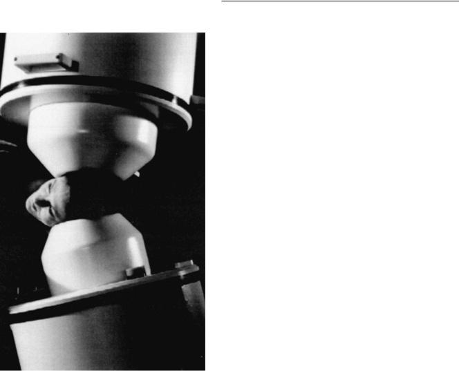
Although Cohen's original MEG recording was performed with a room temperature device, practical MEG recordings currently require the use of superconducting electronics to detect the weak Welds of the human brain. The only available detector sensitive and reliable enough to detect these Welds is the superconducting quantum interference device (SQUID). A low inductance coil (the ªsensorsº in MEG, analogous to EEG electrodes) positioned near the head is inductively coupled to a SQUID. The SQUID is cooled to 4K (or 2269°C) by submersion in a thermally shielded dewar (i.e., a specially constructed ªthermosº with a vacuum space) containing liquid helium. Some of the newest SQUIDS are capable of operating at liquid nitrogen temperature (77 K) and are known as high-temperature devices (Zhang et al., 1993a, b), but they do not currently have the sensitivities required for recording from the brain and are, therefore, not in wide use. Electronic refrigeration systems have also been tried with very limited success with SQUID systems. At present the liquid helium cryogenic systems still remain the industry standard.
Sensor geometries
The issues of noise (i.e., nonspeciWc brain signal combined with nonbrain noise) reduction and sensitivity described in the previous two sections also relate directly to the geometric conWguration of the Xux transformers, or sensors. Several diVerent conWgurations are currently in use around the world, and each has its own particular advantages and disadvantages. The simplest conWguration for magnetic Weld recordings is the magnetometer, a coil of wire typically made of niobium (Fig. 5.5a). Magnetometers are the most sensitive to magnetic Xux of the various types of sensors but as a direct consequence are also sensitive to noise from nonneural sources. As a result, gradiometer conWgurations (Fig. 5.5b±d) are often adopted for noise cancellation.
The Wrst-order axial gradiometer (Fig. 5.5b) consists of a lower coil called a pickup coil and an upper coil called a compensation coil that is wound in the opposite direction and connected in series. This system is designed so that only diVerences (gradients) between the coils will be ampliWed. Consequently, conWguration is quite sensitive to nearby Weld sources, since the high falloV rate for magnetic Welds produces a large diVerence between the two coils. It discriminates eVectively against distant (noise) sources, since they will produce a more uniform Weld at the two

Magnetoencephalography 81
(a)
(b)
(d )
(c )
Fig. 5.5. DiVerent geometric conWgurations of coils:
(a) magnetometer, (b) Wrst-order axial gradiometer, (c) secondorder axial gradiometer, (d) oV-diagonal planar gradiometer, which samples two orthogonal planar gradients (i.e., it is a twochannel device). The arrows indicate directions of winding for coils.
coils. The distance between the pickup coil and the compensation coil is called the baseline and is typically 4±5cm in most systems.
For even greater noise cancellation, second-order or higher gradiometers may be formed. The second-order conWguration shown in Fig. 5.5c essentially consists of two Wrst-order gradiometers connected in opposition, which results in even stronger rejection of distant sources. It is important to remember, however, that although distant sources are often unwanted noise (i.e., nonbrain), deeper brain sources are also increasingly discriminated against as higher-order gradiometers are formed, and this is typically seen as a necessary tradeoV in MEG recordings, particularly in noisier environments such as hospitals.
Figure 5.5d illustrates a planar, or ªWgure eightº con- Wgured, gradiometer (also called a double D, when the opposing coils are ªDº shaped rather than circular). This conWguration results in maximal signals being recorded directly over the current source, rather than to each side as
with the axial geometries discussed previously. This has great intuitive appeal when interpreting the underlying current sources of a given magnetic Weld distribution, particularly when using systems with a small number of sensors. The Neuromag Ltd. MEG systems employ this sensor conWguration in their commercial designs, while those of Canadian Thin Films (CTF) Systems and Biomagnetic Technologies Inc. (BTi) rely on various axial conWgurations. It also has a signiWcant cost advantage over axial systems, since the planar geometry lends itself well to modern thin-Wlm manufacturing techniques.
As digital signal processor technology has evolved, the use of computers to form higher-order-gradients from signals recorded with lower-order sensors has dramatically improved. In these systems, reference sensors are placed some distance from the pickup coils to measure distant source, or noise, activity. Then, either onor oZine, the data acquisition software computes the desired higherorder gradient. CTF Systems was Wrst to use this strategy in their MEG systems, where secondand third-order gradients can be computed from magnetometers or Wrstorder gradiometers. This strategy has two desirable features. First, it allows for extremely sensitive geometries to be employed in the recording while also allowing for good noise cancellation. Second, in some relatively magnetically quiet environments, MEG recordings can be performed in unshielded or poorly shielded rooms (Fig. 5.6).
Sensor arrays
Instrumentation in MEG has evolved signiWcantly since the early 1980s from single channel neuromagnetometer systems to large, high-density whole-head arrays. Several manufacturers (CTF, BTi, Neuromag Ltd.) are currently oVering a variety of systems. Figures 5.7 and 5.8 illustrate two generations of MEG systems.
Large arrays like the whole-head systems from each company oVer the considerable advantages of patient comfort, short data acquisition times, and simultaneous multisite recordings over the smaller array systems, which must be repositioned multiple times during an experiment to provide similar coverage. These systems are conWgured with approximately 150 or more sensors around a helmet designed to Wt a large number of head sizes (Fig. 5.9). Three caveats to the helmet-based systems exist. First, since they are typically designed to accommodate some percentile of adult head sizes (approximately the 98th percentile of head sizes for the BTi instrument; personal communication to author), some people will not Wt well into the helmets. This may be particularly troublesome for studies of individuals with abnormally large heads, such as those with autism
82 |
D. C. Rojas, P. D. Teale and M. L. Reite |
|
|
(a)
(b)
Fig. 5.6. Higher-order gradient formation and recording in unshielded environments. The data illustrate a 100-trial, averaged somatosensory evoked Weld (SEF) produced from stimulation of the right median nerve. Data were collected with the CTF 143channel whole-head MEG system conWgured with Wrst-order gradiometers: (a) Wrst-order averaged data (as collected); (b) synthetic third-order gradient reconstructed from the lowerorder gradiometers (noise cancellation). Note the prominence of the SEF response over the left parietal regions of the image. (Courtesy of Canadian Thin Films, Ltd.)
and fragile X disorder (Davidovitch et al., 1996; Woodhouse et al., 1996; Lainhart et al., 1997). Second, none of the current generation of helmet systems allows for complete coverage of the face, which may be important for recording some brain activities (particularly if the magnetic Weld extrema were seen over the face). Third, fetal MEG recordings would be nearly impossible on such systems, since the mother's abdomen could not Wt into the helmet (specially designed abdominal units are being developed, however). Similarly, infant and nonhuman animal recordings might be extremely diYcult because the subject's heads would be too small, leading to large distances between the head and detectors. (However, Fig. 5.10 shows how a young child can
be placed into one of the current generation instruments.) Finally, the cost of such systems is currently prohibitive to most researchers (approximately US$2.0 million with a shielded room). Taken together, the strengths and weaknesses of whole-head versus partial-coverage arrays should be considered based on the experiments envisaged and the populations of interest to a particular laboratory.
Common data analysis techniques
Signal averaging
As with EEG, the most common data analysis technique in MEG involves signal averaging to improve the SNR. When the signal is time-locked to a sensory or motor event, and several hundred trials are averaged, this produces the commonly seen evoked Weld (analog of the EEG evoked potential; see Fig. 5.11). As with EEG, signal averaging is generally regarded as the sine qua non for extracting neural signals of interest (signal) from the ongoing activity in other brain regions (noise). Other techniques for extracting these small signals from single trials have been applied to EEG data with limited success (e.g., neural network analysis, wavelets, etc.) but their promise is slowly being realized in MEG data analysis as well.
Source analysis
In MEG, as in EEG, in order to understand the relationship between the measured data and the actual neurophysiology of the brain, one must have a model of how one is produced by the other. For MEG and EEG, the most commonly applied model is the single equivalent current dipole (ECD; see also anatomic considerations, above, and Fig. 5.2). As previously described, intraneuronal current Xow in the dendritic tree of a single neuron produces a dipolar magnetic Weld distribution (at least when measured some distance away from the source). Similarly, a small region of tissue with parallel dendritic trees produces a superposition of the dipolar Welds generated by the individual neurons. Therefore, the equivalent part of the ECD model indicates that the single ECD really represents the superimposed activity of many individual dipoles.
The current dipole in the ECD model is characterized as a short length (L) of current (I), and the strength of the dipole, Q, is the product of length and current (Q5 IL, in ampere-meters). The magnetic Weld B (in SI units of tesla) produced by Q at any given point in space is described by:
|
- Q sin 3 |
|
B5 |
0 |
|
4/r 2 |
||
|
where -0 is the permeability of free space, 3 is the angle between Weld point and current, and r is the distance to the
Magnetoencephalography 83
Fig. 5.7. The BTi Magnes II system, conWgured with two 37channel, Wrst-order gradiometer units. The gantry system for the overhead unit of this particular device allows Wve degrees of freedom of movement, but the dewar can only be tilted 45° from vertical to keep the liquid helium from spilling. (Courtesy of Biomagnetic Technologies, Inc.)
point of measurement. This formula indicates that the magnetic Weld is strongest perpendicular (i.e., 35 90°) to the current Xow and diminishes with the square of the distance from the current source (r). The formula is limited in that it describes the magnetic Weld produced by the dipole alone (e.g., as if it were located in a nonconducting space). In fact, there are also extracellular currents (see section on EEG/MEG comparison) that maintain charge equilibrium in the volume. The magnetic Welds associated with these currents, in volumes that exhibit spherical or cylindrical symmetry, are tangential to the surface of the volume and,
therefore, not detected by coils with axes radial to that surface.
The calculation of the magnetic Weld strength at a given point in space is referred to as the forward solution. Just as one may calculate the forward solution for a given current, one may attempt to solve backward from the measured data to determine the current source (the so-called inverse solution, or problem). This is currently the most common data analytic technique employed for MEG data. Unfortunately, the inverse problem is ill posed; it is not possible to solve for the current distribution inside a conductor uniquely from knowledge of the magnetic (or electric) Welds outside that conductor. An inWnite number of source conWgurations can produce the same electromagnetic Weld distribution. However, knowledge of the likely origins of the measured Weld can be used to restrict the possibilities, rejecting biologically implausible solutions. Typically, the inverse solution is realized as an iterative process of changing a dipole's parameters (e.g., strength, orientation, and location) and minimizing the residual error between the forward solution (theoretical Weld) and the measured data through a leastsquares minimization or similar technique (see Figs. 5.12 and 5.13 (p. 82)). A priori hypotheses can be implemented during the Wtting process to reject unlikely source conWgurations. One exciting possibility in MEG/EEG source analysis is that fMRI, SPECT, and/or PET activation regions could be used to restrict the possible locations when such data are available in the same person under the same or similar experimental paradigm (George et al., 1995).
Although the single ECD model is superWcially quite simple, several issues make MEG source analysis complex, including, but not limited to, the model for the conducting medium (also known as the head model). In MEG source analysis, the head is usually modeled as a simple sphere with a single, homogeneously conductive compartment. In reality, of course, the head is not a perfect sphere, and there are at least three or four important layers of diVering conductivity to be considered: brain, meninges, skull, and skin. For superWcial sources, the eVects of the nonhomogeneous medium and skull shape appear to be minimal, but deeper sources, especially those in basal frontal and temporal sites, may need to be modeled in a realistically shaped head model (Hamalainen and Sarvas, 1987, 1989; Menninghaus et al., 1994). Such models are readily available, particularly if MRI data are also available for the subject. In those cases, a realistic head shape with diVerent conductivities can be constructed from boundary element modeling or other similar techniques (Menninghaus et al., 1994).
The single ECD model is often criticized as an oversimpliWcation of the true current distribution within