

Книги по МРТ КТ на английском языке / PLUM AND POSNER S DIAGNOSIS OF STUPOR AND COM-1
.pdf56 Plum and Posner’s Diagnosis of Stupor and Coma
The pupilloconstrictor muscle consists of circumferentially oriented muscle fibers that narrow the pupil when they contract, in the same manner as the drawstring of a purse. The parasympathetic neurons that supply the pupilloconstrictor muscle are located in the ciliary ganglion and in episcleral ganglion cells within the orbit. The preganglionic neurons for pupilloconstriction are located in the oculomotor complex in the brainstem (Edinger-Westphal nucleus) and they arrive in the orbit via the oculomotor or third cranial nerve. The pupilloconstrictor fibers travel in the dorsomedial quadrant of the third nerve, where they are vulnerable to compression by a number of causes (Chapter 3), often before there is clear impairment of the third nerve extraocular muscles. As a result, unilateral loss of pupilloconstrictor tone is of great diagnostic importance in patients with stupor or coma caused by supratentorial mass lesions.
Pharmacology of the Peripheral
Pupillomotor System
Because the state of the pupils is of such importance in the diagnosis of patients with coma, it is sometimes necessary to explore the origin of aberrant responses. Knowledge of the pharmacology of the pupillomotor system is essential to properly interpret the findings.82 The sympathetic preganglionic neurons in the thoracic spinal cord are cholinergic, and they act upon a nicotinic type II receptor on the sympathetic ganglion cells. The sympathetic terminals onto the pupillodilator muscle in the iris are noradrenergic, and they dilate the pupil via a beta-1 adrenergic receptor.
In the presence of a unilateral small pupil, it is possible to determine whether the cause is due to failure of the sympathetic ganglion cells or is preganglionic. In the latter case, the ganglion cells are intact, but not active. The pupil can then be dilated by instilling a few drops of 1% hydroxyamphetamine into the eye, which releases norepinephrine from surviving sympathetic terminals. Because the postsynaptic receptors have become hypersensitive due to the paucity of neurotransmitter being released, there is brisk pupillodilation after instilling the eye drops. Conversely, if the pupil is small due to loss of postganglionic neurons or receptor blockade, hydroxyam-
phetamine will have little if any effect. Postganglionic failure can be differentiated from receptor blockade (e.g., instillation of eyedrops containing a beta blocker such as are used to treat glaucoma) by introduction of 0.1% adrenaline drops, which have direct beta agonist effects. Denervated receptors are hypersensitive and there is brisk pupillary dilation, but a pupil that is small due to a beta blocker does not respond.
The pupilloconstrictor neurons in the oculomotor complex use acetylcholine, and they act on the ciliary and episcleral ganglion cells via a nicotinic II receptor. The parasympathetic ganglion cells, by contrast, activate the pupilloconstrictor muscle via a muscarinic cholinergic synapse. In the presence of a dilated pupil due to an injury to the third nerve or the postganglionic neurons, the hypersensitive receptors will constrict the pupil rapidly in response to a dilute solution of the muscarinic agonist pilocarpine (0.125%). However, if the enlarged pupil is due to atropine, even much stronger solutions of pilocarpine (up to 1.0%) will be unable to constrict the pupil.
CENTRAL PATHWAYS CONTROLLING PUPILLARY RESPONSES
It is important to understand the central pathways that regulate pupillary light responses, because dysfunction in these pathways causes the abnormal pupillary signs seen in patients with coma due to brainstem injury.
Preganglionic sympathetic neurons in the C8-T2 levels of the spinal cord, which regulate pupillodilation, receive inputs from several levels of the brain. The main input driving sympathetic pupillary tone derives from the ipsilateral hypothalamus. Neurons in the paraventricular and arcuate nuclei and in the lateral hypothalamus all innervate the upper thoracic sympathetic preganglionic neurons.83 The orexin/hypocretin neurons in the lateral hypothalamus provide a particularly intense input to this area.84 This input may be important, as the activity of the orexin neurons is greatest during wakefulness, when pupillodilation is maximal.85 The descending hypothalamic input runs through the lateral part of the pontine and medullary brainstem tegmentum, where it is vulnerable to interruption by brainstem injury.7 Electrical stimulation of the descending sympathoexcitatory tract in cats demonstrates
that it runs in a superficial position along the surface of the ventrolateral medulla, just dorsolateral to the inferior olivary nucleus.86 Experience with patients with lateral medullary infarction supports a similar localization in humans. Such patients have a central Horner’s syndrome, which includes not only miosis and ptosis, but also loss of sweating on the entire ipsilateral side of the body. Thus, the sympathoexcitatory pathway remains ipsilateral from the hypothalamus all the way to the spinal cord.
Other brainstem pathways also contribute to pupillodilation. Inputs to the C8-T2 sympathetic preganglionic column arise from a number of brainstem sites, including the Ko¨llikerFuse nucleus, A5 noradrenergic neurons, C1 adrenergic neurons, medullary raphe serotoninergic neurons, and other populations in the rostral ventrolateral medulla that have not been chemically characterized in detail.8 Ascending pain afferents from the spinal cord terminate both in these sites as well as in the periaqueductal gray matter. Brainstem sympathoexcitatory neurons can cause pupillodilation in response to painful stimuli (the ciliospinal reflex).10 They also provide ascending inhibitory inputs to the pupilloconstrictor neurons in the midbrain. As a result, lesions of the pontine tegmentum, which destroy both these ascending inhibitory inputs to the pupilloconstrictor system and the descending excitatory inputs to the pupillodilator system, cause the most severely constricted pupils seen in humans.
Preganglionic parasympathetic neurons are
located in the Edinger-Westphal nucleus in primates.87,88 This complex cell group also
contains peptidergic neurons that mainly provide descending projections to the spinal cord. In rodents and cats, most of the pupilloconstrictor neurons are located outside the Edin- ger-Westphal nucleus, and the nucleus itself mainly consists of the spinally projecting population, so that extrapolation from nonprimate species (where the anatomy and physiology of the system has been most carefully studied) is difficult.
The main input to the Edinger-Westphal nucleus of clinical interest is the afferent limb of the pupillary light reflex. The retinal ganglion cells that contribute to this pathway belong to a special class of irradiance detectors, most of which contain the photopigment me-
Examination of the Comatose Patient |
57 |
lanopsin.89 The same population of retinal ganglion cells that drives the pupillary light reflex also provides inputs to the suprachiasmatic nucleus in the circadian system, and in many cases individual ganglion cells send axonal branches to both systems. Although these ganglion cells are activated by the traditional pathways from rods and cones, they also are directly light sensitive, and as a consequence pupillary light reflexes are preserved in animals and humans with retinal degeneration who lack rods and cones (i.e., are functionally blind). This is in contrast to acute onset of blindness, in which preservation of the pupillary light reflex implies damage to the visual system beyond the optic tracts, usually at the level of the visual cortex.
The brightness-responsive retinal ganglion cells innervate the olivary pretectal nucleus. Neurons in the olivary pretectal nucleus then send their axons through the posterior commissure to the Edinger-Westphal nucleus of both sides.90 The Edinger-Westphal nucleus in humans, as in other species, lies very close to the midline, just dorsal to the main body of the oculomotor nucleus. As a result, lesions that involve the posterior commissure disrupt the light reflex pathway from both eyes, resulting in fixed, slightly large pupils.
Descending cortical inputs can cause either pupillary constriction or dilation, and can either be ipsilateral, contralateral, or bilateral.91 Sites that may produce pupillary responses are found in both the lateral and medial frontal lobes, the occipital lobe, and the temporal lobe. Unilateral pupillodilation has also been reported in patients during epileptic seizures. However, the pupillary response can be either ipsilateral or contralateral to the presumed origin of the seizures. Because so little is known about descending inputs to the pupillomotor system from the cortex and their physiologic role, it is not possible at this point to use pupillary responses during seizure activity to determine the lateralization, let alone localization, of the seizure onset. However, brief, reversible changes in pupillary size may be due to seizure activity rather than structural brainstem injury. We have also seen reversible and asymmetric changes in pupillary diameter in patients with oculomotor dysfunction due to tuberculous meningitis and with severe cases of Guillain-Barre´ syndrome that cause autonomic denervation.
58 Plum and Posner’s Diagnosis of Stupor and Coma
Localizing Value of Abnormal
Pupillary Responses in Patients
in Coma
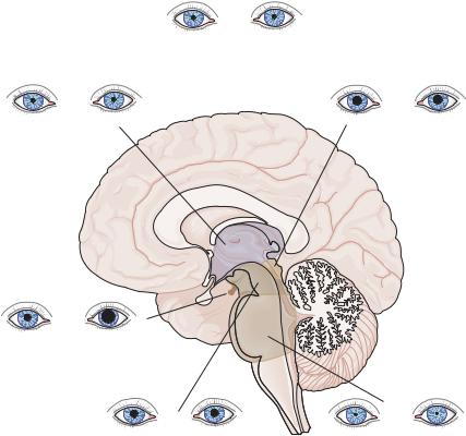
Characteristic pupillary responses are seen with lesions at specific sites in the neuraxis (Figure 2–7).
Diencephalic injuries typically result in small, reactive pupils. Bilateral, small, reactive pupils are typically seen when there is bilateral diencephalic injury or compression, but also are seen in almost all types of metabolic encephalopathy, and therefore this finding is also of limited value in identifying structural causes of coma.
A unilateral, small, reactive pupil accompanied by ipsilateral ptosis is often of great diagnostic value. If there is no associated loss of sweating in the face or the body (even after the patient is placed under a heating lamp that
causes sweating of the contralateral face), then the lesion is likely to be along the course of the internal carotid artery or in the cavernous sinus, superior orbital fissure, or the orbit itself (Raeder’s paratrigeminal syndrome, although in some cases the Horner’s syndrome is merely incomplete). If there is a sweating defect confined to the face (peripheral Horner’s syndrome), the defect must be extracranial (from the T1–2 spinal level to the carotid bifurcation). However, if the loss of sweating involves the entire side of the body (central Horner’s syndrome), it indicates a lesion involving the pathway between the hypothalamus and the spinal cord on the ipsilateral side. Although hypothalamic unilateral injury can produce this finding, lesions of the lateral brainstem tegmentum are a more common cause.
Midbrain injuries may cause a wide range of pupillary abnormalities, depending on the
Diffuse effects of drugs, metabolic encephalopathy, etc.: small, reactive
Diencephalic: |
Pretectal: |
small, reactive |
large, "fixed", hippus |
III nerve (uncall): dilated, fixed
Midbrain: |
Pons: |
midposition, fixed |
pinpoint |
Figure 2–7. Summary of changes in pupils in patients with lesions at different levels of the brain that cause coma. (From Saper, C. Brain stem modulation of sensation, movement, and consciousness. Chapter 45 in: Kandel, ER, Schwartz, JH, Jessel, TM. Principles of Neural Science. 4th ed. McGraw-Hill, New York, 2000, pp. 871–909. By permission of McGraw-Hill.)
nature of the insult. Bilateral midbrain tegmental infarction, involving the oculomotor nerves or nuclei bilaterally, results in fixed pupils, which are either large (if the descending sympathetic tracts are preserved) or midposition (if they are not). However, pupils that are fixed due to midbrain injury may dilate with the ciliospinal reflex. This response distinguishes midbrain pupils from cases of brain death. It is often thought that pupils become fixed and dilated in death, but this is only true if there is a terminal release of adrenal catecholamines. The dilated pupils found immediately after death resolve over a few hours to the midposition, as are seen in patients who are brain dead or who have midbrain infarction.
More distal injury, after the oculomotor nerve leaves the brainstem, is typically unilateral. The oculomotor nerve’s course makes it susceptible to damage by either the uncus of the temporal lobe as it herniates through the tentorial opening (see supratentorial causes of coma, page 103) or an aneurysm of the posterior communicating artery. Either of these lesions may compress the oculomotor nerve from the dorsal direction. Because the pupilloconstrictor fibers lie superficially on the dorsomedial surface of the nerve at this level,92 the first sign of impending disaster may be a unilateral enlarged and poorly reactive pupil. These conditions are discussed in detail in Chapter 3.
Pontine tegmental injury typically results in pinpoint pupils. The pupils can often be seen under magnification to respond to bright light. However, the simultaneous injury to both the descending and ascending pupillodilator pathways causes near maximal pupillary constriction.86 The most common cause is pontine hemorrhage.
Lesions involving the lateral medullary tegmentum, such as Wallenberg’s lateral medullary infarction, may cause an ipsilateral central Horner’s syndrome.
Metabolic and Pharmacologic
Causes of Abnormal
Pupillary Response
Although the foregoing discussion illustrates the importance of the pupillary light response in diagnosing structural causes of coma, it is critical to be able to distinguish structural
Examination of the Comatose Patient |
59 |
causes from metabolic and pharmacologic causes of pupillary abnormalities. Nearly any metabolic encephalopathy that causes a sleepy state may result in small, reactive pupils that are difficult to differentiate from pupillary responses caused by diencephalic injuries. However, the pupillary light reflex is one of the most resistant brain responses during metabolic encephalopathy. Hence, a comatose patient who shows other signs of midbrain depression (e.g., loss of other oculomotor responses) yet retains the pupillary light reflex is likely to have a metabolic disturbance causing the coma.
During or following seizures, one or both pupils may transiently (usually for 15 to 20 minutes, and rarely as long as an hour) be large or react poorly to light. During hypoxia or global ischemia of the brain such as during a cardiac arrest, the pupils typically become large and fixed, due to a combination of systemic catecholamine release at the onset of the ischemia or hypoxia and lack of response by the metabolically depleted brain. If resuscitation is successful, the pupils usually return to a small, reactive state. Pupils that remain enlarged and nonreactive for more than a few minutes after otherwise successful resuscitation are indicative of profound brain ischemia and a poor prognostic sign (see discussion of outcomes from hypoxic/ischemic coma in Chapter 9).
Although most drugs that impair consciousness cause small, reactive pupils, a few produce quite different responses that may help to identify the cause of the coma. Opiates, for example, typically produce pinpoint pupils that resemble those seen in pontine hemorrhage. However, administration of an opioid antagonist such as naloxone results in rapid reversal of both the pupillary abnormality and the impairment of consciousness (naloxone must be given carefully to an opioid-intoxicated patient, because if the patient is opioid dependent, the drug may precipitate acute withdrawal). Chapter 7 discusses the use of naloxone. Muscarinic cholinergic antagonist drugs that cross the blood-brain barrier, such as scopolamine, may cause a confused, delirious state, in combination with large, poorly reactive pupils. Lack of response to pilocarpine eye drops (see above) demonstrates the muscarinic blockade. Glutethimide, a sedative-hypnotic drug that was popular in the 1960s, was notorious for causing large and poorly reactive pupils. Fortunately, it is rarely used anymore.
60 Plum and Posner’s Diagnosis of Stupor and Coma
OCULOMOTOR RESPONSES
The brainstem nuclei and pathways that control eye movements lie in close association with the ascending arousal system. Hence, it is unusual for a patient with a structural cause of coma to have entirely normal eye movements, and the type of oculomotor abnormality often identifies the site of the lesion that causes coma. A key clinical tenet of the coma examination is that, with rare exception (e.g., a comatose patient with a congenital strabismus), asymmetric oculomotor function typically identifies a patient with a structural rather than metabolic cause of coma.
Functional Anatomy of the
Peripheral Oculomotor System
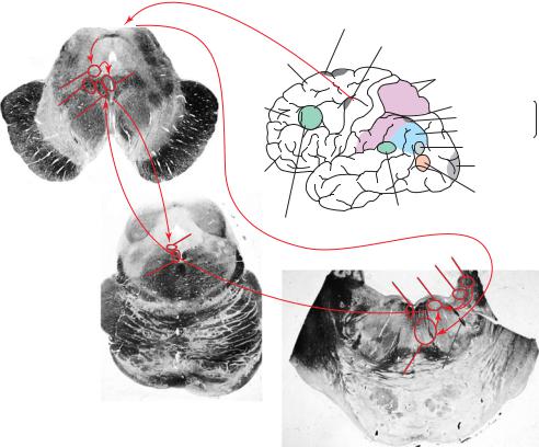
Eye movements are due to the complex and simultaneous contractions of six extraocular muscles controlling each globe. In addition, the muscles of the iris (see above), the lens accommodation system, and the eyelid receive input from some of the same central cell groups and cranial nerves. Each of these can be used to identify the cause of an ocular motor disturbance, and may shed light on the origin of coma (Figure 2–8).93
Lateral movement of the globes is caused by the lateral rectus muscle, which in turn is un-
SCol |
Supplementary Eye Field |
|
|
Frontal Eye Field |
|
||
|
|
||
|
Superior Frontal |
|
|
Illn |
Gyrus |
|
|
|
SUPERIOR PARIETAL LOBULE |
||
|
|
||
RIC |
Middle |
|
|
|
Frontal |
Supramarginal gyrus |
|
RIMLF |
Gyrus |
INFERIOR |
|
MLF |
|
Parietal Eye Field |
PARIETAL |
|
Angular gyrus |
LOBULE |
|
|
|
||
|
|
|
|
|
|
MST |
|
|
Inferior |
Striate Cortex (VI) |
|
|
|
|
|
|
Frontal Gyrus |
|
|
|
|
MT (V5) |
|
|
Vestibular Cortex |
|
|
IVn |
Dorsolateral |
|
|
Prefrontal Cortex |
|
|
|
|
|
SVn |
|
|
|
MVn |
|
MLF |
MLF |
Vln |
|
PPRF
Figure 2–8. A summary diagram showing the major pathways responsible for eye movements. The frontal eye fields (A) provide input to the superior colliculus (SCol) to program saccadic eye movements. The superior colliculus then provides input to a premotor area for causing horizontal saccades (the paramedian pontine reticular formation [PPRF]), which in turn contacts neurons in the abducens nucleus. Abducens neurons (VIn) send axons across to the opposite medial longitudinal fasciculus (MLF) and to the opposite oculomotor nucleus (IIIn) to activate medial rectus motor neurons for the opposite eye. Vertical saccades are controlled by inputs from the superior colliculus to the rostral interstitial nucleus of the MLF (RIMLF) and rostral interstitial nucleus of Cajal (RIC), which act as a premotor area to instruct the neurons in the oculomotor and trochlear (IVn) nuclei to perform a vertical saccade. Vestibular and gaze-holding inputs come to the same ocular motor nuclei from the medial (MVN) and superior (SVN) vestibular nucleus. Note the intimate relationship of these cell groups and pathways with the ascending arousal system.
der the control of the abducens or sixth cranial nerve. The superior oblique muscle and trochlear or fourth cranial nerve have more complex actions. Because the trochlear muscle loops through a pulley, or trochleus, it attaches behind the equator of the globe and pulls it forward rather than back. When the eye turns medially, the action of this muscle is to pull the eye down and in. When the eye is turned laterally, however, the action of the muscle is to intort the eye (rotate it on its axis with the top of the iris moving medially). All of the other extraocular muscles receive their innervation through the oculomotor or third cranial nerve. These include the medial rectus, whose action is to turn the eye inward; the superior rectus, which pulls the eye up and out; and the inferior rectus and oblique, which turn the eye down and out and up and in, respectively. It should be clear from the above that, whereas impairment of mediolateral movements of the eyes mainly indicates imbalance of the two cognate rectus muscles, disturbances of upward or downward movement are far more complex to work out, as they result from dysfunction of the complex set of balanced contractions of the other four muscles. This situation is reflected in the central control of these movements, as will be reviewed below.
The oculomotor nerve exits the brainstem through the medial part of the cerebral peduncle, then travels anteriorly between the superior cerebellar and posterior cerebral arteries. It passes through the tentorial opening and runs adjacent to the posterior communicating artery, where it is subject to injury by posterior communicating artery aneurysms. The nerve then runs through the cavernous sinus and superior orbital fissure to the orbit, where it divides into superior and inferior branches. The superior branch innervates the superior rectus muscle and the levator palpebrae superioris, which raises the eyelid, and the inferior branch supplies the medial and inferior rectus and inferior oblique muscles as well as the ciliary ganglion. The abducens nerve exits from the base of the pons, near the midline. This slender nerve, which is often avulsed when the brain is removed at autopsy, runs along the clivus, through the tentorial opening, into the cavernous sinus and superior orbital fissure, on its way to the lateral rectus muscle. The trochlear nerve is a crossed nerve (i.e., it consists of axons whose cell bodies are on the other side of
Examination of the Comatose Patient |
61 |
the brainstem) and it is the only cranial nerve that exits from the dorsal side of the brainstem. The axons emerge from the anterior medullary vellum just behind the inferior colliculi, then wrap around the brainstem, pass through the tentorial opening, enter the cavernous sinus, and travel through the superior orbital fissure to innervate the superior oblique muscle.
Unilateral or even bilateral abducens palsy is commonly seen as a false localizing sign in patients with increased intracranial pressure. Although the long intracranial course of the nerve is often cited as the cause of its predisposition to injury, the trochlear nerve (which is rarely injured by diffusely increased intracranial pressure) is actually longer,94 and the sharp bend of the abducens nerve as it enters the cavernous sinus may play a more decisive role. From a clinical point of view, however, it is important to remember that isolated unilateral or bilateral abducens palsy does not necessarily indicate a site of injury. The emergence of the trochlear nerve from the dorsal midbrain just behind the inferior colliculus makes it prone to injury by the tentorial edge (which runs along the adjacent superior surface of the cerebellum) in cases of severe head trauma. Thus, trochlear nerve palsy after head trauma does not necessarily represent a focal brainstem injury (although the dorsal brainstem at this level may be damaged by the same process).
The course of all three ocular motor nerves through the cavernous sinus and superior orbital fissure means that they are often damaged in combination by lesions at these sites. Thus, a lesion of all three of these nerves unilaterally indicates injury in the cavernous sinus or superior orbital fissure rather than the brainstem. Head trauma causing a blowout fracture of the orbit may trap the eye muscles, resulting in abnormalities of ocular motility unrelated to any underlying brain injury. The entrapment of the eye muscles is determined by forced duction (i.e., resistance to physically moving the globe) as described below in the examination.
Functional Anatomy of the Central
Oculomotor System
The oculomotor nuclei receive and integrate a large number of inputs that control their
62 Plum and Posner’s Diagnosis of Stupor and Coma
activity and coordinate eye muscle movement to produce normal, conjugate gaze. These afferents arise from cortical, tectal, and tegmental oculomotor systems, as well as directly from the vestibular system and vestibulocerebellum. In principle, these classes of afferents are not greatly different from the types of inputs that control alpha-motor neurons concerned with striated muscles, except the oculomotor muscles do not contain muscle spindles and hence there is no somesthetic feedback.
The oculomotor nuclei are surrounded by areas of the brainstem tegmentum containing
premotor cell groups that coordinate eye movements.93,95,96 The premotor area for regulating
lateral saccades consists of the paramedian pontine reticular formation (PPRF), which is just ventral to the abducens nucleus. The PPRF contains several different classes of neurons with bursting and pausing activities related temporally to horizontal saccades.97 Their main effect is to allow conjugate lateral saccades to the ipsilateral side of space, and when neurons in this area are inactivated by injection of local anesthetic, ipsilateral saccades are slowed or eliminated. In addition, neurons in the dorsal pontine nuclei relay smooth pursuit signals to the flocculus, and the medial vestibular nucleus and flocculus are both important for holding eccentric gaze.98 Inputs from thesesystems converge on the abducens nucleus, which contains two classes of neurons: those that directly innervate the lateral rectus muscle (motor neurons) and those that project through the medial longitudinal fasciculus (MLF) to the opposite medial rectus motor neurons in the oculomotor nucleus. Axons from these latter neurons cross the midline at the level of the abducens nucleus and ascend on the contralateral side of the brainstem to allow conjugate lateral gaze. Thus, pontine tegmental lesions typically result in the inability to move the eyes to the ipsilateral side of space (lateral gaze palsy). Similarly, the premotor area for vertical saccades and gaze holding, respectively, are found in the rostral interstitial nucleus of the MLF and rostral interstitial nucleus of Cajal, which surround the oculomotor nucleus laterally. A premotor area for vergence eye movements is found at the rostral tip of this region, near the midbrain-diencephalic junction. Unilateral lesions of the rostral interstitial nuclei typically reduce vertical saccades as well as causing torsional nystagmus.99,100 Compression of the
midbrain from the tectal surface (e.g., by a pineal tumor) causes loss of vertical eye movements, usually beginning with upgaze.
The PPRF and rostral interstitial nuclei are under the control of descending inputs from the superior colliculus. Each superior colliculus contains a map of the visual world on the contralateral side of space, and electrical stimulation of a specific point in this visual map will command a saccade to the corresponding point in space. In nonmammalian vertebrates, such as frogs, this area is called the optic tectum and is the principal site for directing eye movement; in mammals, it comes largely under the control of the cortical system for directing eye movements.
The cortical descending inputs to the ocular motor system are complex.101 The frontal eye fields (area 8) direct saccadic eye movements to explore behaviorally relevant features of the contralateral side of space. However, it would be incorrect to think of this area as a motor cortex. Unlike neurons in the primary motor cortex, which fire in relation to movements of the limbs in particular directions at particular joints, recordings from area 8 neurons in awake, behaving monkeys indicate that they do not fire during most random saccadic eye movements. However, they are engaged during tasks that require a saccade to a particular part of space only when the saccadic eye movement is part of a behavioral sequence that is rewarded. In this respect, neurons in area 8 are more similar to those in areas of the prefrontal cortex that are involved in planning movements toward the opposite side of space. Area 8 projects widely to both the superior colliculus as well as the premotor areas for vertical and lateral eye movements, and to the ocular motor nuclei themselves.102 Descending axons from area 8 mainly run through the internal medullary lamina of the thalamus to enter the region of the rostral interstitial nucleus of the MLF. They then cross the midline to descend along with the MLF to the contralateral PPRF and abducens nucleus.
In the posterior part of the hemisphere, in the ventrolateral cortex near the occipitoparietal junction, is an area of visual cortex, sometimes called area V5 or area MT, that is im-
portant in judging movement of objects in contralateral space.101,103 Cortex in this region
plays a critical role in following movements originating in that space, including movements
toward the ipsilateral space. Thus, following an object that travels from the left to the right engages the right parietal cortex (area 7) to fix attention on the object, the right area 8 to produce a saccade to pick it up, the right occipital cortex to follow the object to the right, and ultimately the left occipital cortex as well to see the object as it enters the right side of space. Thus, following moving stripes to the right, as in testing optokinetic nystagmus, engages a number of important cortical as well as brainstem pathways necessary to produce eye movements. Hence, although the test is fairly sensitive for picking up oculomotor problems at a cortical and brainstem level, the interpretation of failure of optokinetic nystagmus is a complex process.
In addition to these motor inputs, the ocular motor neurons also receive sensory inputs to guide them. Although there are no spindles in the ocular motor muscles to provide somatic sensory feedback, the ocular motor nuclei depend on two different types of sensory feedback. First, visual feedback allows the rapid correction of errors in gaze. Second, the ocular motor nuclei receive direct and relayed inputs from the vestibular system.104 Because the eyes must respond to changes in head position very quickly to stabilize the visual image on the retina, the direct vestibular input, which identifies angular or linear acceleration of the head, is integrated to providing a signal for rapid correction of eye position. The abducens nucleus is located at the same level as the vestibular complex, and it receives inputs from the medial and superior vestibular nuclei. Additional axons from these nuclei cross the midline and ascend in the contralateral MLF to reach the trochlear and oculomotor nuclei. These inputs from the vestibular system allow both horizontal and vertical eye movements (vestibulo-ocular reflexes) in response to vestibular stimulation.
Another sensory input necessary for the brain to calculate its position in space is head position and movement. Ascending somatosensory afferents, particularly from the neck muscles and vertebral joint receptors, arise from the C2–4 levels of the spinal cord. They ascend through the MLF to reach the vestibular nuclei and cerebellum, where they are integrated with vestibular sensory inputs.
The vestibulocerebellum, including the flocculus, paraflocculus, and nodulus, receives ex-
Examination of the Comatose Patient |
63 |
tensive vestibular input as well as somatosensory and visual afferents.101 The output from the flocculus ensures the accuracy of saccadic eye movements and contributes to pursuit eye movements and the ability to hold an eccentric position of gaze. The vestibulocerebellum is also critical in learning new relationships between eye movements and visual displacement (e.g., when wearing prism or magnification glasses). Lesions of the vestibulocerebellum cause ocular dysmetria (inability to perform accurate saccades), ocular flutter (rapid to-and-
fro eye movements), and opsoclonus (chaotic eye movements).105 It may be difficult to dis-
tinguish less severe cases of vestibulocerebellar function from vestibular dysfunction.
Because the MLF conveys so many classes of input from the pontine level to the midbrain, lesions of the MLF have profound effects on eye movements. After a unilateral MLF lesion, the eye ipsilateral to the lesion cannot follow the contralateral eye in conjugate lateral gaze to the other side of space (an internuclear ophthalmoplegia, a condition that occurs quite commonly in multiple sclerosis and brainstem lacunar infarcts). The abducting eye shows horizontal gaze-evoked nystagmus (slow phase toward the midline, rapid jerks laterally), while the adducting eye stops in the midline (if the lesion is complete) or fails to fully adduct (if it is partial). Bilateral injury to the MLF caudal to the oculomotor complex not only causes a bilateral internuclear ophthalmoplegia, but also prevents vertical vestibuloocular responses or pursuit. Vertical saccades, however, are implemented by the superior colliculus inputs to the rostral interstitial nucleus of Cajal, and are intact. Similarly, vergence eye movements are intact after caudal lesions of the MLF, which allows the paresis of adduction to be distinguished from a medial rectus palsy. More rostral MLF lesions, however, may also damage the closely associated preoculomotor areas for vertical or vergence eye movements.
The Ocular Motor Examination
The examination of the ocular motor system in awake, alert subjects involves testing both voluntary and reflex eye movements. In patients with stupor or coma, testing of reflex eyelid and ocular movements must suffice.99
64 Plum and Posner’s Diagnosis of Stupor and Coma
EYELIDS AND CORNEAL RESPONSES
Begin by noting the position of the eyes and eyelids at rest and observing for spontaneous eye movements. The eyelids at rest in coma, as in sleep, are maintained in a closed position by tonic contraction of the orbicularis oculi muscles. (Patients with long-term impairment of consciousness who enter a persistent vegetative state have alternating cycles of eyes opening and closing; see Chapter 9.) Next, gently raise and then release the eyelids, noting their tone. The eyelids of a comatose patient close smoothly and gradually, a movement that cannot be duplicated by an awake individual simulating unconsciousness. Absence of tone or failure to close either eyelid can indicate facial motor weakness. Blepharospasm, or strong resistance to eyelid opening and then rapid closure, is usually voluntary, suggesting that the patient is not truly comatose. However, lethargic patients with either metabolic or structural lesions may resist eye opening, as do some patients with a nondominant parietal lobe infarct. In awake patients, ptosis may result from either brainstem or hemispheric injury. In patients with unilateral forebrain infarcts, the ptosis is often ipsilateral to hemiparesis.106 In cases of brainstem injury, the ptosis may be part of a Horner’s syndrome (i.e., accompanied by pupilloconstriction), due to injury to the lateral tegmentum, or it may be due to an injury to the oculomotor complex or nerve, in which case it is typically accompanied by pupillodilation. Tonically retracted eyelids (Collier’s sign) may be found in patients with dorsal midbrain or, occasionally, pontine damage.
Spontaneous blinking usually is lost in coma as a function of the depressed level of consciousness and concomitant eye closure. However, in persistent vegetative state, it may return during cycles of eye opening (Chapter 9). Blinking in response to a loud sound or a bright light implies that the afferent sensory pathways are intact to the brainstem, but does not necessarily mean that they are active at a forebrain level. Even patients with complete destruction of the visual cortex may recover reflex blink responses to light,107 but not to threat.108 A unilateral impairment of the speed or depth of the eyelid excursion during blinking occurs in patients with ipsilateral facial paresis.
The corneal reflex can be performed by approaching the eye from the side with a wisp of cotton that is then gently applied to the sclera and pulled across it to touch the corneal surface.109 Eliciting the corneal reflex in coma may require more vigorous stimulation than in an awake subject, but it is important not to touch the cornea with any material that might scratch its delicate surface. Corneal trauma can be completely avoided by testing the corneal reflex with sterile saline. Two to three drops of sterile saline are dropped on the cornea from a height of 4 to 6 inches.109 Reflex closure of both eyelids and elevation of both eyes (Bell’s phenomenon) indicates that the reflex pathways, from the trigeminal nerve and spinal trigeminal nucleus through the lateral brainstem tegmentum to the oculomotor and facial nuclei, remain intact. However, some patients who wear contact lenses may have permanent suppression of the corneal reflex. In other patients with an acute lesion of the descending corticofacial pathways, the blink reflex may be suppressed, but Bell’s phenomenon should still occur. A structural lesion at the midbrain level may result in loss of Bell’s phenomenon, but an intact blink response. A lesion at the midpontine level may not only impair Bell’s phenomenon, but also cause the jaw to deviate to the opposite side (cornealmandibular reflex), a phenomenon that may also occur with eye blink.110
EXAMINATION OF OCULAR
MOTILITY
Hold the eyelids gently in an open position to observe eye position and movements in a comatose patient. A small flashlight or bright ophthalmoscope held about 50 cm from the face and shined toward the eyes of the patient should reflect off the same point in the cornea of each eye if the gaze is conjugate. Most patients with impaired consciousness demonstrate a slight exophoria. If it is possible to obtain a history, ask about eye movements, as a congenital strabismus may be misinterpreted as dysconjugate eye movements due to a brainstem lesion. Observe for a few moments for spontaneous eye movements. Slowly roving eye movements are typical of metabolic encephalopathy, and if conjugate, they imply an intact ocular motor system.
The vestibulo-ocular responses are then tested by rotating the patient’s head (oculocephalic reflexes).99 In patients who may have suffered trauma, it is important first to rule out the possibility of a fracture or dislocation of the cervical spine; until this is done, it may be necessary to skip ahead to caloric testing (see below). The head is rotated first in a lateral direction to either side while holding the eyelids open. This can be done by grasping the head on either side with both hands and using the thumbs to reach across to the eyelids and hold them open. The head movements should be brisk, and when the head position is held at each extreme for a few seconds, the eyes should gradually come back to midposition. Moving the head back to the opposite side then produces a maximal stimulus. The eye movements should be smooth and conjugate. The head is then rotated in a vertical plane (as in head nodding) and the eyes are observed for vertical conjugate movement. During downward head movement, the eyelids may also open (the doll’s head phenomenon).111
The normal response generated by the vestibular input to the ocular motor system is for the eyes to rotate counter to the direction of the examiner’s movement (i.e., turning the head to the right should cause the eyes to deviate to the left). In an awake patient, the voluntary control of gaze overcomes this reflex response. However, in patients with impaired consciousness, the oculocephalic reflex should predominate. This response is often colloquially called the doll’s eye response,111 and normal responses in both horizontal and vertical directions imply intact brainstem pathways from the vestibular nuclei through the lower pontine tegmentum and thence the upper pontine and midbrain paramedian tegmentum (i.e., along the course of the MLF; see below). There may also be a small contribution from proprioceptive afferents from the neck,112 which also travel through the medial longitudinal fasciculus. Because these pathways overlap extensively with the ascending arousal system (see Figure 2–8), it is quite unusual for patients with structural causes of coma to have a normal oculocephalic examination. In contrast, patients with metabolic encephalopathy, particularly due to hepatic failure, may have exaggerated or very brisk oculocephalic responses.
Examination of the Comatose Patient |
65 |
Eye movements in patients who are deeply comatose may respond sluggishly or not at all to oculocephalic stimulation. In such cases, more intense vestibular stimulation may be obtained by testing caloric vestibulo-ocular responses. With appropriate equipment, vestibulo-ocular monitoring can be done using galvanic stimulation and video-oculography.113 However, at the bedside, caloric stimuli and visual inspection are generally used (see Figure 2–9). The ear canal is first examined and, if necessary, cerumen is removed to allow clear visualization that the tympanic membrane is intact. The head of the bed is then raised to about 30 degrees to bring the horizontal semicircular canal into a vertical position so that the response is maximal. If the patient is merely sleepy, the canal may be irrigated with cool water (158C to 208C); this usually induces a brisk response and may occasionally cause nausea and vomiting. Fortunately, in practice, it is rarely necessary to use caloric stimulation in such patients. If the patient is deeply comatose, a maximal stimulus is obtained by using ice water. A large (50 mL) syringe is used, attached to a plastic IV catheter, which is gently advanced until it is near the tympanic membrane. An emesis basin can be placed below the ear, seated on an absorbent pad, to catch the effluent. The ice water is infused at a rate of about 10 mL/minute for 5 minutes, or until a response is obtained. After a response is obtained, it is necessary to wait at least 5 minutes for the response to dissipate before testing the opposite ear. To test vertical eye movements, both external auditory canals are irrigated simultaneously with cold water (causing the eyes to deviate downward) or warm water (causing upward deviation).
The cold water induces a downward convection current, away from the ampulla, in the endolymph within the horizontal semicircular canal. The effect of the current upon the hair cells in the ampulla is to reduce tonic discharge of the vestibular neurons. Because the vestibular neurons associated with the horizontal canal fire fastest when the head is turning toward that side (and thus push the eyes to the opposite side), the result of cold water stimulation is to produce a stimulus as if the head were turning to the opposite side, thus activating the ipsilateral lateral rectus and contralateral medial rectus muscles to drive the