

Книги по МРТ КТ на английском языке / PLUM AND POSNER S DIAGNOSIS OF STUPOR AND COM-1
.pdf36 Plum and Posner’s Diagnosis of Stupor and Coma
47.Rasmussen K, Jacobs BL. Single unit activity of locus coeruleus neurons in the freely moving cat. II. Conditioning and pharmacologic studies. Brain Res 371, 335–344, 1986.
48.Steininger TL, Alam MN, Gong H, et al. Sleepwaking discharge of neurons in the posterior lateral hypothalamus of the albino rat. Brain Res 840, 138– 147, 1999.
49.Wu MF, John J, Boehmer LN, et al. Activity of dorsal raphe cells across the sleep-waking cycle and during cataplexy in narcoleptic dogs. J Physiol 554, 202–215, 2004.
50.Kolta A, Reader TA. Modulatory effects of catecholamines on neurons of the rat visual cortex: single-cell iontophoretic studies. Can J Physiol Pharmacol 67, 615–623, 1989.
51.Sato H, Fox K, Daw NW. Effect of electrical stimulation of locus coeruleus on the activity of neurons in the cat visual cortex. J Neurophysiol 62, 946–958, 1989.
52.Bassant MH, Ennouri K, Lamour Y. Effects of iontophoretically applied monoamines on somatosensory cortical neurons of unanesthetized rats. Neuroscience 39, 431–439, 1990.
53.Lu J, Sherman D, Devor M, et al. A putative flip-flop switch for control of REM sleep. Nature 441, 589– 594, 2006.
54.Saper CB. Diffuse cortical projection systems: anatomical organization and role in cortical function. In: F. Plum, ed. Handbook of Physiology. The Nervous System. V. Bethesda, Md.: American Physiological Society, pp 168–210, 1987.
55.WelchMJ, Meltzer EO, Simons FE. H1-antihistamines and the central nervous system. Clin Allergy Immunol 17, 337–388, 2002.
56.Huang ZL, Qu WM, Li WD, et al. Arousal effect of orexin A depends on activation of the histaminergic system. Proc Natl Acad Sci 98, 9965–9970, 2001.
57.Parmentier R, Ohtsu H, Djebbara-Hannas Z, et al. Anatomical, physiological, and pharmacological characteristics of histidine decarboxylase knock-out mice: evidence for the role of brain histamine in behavioral and sleep-wake control. J Neurosci 22, 7695–7711, 2002.
58.Peyron C, Tighe DK, van den Pol AN, et al. Neurons containing hypocretin (orexin) project to multiple neuronal systems. J Neurosci 18, 9996–10015, 1998.
59.Bittencourt JC, Frigo L, Rissman RA, et al. The distribution of melanin-concentrating hormone in the monkey brain (Cebus apella). Brain Res 804, 140–143, 1998.
60.Bittencourt JC, Presse F, Arias C, et al. The melaninconcentrating hormone system of the rat brain: an immunoand hybridization histochemical characterization. J Comp Neurol 319, 218–245, 1992.
61.Lin CS, Nicolelis MA, Schneider JS, et al. A major direct GABAergic pathway from zona incerta to neocortex. Science 248, 1553–1556, 1990.
62.Lee MG, Hassani OK, Jones BE. Discharge of identified orexin/hypocretin neurons across the sleepwaking cycle. J Neurosci 25, 6716–6720, 2005.
63.Mileykovskiy BY, Kiyashchenko LI, Siegel JM. Behavioral correlates of activity in identified hypocretin/ orexin neurons. Neuron 46, 787–798, 2005.
64.Verret L, Goutagny R, Fort P, et al. A role of melanin-concentrating hormone producing neurons
in the central regulation of paradoxical sleep. BMC Neurosci 4, 19, 2003.
65.Alam MN, Gong H, Alam T, et al. Sleep-waking discharge patterns of neurons recorded in the rat perifornical lateral hypothalamic area. J Physiol 538, 619– 631, 2002.
66.Gelineau JBE. De la narcolepsie. Gaz Hop (Paris) 53, 626–637, 1880.
67.Sakurai T, Amemiya A, Ishii M, et al. Orexins and orexin receptors: a family of hypothalamic neuropeptides and G protein-coupled receptors that regulate feeding behavior. Cell 92, 573–585, 1998.
68.de Lecea L, Kilduff TS, Peyron C, et al. The hypocretins: hypothalamus-specific peptides with neuroexcitatory activity. Proc Natl Acad Sci 95, 322–327, 1998.
69.Willie JT, Chemelli RM, Sinton CMYM. To eat or to sleep? Orexin in the regulation of feeding and wakefulness. Annu Rev Neurosci 24, 429–458, 2001.
70.Chimelli RM, Willie JT, Sinton CM, et al. Narcolepsy in orexin knockout mice: molecular genetics of sleep regulation. Cell 98, 437–451, 1999.
71.Lin L, Faraco J, Li R, et al. The sleep disorder canine narcolepsy is caused by a mutation in the hypocretin (orexin) receptor 2 gene. Cell 98, 365–376, 1999.
72.Peyron C, Faraco J, Rogers W, et al. A mutation in a case of early onset narcolepsy and a generalized absence of hypocretin peptides in human narcoleptic brains. Nat Med 6, 991–997, 2000.
73.Ripley B, Overeem S, Fujiki N, et al. CSF hypocretin/ orexin levels in narcolepsy and other neurological conditions. Neurology 57, 2253–2258, 2001.
74.Thannickal TC, Moore RY, Nienhuis R, et al. Reduced number of hypocretin neurons in human narcolepsy. Neuron 27, 469–474, 2000.
75.Marcus JN, Aschkenasi CJ, Lee CE, et al. Differential expression of orexin receptors 1 and 2 in the rat brain. J Comp Neurol 435, 6–25, 2001.
76.Saper CB. Organization of cerebral cortical afferent systems in the rat. II. Magnocellular basal nucleus. J Comp Neurol 222, 313–342, 1984.
77.Zaborsky L, Brownstein MJ, Palkovits M. Ascending projections to the hypothalamus and limbic nuclei from the dorsolateral pontine tegmentum: a biochemical and electron microscopic study. Acta Morphol Acad Sci Hung 25, 175–188, 1977.
78.Zaborsky L, Cullinan WE, Luine VN. Catecholami- nergic-cholinergic interaction in the basal forebrain. Prog Brain Res 98, 31–49, 1993.
79.Lee MG, Hassani OK, Alonso A, et al. Cholinergic basal forebrain neurons burst with theta during waking and paradoxical sleep. J Neurosci 25, 4365– 4369, 2005.
80.Richardson RT, DeLong MR. Nucleus basalis of Meynert neuronal activity during a delayed response task in monkey. Brain Res 399, 364–368, 1986.
81.Richardson RT, DeLong MR. Context-dependent responses of primate nucleus basalis neuron in a go/ no-go-go task. J Neurol Sci 10, 2528–2540, 1990.
82.Sherin JE, Shiromani PJ, McCarley RW, et al. Activation of ventrolateral preoptic neurons during sleep. Science 271, 216–219, 1996.
83.Sherin JE, Elmquist JK, Torrealba F, et al. Innervation of histaminergic tuberomammilary neurons by GABAergic and alaninergic neurons in the ven-
Pathophysiology of Signs and Symptoms of Coma |
37 |
trolateral preoptic nucleus of the rat. J Neurosci 18, 4705–4721, 1998.
84.Saper CB, Chou TC, Scammell TE. The sleep switch: hypothalamic control of sleep and wakefulness. Trends Neurosci 24, 726–731, 2001.
85.Gaus SE, Strecker RE, Tate BA, et al. Ventrolateral preoptic nucleus contains sleep-active, galaninergic neurons in multiple mammalian species. Neuroscience 115, 285–294, 2002.
86.Sallanon M, Denoyer M, Kitahama K, et al. Longlasting insomnia induced by preoptic neuron lesions and its transient reversal by muscimol injection into the posterior hypothalamus in the cat. Neuroscience 32, 669–683, 1989.
87.Lu J, Greco MA, Shiromani P, et al. Effect of lesions of the ventrolateral preoptic nucleus on NREM and REM sleep. J Neurosci 20, 3820–3842, 2000.
88.Lu J, Bjorkum AA, Xu M, et al. Selective activation of the extended ventrolateral preoptic nucleus during rapid eye movement sleep. J Neurosci 22, 4568– 4576, 2002.
89.Hara J, Beuckmann CT, Nambu T, et al. Genetic ablation of orexin neurons in mice results in narcolepsy, hypophagia, and obesity. Neuron 30, 345–354, 2001.
90.Lydic R, Douglas CL, Baghdoyan HA. Microinjection of neostigmine into the pontine reticular formation of C57BL/6J mouse enhances rapid eye movement sleep and depresses breathing. Sleep 25, 835–841, 2002.
91.Lorente de No R. Cerebral cortex: architecture, intracortical connections, motor projections. In: JF Fulton, ed. Physiology of the Nervous System. New York: Oxford University Press, pp 291–340, 1938.
92.Hubel DH, Wiesel TN. Shape and arrangement of columns in cat’s striate cortex. J Physiol 165, 559– 568, 1963.
93.McCasland JS, Woolsey TA. High-resolution 2- deoxyglucose mapping of functional cortical columns in mouse barrel cortex. J Comp Neurol 278, 555– 569, 1988.
94.Gilbert CD, Wiesel TN. Columnar specificity of intrinsic horizontal and corticocortical connections in cat visual cortex. J Neurosci 9, 2432–2442, 1989.
95.Hubel DH, Wiesel TN. The period of susceptibility to the physiological effects of unilateral eye closure in kittens. J Physiol 206, 419–436, 1970.
96.Frank MG, Issa NP, Stryker MP. Sleep enhances plasticity in the developing visual cortex. Neuron 30, 275– 287, 2001.
97.Hardcastle VG. Consciousness and the neurobiology of perceptual binding. Semin Neurol 17, 163–170, 1997.
98.Revonsuo A. Binding and the phenomenal unity of consciousness. Conscious Cogn 8, 173–185, 1999.
99.Nishikawa T, Okuda J, Mizuta I, et al. Conflict of intentions due to callosal disconnection. J Neurol Neurosurg Psychiatry 71, 462–471, 2001.
100.Alexander GE, DeLong MR, Strick PL. Parallel organization of functionally segregated circuits linking basal ganglia and cortex. Annu Rev Neurosci 9, 357– 381, 1986.
101.Alexander GE, Crutcher MD, DeLong MR. Basal ganglia-thalamocortical circuits: parallel substrates for motor,oculomotor,‘‘prefrontal’’and‘‘limbic’’functions. Prog Brain Res 119–146, 1990.
102.Wyrtzes LM, Chatrian GE, Shaw CM, et al. Acute failure of forebrain with sparing of brain-stem function. Electroencephalographic, multimodality evokedpotential, and pathologic findings. Arch Neurol 46, 93–97, 1989.
103.van der Knaap MS, Smit LS, Nauta JJ, et al. Cortical laminar abnormalities—occurrence and clinical significance. Neuropediatrics 24, 143–148, 1993.
104.Adams JH, Graham DI, Jennett B. The neuropathology of the vegetative state after an acute brain insult. Brain 123, 1327–1338, 2000.
105.Caplan LR. ‘‘Top of the basilar’’ syndrome. Neurology 30, 72–79, 1980.
106.Wechler B, Dell’Isola B, Vidailhet M, et al. MRI in 31 patients with Behcet’s disease and neurological involvement: prospective study with clinical correlation. J Neurol Neurosurg Psychiatry 56, 793–798, 1993.
107.Park-Matsumoto YC, Ogawa K, Tazawa T, et al. Mutism developing after bilateral thalamo-capsular lesions by neuro-Behcet disease. Acta Neurol Scand 91, 297–301, 1995.
108.Kinney HC, Korein J, Panigrahy A, et al. Neuropathological findings in the brain of Karen Ann Quinlan. The role of the thalamus in the persistent vegetative state. N Engl J Med 330 (21), 1469–1475, 1994.
109.Reeves AG, Plum F. Hyperphagia, rage, and demential accompanying a ventromedial hypothalamic neoplasm. Arch Neurol 20, 616–624, 1969.
110.Parvizi J, Damasio AR. Neuroanatomical correlates of brainstem coma. Brain 126, 1524–1536, 2003.

Chapter 2
Examination of the
Comatose Patient
OVERVIEW
HISTORY
GENERAL PHYSICAL EXAMINATION
LEVEL OF CONSCIOUSNESS
ABC: AIRWAY, BREATHING,
CIRCULATION
Circulation
Respiration
PUPILLARY RESPONSES
Examine the Pupils and Their Responses
Pathophysiology of Pupillary Responses:
Peripheral Anatomy of the Pupillomotor
System
Pharmacology of the Peripheral
Pupillomotor System
Localizing Value of Abnormal Pupillary
Responses in Patients in Coma
Metabolic and Pharmacologic Causes
of Abnormal Pupillary Response
OCULOMOTOR RESPONSES
Functional Anatomy of the Peripheral
Oculomotor System
OVERVIEW
Coma, indeed any alteration of consciousness, is a medical emergency. The physician encountering such a patient must begin examination
Functional Anatomy of the Central
Oculomotor System
The Ocular Motor Examination
Interpretation of Abnormal
Ocular Movements
MOTOR RESPONSES
Motor Tone
Motor Reflexes
Motor Responses
FALSE LOCALIZING SIGNS IN PATIENTS
WITH METABOLIC COMA
Respiratory Responses
Pupillary Responses
Ocular Motor Responses
Motor Responses
MAJOR LABORATORY DIAGNOSTIC AIDS
Blood and Urine Testing
Computed Tomography Imaging
and Angiography
Magnetic Resonance Imaging
and Angiography
Magnetic Resonance Spectroscopy
Neurosonography
Lumbar Puncture
Electroencephalography and
Evoked Potentials
and treatment simultaneously. The examination must be thorough, but brief. The examination begins by informally assessing the patient’s level of consciousness. First, the physician addresses the patient verbally. If the patient does
38

not respond to the physician’s voice, the physician may speak more loudly or shake the patient. When this fails to produce a response, the physician begins a more formal coma evaluation.
The examiner must systematically assess the arousal pathways. To determine if there is a structural lesion involving those pathways, it is necessary also to examine the function of brainstem sensory and motor pathways that are adjacent to the arousal system. In particular, because the oculomotor circuitry enfolds and surrounds most of the arousal system, this part of the examination is particularly informative. Fortunately, the examination of the comatose patient can usually be accomplished very quickly because the patient has such a limited range of responses.However,theexaminermustbecome conversant with the meaning of the signs elicited in that examination, so that decisions that may save the patient’s life can then be made quickly and accurately.
The evaluation of the patient with a reduced level of consciousness, like that of any patient, requires a history (to the extent possible), physical examination, and laboratory evaluation. These are considered, in turn, in this chapter. However, as soon as it is determined that a patient has a depressed level of consciousness, the next step is to ensure that the patient’s brain is receiving adequate blood and oxygen.
The emergency treatment of the comatose patient is detailed in Chapter 7. The physiology and pathophysiology of the cerebral circulation and of respiration are considered in the paragraphs below.
HISTORY
In patients with nervous system dysfunction, the history is the most important part of the examination (Table 2–1). Of course, patients with coma or diminished states of consciousness by definition are not able to give a history. Thus, the history must be obtained if possible from relatives, friends, or the individuals, usually the emergency medical personnel, who brought the patient to the hospital.
The onset of coma is often important. In a previously healthy, young patient, the sudden onset of coma may be due to drug poisoning, subarachnoid hemorrhage, or head trauma; in the elderly, sudden coma is more likely caused
Examination of the Comatose Patient |
39 |
Table 2–1 Examination of the
Comatose Patient
History ( from Relatives, Friends, or Attendants)
Onset of coma (abrupt, gradual)
Recent complaints (e.g., headache, depression, focal weakness, vertigo)
Recent injury
Previous medical illnesses (e.g., diabetes, renal failure, heart disease)
Previous psychiatric history
Access to drugs (sedatives, psychotropic drugs)
General Physical Examination
Vital signs Evidence of trauma
Evidence of acute or chronic systemic illness Evidence of drug ingestion (needle marks,
alcohol on breath)
Nuchal rigidity (assuming that cervical trauma has been excluded)
Neurologic Examination
Verbal responses Eye opening Optic fundi Pupillary reactions
Spontaneous eye movements Oculocephalic responses (assuming cervical
trauma has been excluded) Oculovestibular responses Corneal responses Respiratory pattern
Motor responses Deep tendon reflexes Skeletal muscle tone
by cerebral hemorrhage or infarction. Most patients with lesions compressing the brain either have a clear history of trauma (e.g., epidural hematoma; see Chapter 4) or a more gradual rather than abrupt impairment of consciousness. Gradual onset is also true of most patients with metabolic disorders (see Chapter 5).
The examiner should inquire about previous medical symptoms or illnesses or any recent trauma. A history of headache of recent onset points to a compressive lesion, whereas the history of depression or psychiatric disease may suggest drug intoxication. Patients with known diabetes, renal failure, heart disease, or other chronic medical illness are more likely to be suffering from metabolic disorders or perhaps brainstem infarction. A history of premonitory signs, including focal weakness such as dragging
40 Plum and Posner’s Diagnosis of Stupor and Coma
of the leg or complaints of unilateral sensory symptoms or diplopia, suggests a cerebral or brainstem mass lesion.
GENERAL PHYSICAL
EXAMINATION
The general physical examination is an important source of clues as to the cause of unconsciousness. After stabilizing the patient (Chapter 7), one should search for signs of head trauma. Bilateral symmetric black eyes suggest basal skull fracture, as does blood behind the tympanic membrane or under the skin overlying the mastoid bone (Battle’s sign). Examine the neck with care; if there is a possibility of trauma, the neck should be immobilized until cervical spine instability has been excluded by imaging. Resistance to neck flexion in the presence of easy lateral movement suggests meningeal inflammation such as meningitis or subarachnoid hemorrhage. Flexion of the legs upon flexing the neck (Brudzinski’s sign) confirms meningismus. Examination of the skin is also useful. Needle marks suggest drug ingestion. Petechiae may suggest meningitis or intravascular coagulation. Pressure sores or bullae indicate that the patient has been unconscious and lying in a single position for an extended period of time, and are especially frequent in patients with barbiturate overdosage.1
LEVEL OF CONSCIOUSNESS
After conducting the brief history and examination as outlined above and stabilizing the patients’ vital functions, the examiner should
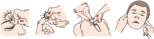
conduct a formal coma evaluation. In assessing the level of consciousness of the patient, it is necessary to determine the intensity of stimulation necessary to arouse a response and the quality of the response that is achieved. When the patient does not respond to voice or vigorous shaking, the examiner next provides a source of pain to arouse the patient. Several methods for providing a sufficiently painful stimulus to arouse the patient without causing tissue damage are illustrated in Figure 2–1. It is best to begin with a modest, lateralized stimulus, such as compression of the nail beds, the supraorbital ridge, or the temporomandibular joint. These give information about the lateralization of motor response (see below), but must be repeated on each side in case there is a focal lesion of the pain pathways on one side of the brain or spinal cord. If there is no response to the stimulus, a more vigorous midline stimulus may be given by the sternal rub. By vigorously pressing the examiner’s knuckles into the patient’s sternum and rubbing up and down the chest, it is possible to create a sufficiently painful stimulus to arouse any subject who is not deeply comatose.
The response of the patient is noted and graded. The types of motor responses seen are considered in the section on motor responses (page 73). However, the level of response is important to the initial consideration of the depth of impairment of consciousness. In descending order of arousability, a sleepy patient who responds to being addressed verbally or light shaking, or one who responds verbally to more intense mechanical stimulation, is said to be lethargic or obtunded. A patient whose best response to deep pain is to attempt to push the examiner’s arm away is considered to be stuporous, with localizing responses. Patients who
A B C D
Figure 2–1. Methods for attempting to elicit responses from unconscious patients. Noxious stimuli can be delivered with minimal trauma to the supraorbital ridge (A), the nail beds or the fingers or toes (B), the sternum (C) or the temporomandibular joints (D).

Box 2–1 Coma Scales
A number of different scales have been devised for scoring patients with coma. The value of these is in providing a simple estimate of the prognosis for different groups of patients. Obviously, this is related as much to the cause of the coma (when known) as to the current status of the examination. Teasdale and Jennett’s Glasgow Coma Scale (GCS),2 devised to categorize patients with head trauma, is reproduced below. Unfortunately, when used by emergency room physicians, interrater agreement is only moderate.3 Two simple scales, ACDU (alert, confused, drowsy, unresponsive) and AVPU (alert, response to voice, response to pain, unresponsive)4 are about as accurate as the GCS and much easier to use.4 The ACDU scale appears better at identifying early deterioration in level of consciousness. A recently validated coma scale, the FOUR scale (full outline of unresponsiveness), provides more neurologic detail than the GCS. However, no scale is adequate for all patients; hence, the best policy in recording the results of the coma examination is simply to describe the findings.
Nevertheless, the GCS is widely used, and still is probably the best for most trauma patients.5 It is useful to obtain GCS scores, which can be compared against large databases to evaluate prognosis for specific etiologies of coma (see Chapter 9).
FOUR Score (Full Outline of Unresponsiveness)109
Eye Response
4 |
¼ eyelids open or opened, tracking, or blinking to command |
3 |
¼ eyelids open but not tracking |
2 |
¼ eyelids closed but open to loud voice |
1 |
¼ eyelids closed but open to pain |
0 |
¼ eyelids remain closed with pain |
Motor Response |
|
4 |
¼ thumbs-up, fist, or peace sign |
3 |
¼ localizing to pain |
2 |
¼ flexion response to pain |
1 |
¼ extension response to pain |
0 |
¼ no response to pain or generalized myoclonus status |
Brainstem Reflexes |
|
4 |
¼ pupil and corneal reflexes present |
3 |
¼ one pupil wide and fixed |
2 |
¼ pupil or corneal reflexes absent |
1 |
¼ pupil and corneal reflexes absent |
0 |
¼ absent pupil, corneal, and cough reflex |
Respiration |
|
4 |
¼ not intubated, regular breathing pattern |
3 |
¼ not intubated, Cheyne-Stokes breathing pattern |
2 |
¼ not intubated, irregular breathing |
1 |
¼ breathes above ventilator rate |
0 |
¼ breathes at ventilator rate or apnea |
(continued)
41

42 Plum and Posner’s Diagnosis of Stupor and Coma
Box 2–1 Coma Scales (cont.)
Glasgow Coma Scale
Eye Response
4 ¼ eyes open spontaneously
3 ¼ eye opening to verbal command
2 ¼ eye opening to pain
1 ¼ no eye opening
Motor Response
6 ¼ obeys commands
5 ¼ localizing pain
4 ¼ withdrawal from pain
3 ¼ flexion response to pain
2 ¼ extension response to pain
1 ¼ no motor response
Verbal Response
5 ¼ oriented
4 ¼ confused
3 ¼ inappropriate words
2 ¼ incomprehensible sounds
1 ¼ no verbal response
A GCS score of 13 or higher indicates mild brain injury, 9 to 12 moderate brain injury, and 8 or less severe brain injury.
AVPU |
ACDU |
Is the patient |
Is the patient |
Alert and oriented? |
Alert and oriented? |
Responding to voice? |
Confused? |
Responding to pain? |
Drowsy? |
Unresponsive? |
Unresponsive? |
make only nonspecific motor responses (wincing, restlessness, withdrawal reflexes) without a directed attempt to defend against the stimulus are considered to have a nonlocalizing response and are comatose. Patients who fail to respond at all are in the deepest stage of coma.
This rough grading system, from verbal responsiveness, to localizing responses, to nonlocalizing responses, to no response, is all that is needed for an initial assessment of the depth of unresponsiveness that can be used to follow the progress of the patient. If the initial evaluation of the level of consciousness demonstrates impairment, it is essential to progress through the next steps of the coma examina-
tion as rapidly as possible to safeguard that patient’s life. More elaborate coma scales are described in Box 2–1, but many of these depend upon the results of later stages in the examination, and it is never justified to delay attending to the basics of airway, breathing, and circulation while performing a more elaborate scoring evaluation.
ABC: AIRWAY, BREATHING, CIRCULATION
It is critical to ensure that the patient’s airway is maintained, that he or she is breathing ad-
equately, and that there is sufficient arterial perfusion pressure. The first goal must be to correct any of these conditions if they are found inadequate (Chapter 7). In addition, blood pressure, heart rate, and respiration may provide valuable clues to the cause of coma.
Circulation
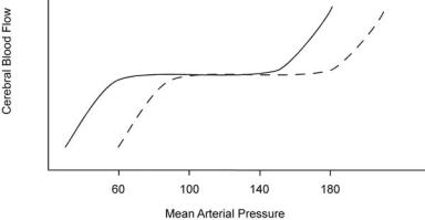
It is critical first to ensure that the brain is receiving adequate blood flow. Cerebral perfusion pressure is the systemic blood pressure minus the intracranial pressure. The physician can measure blood pressure but in the initial examination can only estimate intracranial pressure. Over a wide range of blood pressures, cerebral perfusion remains stable because the brain autoregulates its blood flow by mechanisms described in the paragraphs below and illustrated in Figure 2–2. If the blood pressure falls too low or becomes too high, autoregulation fails and cerebral perfusion follows perfusion pressure passively; that is, it falls as the blood pressure falls and rises as the blood pressure rises. In this situation, both too low (ischemia) and too high (hypertensive encephalopathy; see Chapter 5) a blood pressure can damage the brain. To ensure adequate
Examination of the Comatose Patient |
43 |
brain perfusion, the physician should attempt to maintain the blood pressure at a level normal for the individual patient. For example, a patient with chronic hypertension autoregulates at a higher level than a normotensive patient. Lowering the blood pressure to a ‘‘normal level’’ may deprive the brain of an adequate blood supply (see Figure 2–2). Conversely, the cerebral blood flow (CBF) in children and pregnant women, who normally run low blood pressures, is regulated at lower levels and may develop excessive perfusion if the blood pressure is raised (e.g., pre-eclampsia).
The perfusion pressure of the brain may be influenced by the position of the head. In a normal individual, as the head is raised, the systemic arterial pressure is maintained by blood pressure reflexes. At the same time, the arterial perfusion pressure to the head is reduced by the distance the head is raised above the heart, but the intracranial pressure is also reduced because of the improved venous and cerebrospinal fluid (CSF) drainage. The net effect is that there is very little change in brain perfusion pressure or CBF. On the other hand, in a patient with stenosis of a carotid or vertebral artery, the perfusion pressure for that vessel may be much lower than systemic arterial pressure. If the head of the bed is raised,
Figure 2–2. Cerebral autoregulation in hypertension. Schematic representation of autoregulation of cerebral blood flow (CBF) in normotensive (solid line) and hypertensive (dashed line) subjects. In both groups, within a range of about 100 mm Hg, increases or decreases in mean arterial pressure are associated with maintenance of CBF due to appropriate changes in arteriolar resistance. Changes in pressure outside this range are eventually associated with loss of autoregulation, leading to a reduction (with hypotension) or an elevation (with marked hypertension) in CBF. Note that hypertensive encephalopathy (increased blood flow with pressures exceeding the autoregulatory range) may occur with a mean arterial pressure below 200 mm Hg in the normotensive individual, but may require a much higher mean arterial pressure in patients who have sustained hypertension. Conversely, lowering blood pressure to the ‘‘normal range’’ of a mean arterial pressure of 80 mm Hg (equivalent to 120/60) may produce a clinically significant fall in CBF, particularly if there is a preexisting cerebrovascular stenosis.
44 Plum and Posner’s Diagnosis of Stupor and Coma
perfusion pressure may fall below the threshold for autoregulation, and blood flow may be diminished below the level needed to support neurologic function. Such patients may show improvement in neurologic function when the head of the bed is flat. Conversely, in cases of head trauma where there is increased intracranial pressure, it may be important to raise the head of the bed 15 to 30 degrees to improve venous drainage to maximize cerebral perfusion pressure.6 Similarly, it is necessary to remove tight neckwear and ensure that a cervical spine collar is not applied too tightly to a victim of head injury to avoid diminishing venous outflow from the brain.
In a patient with impaired consciousness, the blood pressure can give important clues to the level of the nervous system that has been damaged. Damage to the descending sympathetic pathways that support blood pressure may result in a fall to levels seen after spinal transaction (mean arterial pressure about 60 to 70 mm Hg). Blood pressure is supported by a descending sympathoexcitatory pathway from the rostral ventrolateral medulla to the spinal cord, and so damage along the course of this pathway can result in spinal levels of blood pressure. The hypothalamus in turn provides a descending sympathoexcitatory input to the medulla and the spinal cord.7,8 As a consequence, bilateral diencephalic lesions result in a fall in sympathetic tone, including meiotic pupils (see below), decreased sweating responses, and a generally low level of arterial pressure.9
However, persistent hypotension below these levels in a comatose patient is almost never caused by an acute neurologic injury. One of the most common mistakes seen in evaluation of a comatose patient with a mean arterial pressure below 60 mm Hg is the assumption that a neurologic event may have caused the hypotension. This is almost never the case. A mean arterial pressure at or above 60 mm Hg is generally sufficient in a supine patient to support cerebral and systemic function. On the other hand, acute hypotension, due to cardiogenic or vasomotor shock, is a common cause of loss of consciousness and a threat to the patient’s life. Thus, the initial evaluation of a comatose patient with low blood pressure should focus on identifying the cause of and correcting the hypotension.
On the other hand, lesions that result in stimulation of the sympathoexcitatory system may cause an increase in blood pressure. For example, pain is a major ascending sympathoexcitatory stimulus, which acts via direct collaterals from the ascending spinothalamic tract into the rostral ventrolateral medulla. The elevation of blood pressure in response to a painful stimulus applied to the body (pinch of skin, ster-
nal rub) is evidence of intact medullospinal connections.10,11 In a patient who is still semi-
wakeful after subarachnoid hemorrhage, blood pressure may be elevated as a response to headache pain. Each of these conditions is associated with a rise in heart rate as well.
Direct pressure to the floor of the medulla can activate the Cushing reflex, an increase in blood pressure and a decrease in heart rate.12 In children, the Cushing reflex may be seen when there is a generalized increased intracranial pressure, even above the tentorium. However, the more rigid compartmentalization of intracranial contents in adults usually prevents this phenomenon unless the expansile mass is in the posterior fossa.
Activation of descending sympathoexcitatory pathways from the forebrain may also elevate blood pressure. Irritative lesions of the hypothalamus, such as occur with subarachnoid hemorrhage, may result in an excess hypothalamic input to the sympathetic and parasympathetic control systems.13 This condition can trigger virtually any type of cardiac arrhythmia, from sinus pause to supraventricular tachycardia to ventricular fibrillation.14 However, the most common finding in subarachnoid hemorrhage is a pattern of subendocardial ischemia. Such patients may in fact have enzyme evidence of myocardial infarction, and at autopsy demonstrate contraction band necrosis of the myocardium.15
Sympathoexcitation is also seen in patients who are delirious. The infralimbic and insular cortex and the central nucleus of the amygdala provide important inputs to sympathoexcitatory areas of the hypothalamus and the medulla.8 Activation of these areas due to misperception of stimuli in the environment causing emotional responses such as fear or anger may result in hypertension, tachycardia, and enlarged pupils.
Stokes-Adams attacks are periods of brief loss of consciousness due to lack of adequate
cerebral perfusion. These almost always occur in an upright position. In recumbent positions, when the head is at the same height as the heart, it takes a much steeper fall in blood pressure (below 60 to 70 mm Hg mean pressure) to cause loss of consciousness. The fall in blood pressure during a Stokes-Adams attack may reflect a failure of the baroreceptor reflex arc on assuming an upright posture (in which case it can be reproduced by testing orthostatic responses). Alternatively, hyperactivity of the baroreceptor reflex nerves may occasionally cause hypotension (e.g., in patients with carotid sinus hypersensitivity or glossopharyngeal neuralgia, where brief bursts of activity in barore-
ceptor nerves trigger a rapid fall in heart rate and blood pressure).16,17 In other patients, the
fall in blood pressure may be caused by an intermittent failure of the pump (i.e., cardiac arrhythmia). Thus, careful cardiologic evaluation is required if a neurologic cause is not identified.
PATHOPHYSIOLOGY
The brain ordinarily tightly controls the circulation to provide an adequate level of cerebral perfusion. It does this in two ways. First,
across a wide range of arterial blood pressures, it autoregulates its own blood flow.18–21 The
mechanism for this remarkable stability of blood flow is not entirely understood, although it appears to be due to intrinsic innervation of the cerebral blood vessels and may also be regulated by local metabolism.20,22 In general, local increases in CBF correspond to increases in local metabolic rate, allowing the use of blood flow (in positron emission tomography [PET] imaging) or local blood volume (in functional magnetic resonance imaging [MRI]) to approximate neuronal activity. However, there are also neuronal networks that regulate cerebral perfusion distinct from metabolic need. The two systems normally act in concert to ensure sufficient blood supply to allow normal cerebral function over a wide range of blood pressures but are dysregulated following some brain injuries.
Second, the brain acts through the autonomic nervous system to acutely adjust systemic arterial pressure in order to maintain a pressure head that is within the range that allows cerebral autoregulation. Blood pressure is the product of the cardiac output times the total vascular peripheral resistance. Cardiac
Examination of the Comatose Patient |
45 |
output in turn is the product of heart rate and stroke volume. Both heart rate and stroke volume are increased by beta-1 adrenergic stimulation from sympathetic nerves (or adrenal catechols), which play a key role in regulating cardiac output. Heart rate is slowed by muscarinic cholinergic action of the vagus nerve, and hence, increased vagal tone decreases cardiac output. Peripheral resistance is regulated mainly by the level of alpha-1 adrenergic tone in small arterioles, the most important resistance vessels. Therefore, the blood pressure is regulated by the balance of sympathetic tone, which increases both cardiac output and vasoconstrictor tone, versus parasympathetic tone, which slows heart rate and therefore decreases cardiac output. The cardiac vagal tone is maintained by the nucleus ambiguus in the medulla, which contains most of the cardiac parasympathetic preganglionic neurons.23 Sympathetic vascular and cardiac sympathetic tone is set by neurons in the rostral ventrolateral medulla that provide a tonic activating input to the sympathetic preganglionic neurons in the thoracic spinal cord.24
When in a lying position, the brain is at the same level as the heart, but as one rises, the brain elevates to a position 20 to 30 cm above the heart. This drop in perfusion pressure (arterial pressure minus intracranial pressure) is equivalent to 15 to 23 mm Hg, and it may be sufficient to cause a drop in cerebral perfusion pressure that would make it difficult to maintain CBF necessary to allow conscious brain function.
To defend against such a precipitous fall in perfusion pressure, the brain maintains reflex mechanisms to compensate for the hydrodynamic consequences of gravity. The level of arterial pressure is measured at two sites, the aortic arch (by the aortic depressor nerve, a branch of the vagus nerve) and the carotid bifurcation (by the carotid sinus nerve, a branch of the glossopharyngeal nerve). These two nerves terminate in the brain in the nucleus of the solitary tract, which is the main relay for all visceral sensory information in the brain.25,26 The nucleus of the solitary tract then provides an excitatory input to the caudal ventrolateral medulla.27
The caudal ventrolateral medulla in turn provides an ascending inhibitory input to the tonic vasomotor neurons in the rostral ventrolateral