

Книги по МРТ КТ на английском языке / PLUM AND POSNER S DIAGNOSIS OF STUPOR AND COM-1
.pdf76 Plum and Posner’s Diagnosis of Stupor and Coma
patients with coma due to an acidotic condition (e.g., diabetic ketoacidosis or intoxication with ethylene glycol). The low blood pH drives the deep respiratory efforts, which reduce the PCO2 in the blood, thus producing a compensatory respiratory alkalosis. This must be distinguished from sepsis, hepatic encephalopathy, or cardiac dysfunction, conditions that often cause a primary respiratory alkalosis, with compensatory metabolic acidosis.143–145 The nature of the primary insult is determined by whether the blood pH is low (metabolic acidosis with respiratory compensation) or high (primary respiratory alkalosis).
lack of pupillary responses persists. Signs of major motor seizure, such as tongue biting or incontinence, or a transient metabolic acidosis are helpful in alerting the examiner to the possibility of a recent seizure. In addition, because the seizure usually results in the release of adrenalin, the pupils typically are large after a seizure.
Very deep coma due to sedative intoxication may suppress all brainstem responses, including pupillary light reactions, and simulate brain death (see Chapter 6). For this reason, it is critical to do urinary and blood toxic and drug screening on any patient who is so deeply comatose as to lack pupillary responses.
Pupillary Responses
A key problem with interpreting pupillary responses is that either metabolic coma or diencephalic level dysfunction may cause bilaterally small and symmetric, reactive pupils. Thus, a patient with small pupils and little in the way of focal neurologic impairment may still have impairment that can be attributed to either a diencephalic lesion or to symmetric forebrain compression (e.g., by bilateral subdural hematomas). As a result, it is generally necessary to do an imaging study (see below) within the first few hours in most comatose patients, even if the cause is believed to be metabolic.
Very small pupils may be indicative of pontine level dysfunction, often indicating an acute destructive lesion such as a hemorrhage. However, similar pinpoint but reactive pupils may be seen in opiate intoxication. Hence, in patients who present with pinpoint pupils and coma, it is necessary to administer an opiate antagonist such as naloxone to reverse potential opiate overdose. (Because an opioid antagonist can elicit severe withdrawal symptoms in a physically dependent patient, the drug should be diluted and delivered slowly, stopping as soon as one notes the pupils to enlarge and the patient to arouse. See Chapter 7 for details.)
Unreactive pupils usually indicate structural disease of the nervous system, but pupils may become unreactive briefly after a seizure. When a patient is seen who may have had an unobserved seizure within the past 30 minutes or so, it is necessary to re-examine the patient 15 to 30 minutes later to make sure that the
Ocular Motor Responses
Typical oculocephalic responses, as seen in a comatose patient with an intact brainstem, are not seen in awake subjects, whose voluntary eye movements supersede the brainstem vestibular responses. In fact, brainstem oculocephalic responses (as if the eyes were fixed on a point in the distance) are nearly impossible for an awake patient to simulate voluntarily, and therefore are a useful differential point in identifying psychogenic unresponsiveness. On the other hand, oculocephalic responses may become particularly brisk in patients with hepatic coma.
Certain drugs may eliminate oculocephalic and even caloric vestibulo-ocular responses. Acute administration of phenytoin quite often has this effect, which may persist for 6 to 12 hours.146 Occasionally, patients who have ingested an overdose of various tricyclic antidepressants may also have absence of vestibuloocular responses.147 Patients in very deep metabolic coma, particularly with sedative drugs, may also eventually lose oculovestibular responses.
Ophthalmoplegia is also seen in combination with areflexia and ataxia in the Miller Fisher variant of Guillain-Barre´ syndrome. While such patients usually do not have impairment of consciousness, the Miller Fisher syndrome occasionally occurs in patients who also have autoimmune brainstem encephalitis (Bickerstaff’s encephalitis), with impairment of consciousness, and GQ1b autoantibodies.148 In such cases, the relationship of the loss of eye movements to the impairment of conscious-
ness may be confusing, and the prognosis may be much better than would be indicated by the lack of these brainstem reflexes, particularly if the patient receives early plasmapheresis or intravenous immune globulin. If breathing is also affected by the Guillain-Barre´ syndrome, the picture may even simulate brain death.149 This condition must be considered among the reversible causes of coma that require exclusion before brain death is declared (see Chapter 8).
Isolated unilateral or bilateral abducens palsy may be seen in some patients with increased intracranial pressure, even due to nonfocal causes such as pseudotumor cerebri.150 It may also occur with low CSF pressure, with a spontaneous leak, or after lumbar puncture.151 In rare cases the trochlear nerve may also be involved.152
Motor Responses
Patients with metabolic coma may have paratonia and/or extensor plantar responses. However, spastic rigidity should not be present. Rarely, patients with metabolic causes of coma, particularly hypoglycemia,153 will present with asymmetric motor responses or even hemiplegia (see Chapter 5). Some have suggested that the focal signs represent the unmasking of subclinical neurologic impairment. It is true that most metabolic causes of coma may exacerbate a pre-existing neurologic focal finding, but the presence and even the distribution of focal findings in patients with hypoglycemia may vary from one episode to the next, so that the evidence for a structural cause is not convincing. Furthermore, focal signs caused by hypoglycemia are more common in children than adults, again suggesting the absence of an underlying structural lesion. Similarly, focal deficits are observed with hypertensive encephalopathy, but in this case imaging usually identifies brain edema consistent with these focal neurologic deficits. Cortical blindness is the most common of these deficits; edema of the occipital white matter is seen on magnetic resonance images, the so-called posterior leukoencephalopathy syndrome.154 A number of severe metabolic causes of coma, especially hepatic coma, may also cause either decerebrate or decorticate posturing. In general, although it is important to be alert to the pos-
Examination of the Comatose Patient |
77 |
sibility of false localizing signs in patients with metabolic causes of coma, unless a structural lesion can be ruled out, it is still usually necessary to proceed as if the coma has a structural cause, until proven otherwise.
MAJOR LABORATORY
DIAGNOSTIC AIDS
The neurologic examination, as described above, is the cornerstone for the diagnosis of stupor and coma. It can be done at the bedside within a matter of a few minutes, and it provides critical diagnostic clues to determine the tempo of the further evaluation. If focal findings are seen, it may be necessary to institute treatment even before the remainder of the diagnostic testing can be completed. The same may be true for some types of metabolic coma, such as meningitis or hypoglycemia. On the other hand, if the evidence from a nonfocal examination points toward a diffuse metabolic encephalopathy, the examiner usually has time to employ additional diagnostic tools.
Blood and Urine Testing
Because of the propensity for some metabolic comas to cause focal neurologic signs, it is important to perform basic blood and urine testing on virtually every patient who presents with coma. It is important to draw blood for glucose and electrolytes, and to do toxic and drug screening almost immediately. The blood should not be drawn in a limb with a running intravenous line, as this may alter the glucose or electrolytes. Blood gases should be drawn if there is any suspicion of respiratory insufficiency or acid-base abnormality. Urine can then be collected for urinalysis and screening for toxic substances or drugs (which may no longer be detectable in the bloodstream). In a woman of reproductive age, pregnancy testing should also be done as this may affect the evaluation (e.g., MRI scan may be preferable to CT, if there is a choice). A bedside measurement of blood glucose is sufficiently accurate to rule out hypoglycemia and obviate the need for giving glucose. However, if glucose is given, 100 mg of thiamine should be given as well to prevent precipitating Wernicke encephalopathy (see Chapter 5).
78 Plum and Posner’s Diagnosis of Stupor and Coma
Computed Tomography Imaging
and Angiography
CT scanning is now ubiquitous, and it should be applied to any patient who does not have an immediately obvious source of coma (e.g., a hypoglycemic patient who arouses with injection of IV glucose). However, it is still necessary to complete the examination first, as a patient who is in incipient uncal herniation, or whose fourth ventricle is compressed by a mass lesion, may die even during the few minutes it takes to get a scan, and may need to be treated emergently first. Similarly, for comatose patients in whom meningitis is suspected, it is now standard practice to give IV antibiotics first, before taking the patient for a CT scan, to rule out a mass lesion prior to doing a lumbar puncture (but see discussion on lumbar puncture below and on meningitis in Chapters 4 and 5).
Emergency CT scans done for diagnostic purposes in patients with a depressed level of consciousness may appear to be simple to interpret. This is certainly the case for large, acute hemorrhages or extensive infarcts. However, subacute infarction may become isodense with brain during the second week, and hemorrhage may be isodense during the third week after onset. Acute infarcts may be difficult to identify, and if there is bilateral edema, it may be quite difficult to distinguish from ‘‘hypernormal brain’’ (i.e., small ventricles and general decrease in prominence of the sulci, which may be seen in young normal brains, particularly if the scan is not of good quality).
In such cases, it may be useful either to obtain a CT scan with contrast, or to have an MRI scan done (see below). Current-generation CT scanners are fast enough that it is rarely necessary to sedate a patient to eliminate motion artifact. However, many MRI examinations still take significantly longer, and they may be compromised if the patient moves. Such patients may be sedated with a short-acting benzodiazepine, which can be reversed if necessary with flumazenil. However, conscious sedation should only be done under the continuous supervision of a physician who is capable of intubating the patient if respirations are depressed or compromised.
Computed tomography angiography (CTA) involves reconstruction of images of the in-
tracranial circulation from images acquired during an intravenous bolus injection of contrast dye. Perfusion CT may also identify areas of decreased perfusion, even in cases where the plain CT does not yet show an infarct (see Figure 2–11). CTA is highly accurate for demonstrating occlusions or stenoses of intracranial vessels, but does not give the resolution of conventional direct imaging angiography. The images can be acquired quickly and the method is applicable to patients (see below) who may not be eligible for magnetic resonance angiography (MRA). However, extracting the vascular images currently requires more user interaction and takes longer than MRA. The use of large amounts of contrast dye can also be a drawback if the patient’s history of dye reaction and renal function are not available.
Magnetic Resonance Imaging
and Angiography
MRI scans take substantially longer than CT scans, and they are often less available for emergency scanning. Hence, they are less often used for primary scanning of patients with coma. However, in many cases, it is necessary to obtain an MRI scan if a significant question remains about the origin of the coma after the CT imaging. Diffusion-weighted imaging may demonstrate an infarct that otherwise cannot be documented acutely. Additional sequences that measure the apparent diffusion coefficient of water in the brain (ADC mapping) and perfusion with blood can be used in cases where the standard diffusion imaging is confounded by background T2 bright lesions. This in turn may lead to a lifesaving intervention (e.g., intraarterial tPA in the case of basilar artery occlusion). MRA may also demonstrate arterial occlusion noninvasively, and MR venography may identify a dural sinus thrombosis. While T1 and T2 MRI sequences are not as sensitive as CT scanning for identifying acute blood, the combination of fluid-attenuated inversion recovery (FLAIR) and gradient echo T2* sequences is at least as sensitive in acute subarachnoid hemorrhage and may be more sensitive if the bleeding is subacute.155
On the other hand, MR scanning has significant limitations for its use in many comatose patients. Because MRI scanners use a
Examination of the Comatose Patient |
79 |
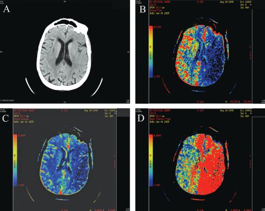
Figure 2–11. A series of computed tomography (CT) scans through the brain of a patient with a left internal carotid occlusion. Note that in the noncontrast CT scan in panel (A), there is loss of the gray-white differentiation and effacement of the sulci over the middle cerebral artery distribution on the left. Panel (B) shows the perfusion blood flow map, indicating that there is very low flow within the left middle cerebral artery distribution, but that there is also impairment of blood flow in both anterior cerebral arteries, consistent with loss of the contribution from the left internal carotid artery. Although the blood volume (C) is relatively normal in these areas, mean transit time (D) is also abnormal, indicating that tissue in the anterior cerebral distributions is at risk of infarction.
high magnetic field, they are not compatible with certain types of implants in patients, including cardiac pacemakers and deep brain stimulators. Patients who require mechanical ventilation must either be ventilated by hand during the scan or placed on a specialized MR-compatible ventilator. In addition, most sequences take substantially longer than CT scans, so that clear images require that the patient not move.
MRA can reveal most stenoses or occlusions of cerebral blood vessels. It requires only a few additional minutes during a conventional MRI scanning session, and the images are extracted by computer and therefore can be recovered very quickly. However, the MRA is very flow
dependent, and tends to exaggerate the degree of stenosis in areas of slow flow.
Magnetic Resonance Spectroscopy
Magnetic resonance spectroscopy (MRS)156 is becoming increasingly important in the diagnosis and prognosis of patients with a variety of illnesses that cause delirium, stupor, or coma (Figure 5–7). The technique identifies neurochemicals in regions of both normal and abnormal brain. Although special techniques allow the identification of as many as 80 brain metabolites, most clinical centers using standard MRI machines perform proton (1H) MRS
80 Plum and Posner’s Diagnosis of Stupor and Coma
that can identify about 13 brain metabolites (see Figure 5–7, page 226).
Myo-inositol (mI) is a sugar-like molecule present in astrocytes. It helps to regulate cell volume. Its presence serves as a marker of astrocytes. The metabolite is elevated in a number of disorders including hyperosmolar states, progressive multifocal leukoencephalopathy, renal failure, and diabetes. Levels are decreased in hyponatremia, chronic hepatic encephalopathy, tumor, and stroke.
Creatine (Cr) is actually the sum of creatine and phosphocreatine, a reliable marker of energy metabolism in both neurons and astrocytes. The total creatine peak remains constant, allowing other peaks to be calculated as ratios to the height of the creatine peak.
N-Acetylaspartate (NAA) is an amino acid derivative synthesized in neurons and transported down axons. It marks the presence of viable neurons, axons, and dendrites. Its levels may be increased in hyperosmolar states and are decreased in almost any disease that causes destruction of neurons or their processes.
The choline (Cho) peak represents several membrane components, primarily phosphocholine and glycerophosphocholine. Choline is found in higher concentration in glial cells and is thus higher in white matter than gray matter. It is increased in tumors (particularly relative to NAA), strokes, and hyperosmolar states. It is decreased in liver disease and hyponatremia.
Glutamate/glutamine (Glx) represents a mixture of amino acids and amines involved in excitatory and inhibitory transmission as well as products of the Krebs cycle and mitochondrial redox systems. The peak is elevated in hypoxic encephalopathy and in hyperosmolar states; it is diminished in hyponatremia.
Lactate (Lac), not visible in normal brain, is a product of anaerobic glycolysis and is thus increased in hypoxic/ischemic encephalopathy, diabetic acidosis, stroke, and recovery from cardiac arrest. It is also increased in highly aggressive tumors.
A lipid peak is not present in normal brain but is identified in areas of brain necrosis, particularly in rapidly growing tumors. Cerebral fat embolism (see Chapter 5) can also cause a lipid peak.157
The clinical use of some of these spectra in stuporous or comatose patients is discussed in Chapter 5.
Neurosonography
Intracranial Doppler sonography identifies flow of blood in arteries, particularly the middle cerebral artery. The absence of flow in the brain has been used to confirm brain death, particularly in patients who have received sed-
ative drugs that may alter some of the clinical findings (see Chapter 8).158,159 The tech-
nique is also useful for following patients with strokes, head injuries, and hypoxic/ischemic encephalopathy.160,161 The injection of gasfilled microbubbles enhances the sonographic echo and provides better delineation of blood flow, occlusions, pseudo-occlusions, stenosis, and collateral circulation.162
Doppler studies of the extracranial carotid circulation are frequently done as a routine part of stroke evaluation at many centers. However, this is rarely helpful for patients in coma. If the coma is due to a reversible stenosis or occlusion of a single vessel, it almost always will be in the vertebrobasilar, not the carotid, circulation. If the patient is going to receive an MRI scan, the MRA of the cervical vessels, which examines both the carotid and the vertebrobasilar circulation, is generally more revealing.
Lumbar Puncture
Although often overlooked in the technologic era, the examination of the CSF still plays a central role in neurologic diagnosis, particularly in patients with a depressed level of consciousness. Once an imaging study has been performed, it is necessary to proceed with lumbar puncture as soon as possible for patients with no clear diagnosis. Rare patients in whom subarachnoid hemorrhage was not detected on imaging may demonstrate blood in the CSF. Similarly, occasional patients with bacterial meningitis or viral encephalitis may present with a depressed level of consciousness (sometimes after a missed seizure), and may not yet have sufficient meningismus to make the diagnosis of meningitis clear from examination. This may be particularly difficult to determine in patients who have underlying rigidity of the cervical spine (evidenced by resistance to lateral as well as flexion movements

of the neck). Nevertheless, it is imperative to identify infection as early as possible to allow the administration of antibiotics or antiviral agents.
Patient 2–2
A 73-year-old woman who was on 10 mg/day of prednisone for her ulcerative colitis had a 2-day history of presumed gastroenteritis, with fever, nausea, and vomiting. She awoke on the third day and found it difficult to walk to the bathroom. By the afternoon she had difficulty swallowing, her voice was hoarse, and her left limbs were clumsy. She was brought to the hospital by ambulance, and examination in the emergency department disclosed a lethargic patient who could be easily wakened. Pupils were equal and constricted from 3 to 2 mm with light, but the left eye was lower than the right, she complained of skewed diplopia, and there was difficulty maintaining gaze to the left. There was left-sided facial numbness and lower motor neuron facial weakness. Hearing was intact, but her voice was hoarse. The tongue deviated to the right and there was distal weakness in her arms, and the left limbs were clumsy on fine motor tasks and showed dysmetria.
MRI scan showed a left pontomedullary lesion surrounded by edema, which was bright on diffusion-weighted imaging, and she was diagnosed as having a brainstem infarct. However, despite normal MRA of the vertebrobasilar system, her deficits progressed over the next day. A senior neuroradiologist noticed some enhancement at the periphery of the lesion on review of the MRI scan, and suggested an abscess. Lumbar puncture disclosed 47 white blood cells/mm3 and elevated protein, and she recovered after being treated for
Listeria monocytogenes. An MRI scan much later in her course, disclosing a multioculated abscess, is shown in Figure 4–13.
Comment. This case demonstrates the importance of examining the spinal fluid, even when a presumptive diagnosis of vascular disease is entertained. This is particularly true in patients with fever, elevated white blood cell count, or stiff neck, where infectious disease is a consideration. However, every patient with an undetermined cause of coma requires lumbar puncture as part of the routine evaluation.
Examination of the Comatose Patient |
81 |
The timing of lumbar puncture with respect to CT scanning is discussed in Chapters 4 and 5. However, in some circumstances, scanning may not be not immediately available. In these cases it is common to give antibiotics immediately and then do imaging and lumbar puncture up to a few hours later. However, once the antibiotics have penetrated the CSF, the ability to grow a bacterial pathogen and identify its susceptibilities may be permanently compromised. Hence, deferring lumbar puncture in such cases until after the scanning procedure may do the patient harm. For this reason, when the evidence for meningitis is compelling, it may be necessary to do the lumbar puncture without benefit of prior imaging. As discussed in Chapters 4 and 5, the danger of this procedure is greatly overestimated. If the examination is nonfocal, and there is no evidence of papilledema on funduscopy, it is extremely rare to precipitate brain herniation by lumbar puncture. The benefit of establishing the exact microbial diagnosis far outweighs the risk of herniation.
A critical but often overlooked component of the lumbar puncture is to measure and record the opening pressure. Elevated pressure may be a key sign that leads to diagnosis of venous sinus thrombosis, cerebral edema, or other serious conditions that can cause coma. In addition to the routine cell count, protein, and glucose, CSF should be obtained for full cultures, including tuberculosis and fungal agents; serology and polymerase chain reaction (PCR) for specific agents such as syphilis, Lyme disease, and herpes encephalitis; and cytology, as cancer or leukemia sometimes may present with meningeal and subarachnoid infiltration. It is a good practice to set aside several milliliters of refrigerated CSF in case additional studies become necessary. This entire group of tests typically requires about 20 mL of CSF, an amount that the choroid plexus in the brain restores within about an hour.
One common problem is that the lumbar tap may be traumatic, yielding bloody CSF. This may make it difficult to determine the underlying numbers of both red and white blood cells in the CSF. If the cells come from the blood (rather than the white cells being elevated within the CSF, e.g., due to infection), the proportion of the red and white cells should remain the same as in the blood (usually
82 Plum and Posner’s Diagnosis of Stupor and Coma
500 to 1,000 red cells per one white cell). If the tap is bloody, many clinicians send fluid from both tubes 1 and 4 for cell count. A falling count indicates that the tap was traumatic, but it does not tell you what the underlying CSF counts were compared with the count in tube 4. Nor does lack of a falling cell count indicate that the blood was there before the tap (the tip of the needle may be partially within or adjacent to a bleeding vein). An alternative approach is to examine the CSF for xanthochromia. However, CSF may be stained yellow due to high protein or bilirubin. Examination of the red blood cells under the microscope immediately after the tap may be helpful. Fresh red cells have the typical doughnut-shaped morphology, whereas crenelated cells indicate that they have been in the extravascular space for some time. Similarly, if the CSF sample is spun in a centrifuge until there are no red blood cells in the supernatant, the fluid can be tested for blood products with a urine dipstick. A positive test indicates breakdown of red blood cells, which typically takes at least 6 hours to occur after a subarachnoid hemorrhage, and demonstrates that the blood was there before the tap.
Electroencephalography and
Evoked Potentials
Electroencephalography (EEG) is useful as an objective electrophysiologic assay of cortical function in patients who do not respond to normal sensory stimuli. A typical waking EEG is dominated anteriorly by low-voltage beta activity (faster than 13 Hz). During periods of quiet wakefulness, the EEG may slow into the alpha range (8 to 13 Hz) and the wave activity may be more rhythmic and symmetric. As the patient becomes more drowsy, higher voltage theta rhythms (4 to 7 Hz) become dominant; delta activity (1 to 3 Hz) predominates in patients who are deeply asleep or comatose. The EEG provides a rough but fairly accurate estimate of the degree to which a patient who is unresponsive may be simply uncooperative.
On the other hand, occasional patients with coma due to brainstem injury show an alpha EEG pattern. The alpha activity in such patients is usually more regular and less variable
than in an awake patient, and it is not inhibited by opening the eyes.163 It may be possible to
drive the EEG by photic stimulation in alpha coma. Certain types of metabolic encephalopathy may also have characteristic EEG changes. For example, triphasic waves are often seen in patients with hepatic encephalopathy, but
can be seen in other metabolic disorders that cause coma.163,164
The EEG is most helpful in diagnosing impairment of consciousness due to nonconvulsive status epilepticus.165 Such patients may lack the usual behavioral signs of complex partial seizures, such as lip smacking or blinking, and may present as merely confused, drowsy, or even stuporous or comatose. Some patients may demonstrate twitching movements of the eyelids or extremities, but others give no external sign of epileptic activity. In one series, 8% of comatose patients were found to be suffering from nonconvulsive status epilepticus.166 When the EEG shows continuous epileptic activity, the diagnosis is easy and anticonvulsants are required. However, nonconvulsive status epilepticus may occur in patients without characteristic EEG changes,167 probably because the seizure activity is mainly in areas such as the medial temporal lobes that are not sampled by the surface electrodes. Accordingly, if one suspects that the patient’s loss of consciousness is a result of nonconvulsive status epilepticus, it is probably wise to administer a short-acting benzodiazepine and observe the patient’s response. If the patient improves, antiepileptic drugs should be administered. Unfortunately, some patients with a clinical and electroencephalographic diagnosis of nonconvulsive status epilepticus do not respond to anticonvulsant drugs, because the underlying process causing the seizure activity is too severe to be suppressed by routine doses of drugs. Such patients are sometimes treated by large intravenous doses of gamma-aminobutyric acid agonist drugs, such as barbiturates or propofol, which at sufficiently high dosage can suppress all brain activity. However, unless the underlying brain process can be reversed, the prognosis of patients with nonconvulsive status epilepticus who do not awaken after anticonvulsant treatment is poor168 (see also Seizures in Chapter 5).
Evoked potentials may also be used to test the integrity of brainstem and forebrain pathways in comatose patients. Although they do not provide reliable information on the location of a lesion in the brainstem, both auditoryand somatosensory-evoked potentials, and cor-
tical event-related potentials, can provide information on the prognosis of patients in coma.169 This use will be discussed in greater detail in Chapter 8.
REFERENCES
1.Dunn C, Held JL, Spitz J, et al. Coma blisters: report and review. Cutis 45 (6), 423–426, 1990.
2.Teasdale G, Jennett B. Assessment and prognosis of coma after head injury. Acta Neurochir (Wien) 34 (1–4), 45–55, 1976.
3.Gill MR, Reiley DG, Green SM. Interrater reliability of Glasgow Coma Scale scores in the emergency department. Ann Emerg Med 43, 215–223, 2004.
4.McNarry AF, Goldhill DR. Simple bedside assessment of level of consciousness: comparison of two simple assessment scales with the Glasgow Coma scale. Anaesthesia 59, 34–37, 2004.
5.Servadei F. Coma scales. Lancet 367 (9510), 548– 549, 2006.
6.Ropper AH, O’Rourke D, Kennedy SK. Head position, intracranial pressure, and compliance. Neurology 32 (11), 1288–1291, 1982.
7.Saper CB, Loewy AD, Swanson LW, et al. Direct hypothalamo-autonomic connections. Brain Res 117 (2), 305–312, 1976.
8.Saper CB. Central autonomic system. In Paxinos G. ed. The Rat Nervous System. Elsevier Academic Press, San Diego, pp 761–796, 2004.
9.Rossetti AO, Reichhart MD Bogousslavsky J. Central Horner’s syndrome with contralateral ataxic hemiparesis: a diencephalic alternate syndrome. Neurology 61 (3), 334–338, 2003.
10.Reeves AG, Posner JB. The ciliospinal response in man. Neurology 19, 1145–1152, 1969.
11.Vassend O, Knardahl S. Cardiovascular responsiveness to brief cognitive challenges and pain sensitivity in women. Eur J Pain 8 (4), 315–324, 2004.
12.Zidan AH, Girvin JP. Effect on the Cushing response of different rates of expansion of a supratentorial mass. J Neurosurg 49 (1), 61–70, 1978.
13.Kawahara E, Ikeda S, Miyahara Y, et al. Role of autonomic nervous dysfunction in electrocardio-graphic abnormalities and cardiac injury in patients with acute subarachnoid hemorrhage. Circ J 67 (9), 753– 756, 2003.
14.Lorsheyd A, Simmers TA Robles De Medina EO. The relationship between electrocardiographic abnormalities and location of the intracranial aneurysm in subarachnoid hemorrhage. Pacing Clin Electrophysiol 26 (8), 1722–1728, 2003.
15.McLaughlin N, Bojanowski MW, Girard F, et al. Pulmonary edema and cardiac dysfunction following subarachnoid hemorrhage. Can J Neurol Sci 32 (2), 178–185, 2005.
16.Ferrante L, Artico M, Nardacci B, Fraioli B, Cosentino F, Fortuna A. Glossopharyngeal neuralgia with cardiac syncope. Neurosurgery 36, 58–63, 1995.
17.Cole CR, Zuckerman J Levine BD. Carotid sinus ‘‘irritability’’ rather than hypersensitivity: a new name for an old syndrome? Clin Auton Res 11(2), 109– 113, 2001.
Examination of the Comatose Patient |
83 |
18.Paulson OB, Strandgaard S Edvinsson L. Cerebral autoregulation. Cerebrovasc Brain Metab Rev 2(2), 161–192, 1990.
19.Strandgaard S, Paulson OB. Regulation of cerebral blood flow in health and disease. J Cardiovasc Pharmacol 19 (Suppl 6), S89–S93, 1992.
20.Wahl M, Schilling L. Regulation of cerebral blood flow—a brief review. Acta Neurochir Suppl (Wien) 59, 3–10, 1993.
21.Schondorf R, Benoit J, Stein R. Cerebral autoregulation in orthostatic intolerance. Ann N Y Acad Sci 940, 514–526, 2001.
22.Sato A, Sato Y, Uchida S. Regulation of cerebral cortical blood flow by the basal forebrain cholinergic fibers and aging. Auton Neurosci 96 (1), 13–19, 2002.
23.Bieger D, Hopkins DA. Viscerotopic representation of the upper alimentary tract in the medulla oblongata in the rat: the nucleus ambiguus. J Comp Neurol 262 (4), 546–562, 1987.
24.Ross CA, Ruggiero DA, Park DH, et al. Tonic vasomotor control by the rostral ventrolateral medulla: effect of electrical or chemical stimulation of the area containing C1 adrenaline neurons on arterial pressure, heart rate, and plasma catecholamines and vasopressin. J Neurosci 4(2), 474–494, 1984.
25.Panneton WM, Loewy AD. Projections of the carotid sinus nerve to the nucleus of the solitary tract in the cat. Brain Res 191 (1), 239–244, 1980.
26.Ciriello J. Brainstem projections of aortic baroreceptor afferent fibers in the rat. Neurosci Lett 36 (1), 37–42, 1983.
27.Ross CA, Ruggiero DA, Reis DJ. Projections from the nucleus tractus solitarii to the rostral ventrolateral medulla. J Comp Neurol 242 (4), 511–534, 1985.
28.Blessing WW, Reis DJ. Inhibitory cardiovascular function of neurons in the caudal ventrolateral medulla of the rabbit: relationship to the area containing A1 noradrenergic cells. Brain Res 253 (1–2), 161– 171, 1982.
29.Smith JC, Ellenberger HH, Ballanyi K, et al. PreBotzinger complex: a brainstem region that may generate respiratory rhythm in mammals. Science 254 (5032), 726–729, 1991.
30.Gray PA, Janczewski WA, Mellen N, et al. Normal breathing requires preBotzinger complex neuroki- nin-1 receptor-expressing neurons. Nat Neurosci 4, 927–930, 2001.
31.Wallach JH, Loewy AD. Projections of the aortic nerve to the nucleus tractus solitarius in the rabbit. Brain Res 188 (1), 247–251, 1980.
32.Torrealba F, Claps A. The carotid sinus connections: a WGA-HRP study in the cat. Brain Res 455 (1), 134–143, 1988.
33.Kalia M, Richter D. Rapidly adapting pulmonary receptor afferents: I. Arborization in the nucleus of the tractus solitarius. J Comp Neurol 274 (4), 560– 573, 1988.
34.Feldman JL, Ellenberger HH. Central coordination of respiratory and cardiovascular control in mammals. Annu Rev Physiol 50, 593–606, 1988.
35.Weston MC, Stornetta RL, Guyenet PG. Glutamatergic neuronal projections from the marginal
84 Plum and Posner’s Diagnosis of Stupor and Coma
layer of the rostral ventral medulla to the respiratory centers in rats. J Comp Neurol 473 (1), 73–85, 2004.
36.Richerson GB. Serotonergic neurons as carbon dioxide sensors that maintain pH homeostasis. Nat Rev Neurosci 5(6), 449–461, 2004.
37.Chamberlin NL, Saper CB. Topographic organization of respiratory responses to glutamate microstimulation of the parabrachial nucleus in the rat. J Neurosci 14 (11 Pt 1), 6500–6510, 1994.
38.Chamberlin NL, Saper CB. A brainstem network mediating apneic reflexes in the rat. J Neurosci 18 (15), 6048–6056, 1998.
39.Meah MS, Gardner WN. Post-hyperventilation apnoea in conscious humans. J Physiol 477 (Pt 3), 527– 538, 1994.
40.Jennett S, Ashbridge K, North JB. Post-hyperventi- lation apnoea in patients with brain damage. J Neurol Neurosurg Psychiatry 37 (3), 288–296, 1974.
41.Cherniack NS, Longobardo G, Evangelista CJ. Causes of Cheyne-Stokes respiration. Neurocrit Care 3(3), 271–279, 2005.
42.Lange RL, Hecht HH. The mechanism of CheyneStokes respiration. J Clin Invest 41, 42–52, 1962.
43.Murdock DK, Lawless CE, Loeb HS, et al. The effect of heart transplantation on Cheyne-Stokes respiration associated with congestive heart failure. J Heart Transplant 5(4), 336–337, 1986.
44.Hudgel DW, Devadatta P, Quadri M, et al. Mechanism of sleep-induced periodic breathing in convalescing stroke patients and healthy elderly subjects. Chest 104 (5), 1503–1510, 1993.
45.Rubin AE, Gottlieb SH, Gold AR, et al. Elimination of central sleep apnoea by mitral valvuloplasty: the role of feedback delay in periodic breathing. Thorax 59 (2), 174–176, 2004.
46.Vespa PM, Bleck TP. Neurogenic pulmonary edema and other mechanisms of impaired oxygenation after aneurysmal subarachnoid hemorrhage. Neurocrit Care 1(2), 157–170, 2004.
47.Simon RP. Neurogenic pulmonary edema. Neurol Clin 11(2), 309–323, 1993.
48.Tarulli AW, Lim C, Bui JD, et al. Central neurogenic hyperventilation: a case report and discussion of pathophysiology. Arch Neurol 62 (10), 1632–1634, 2005.
49.Shams PN, Waldman A, Plant GT. B cell lymphoma of the brain stem masquerading as myasthenia. J Neurol Neurosurg Psychiatry 72, 271–273, 2002.
50.Rodriguez M, Baele PL, Marsh HM, et al. Central neurogenic hyperventilation in an awake patient with brainstem astrocytoma. Ann Neurol 11, 625–628, 1982.
51.Siderowf AD, Balcer LJ, Kenyon LC, et al. Central neurogenic hyperventilation in an awake patient with a pontine glioma. Neurology 46, 1160–1162, 1996.
52.Hilaire G, Pasaro R. Genesis and control of the respiratory rhythm in adult mammals. News Physiol Sci 18, 23–28, 2003.
53.El Khatib MF, Kiwan RA, Jamaleddine GW. Buspirone treatment for apneustic breathing in brain stem infarct. Respir Care 48, 956–958, 2003.
54.Bassetti C, Aldrich MS, Quint D. Sleep-disordered breathing in patients with acute supraand infra-
tentorial strokes. A prospective study of 39 patients. Stroke 28, 1765–1772, 1997.
55.Pang KP, Terris DJ. Screening for obstructive sleep apnea: an evidence-based analysis. Am J Otolaryngol 27 (2), 112–118, 2006.
56.Iber C. Sleep-related breathing disorders. Neurol Clin 23(4), 1045–1057, 2005.
57.Schlaefke ME, Kille JF, Loeschcke HH. Elimination of central chemosensitivity by coagulation of a bilateral area on the ventral medullary surface in awake cats. Pflugers Arch 378 (3), 231–241, 1979.
58.Fodstad H. Pacing of the diaphragm to control breathing in patients with paralysis of central nervous system origin. Stereotact Funct Neurosurg 53 (4), 209–222, 1989.
59.Bogousslavsky J, Khurana R, Deruaz JP, et al. Respiratory failure and unilateral caudal brainstem infarction. Ann Neurol 28 (5), 668–673, 1990.
60.Auer RN, Rowlands CG, Perry SF, et al. Multiple sclerosis with medullary plaques and fatal sleep apnea (Ondine’s curse). Clin Neuropathol 15 (2), 101– 105, 1996.
61.Manconi M, Mondini S, Fabiani A, et al. Anterior spinal artery syndrome complicated by the Ondine curse. Arch Neurol 60 (12), 1787–1790, 2003.
62.Polatty RC, Cooper KR. Respiratory failure after percutaneous cordotomy. South Med J 79 (7), 897– 899, 1986.
63.Amiel J, Laudier B, ttie-Bitach T, et al. Polyalanine expansion and frameshift mutations of the pairedlike homeobox gene PHOX2B in congenital central hypoventilation syndrome. Nat Genet 33 (4), 459– 461, 2003.
64.Stankiewicz JA, Pazevic JP. Acquired Ondine’s curse. Otolaryngol Head Neck Surg 101 (5), 611– 613, 1989.
65.Ezure K, Tanaka I. Convergence of central respiratory and locomotor rhythms onto single neurons of the lateral reticular nucleus. Exp Brain Res 113 (2), 230–242, 1997.
66.Daquin G, Micallef J, Blin O. Yawning. Sleep Med Rev 5(4), 299–312, 2001.
67.Argiolas A, Melis MR. The neuropharmacology of yawning. Eur J Pharmacol 343 (1), 1–16, 1998.
68.Launois S, Bizec JL, Whitelaw WA, et al. Hiccup in adults: an overview. Eur Respir J 6, 563–575, 1993.
69.Straus C, Vasilakos K, Wilson RJ, et al. A phylogenetic hypothesis for the origin of hiccough. Bioessays 25, 182–188, 2003.
70.Cersosimo RJ, Brophy MT. Hiccups with high dose dexamethasone administration—a case report. Cancer 82, 412–414, 1998.
71.LeWitt PA, Barton NW, Posner JB. Hiccup with dexamethasone therapy. Letter to the editor. Ann Neurol 12, 405–406, 1982.
72.Souadjian JV, Cain JC. Intractable hiccup. Etiologic factors in 220 cases. Postgrad Med 43, 72–77, 1968.
73.Walker P, Watanabe S, Bruera E. Baclofen, a treatment for chronic hiccup. J Pain Symptom Manage 16, 125–132, 1998.
74.Friedman NL. Hiccups: a treatment review. Pharmacotherapy 16, 986–995, 1996.
75.Furukawa N, Hatano M, Fukuda H. Glutaminergic vagal afferents may mediate both retching and gastric
adaptive relaxation in dogs. Auton Neurosci 93 (1–2), 21–30, 2001.
76.Balaban CD. Vestibular autonomic regulation (including motion sickness and the mechanism of vomiting). Curr Opin Neurol 12 (1), 29–33, 1999.
77.Hornby PJ. Central neurocircuitry associated with emesis. Am J Med 111 (Suppl 8A), 106S–112S, 2001.
78.Yamamoto H, Kishi T, Lee CE, et al. Glucagon-like peptide-1-responsive catecholamine neurons in the area postrema link peripheral glucagon-like peptide- 1 with central autonomic control sites. J Neurosci 23(7), 2939–2946, 2003.
79.Chen CJ, Scheufele M, Sheth M, et al. Isolated relative afferent pupillary defect secondary to contralateral midbrain compression. Arch Neurol 61, 1451–1453, 2004.
80.Hornblass A. Pupillary dilatation in fractures of the floor of the orbit. Ophthalmic Surg 10(11), 44–46, 1979.
81.Antonio-Santos AA, Santo RN, Eggenberger ER. Pharmacological testing of anisocoria. Expert Opin Pharmacother 6 (12), 2007–2013, 2005.
82.McLeod JG, Tuck RR. Disorders of the autonomic nervous system: part 2. Investigation and treatment. Ann Neurol 21(6), 519–529, 1987.
83.Zhang YH, Lu J, Elmquist JK, et al. Lipopolysaccharide activates specific populations of hypothalamic and brainstem neurons that project to the spinal cord. J Neurosci 20 (17), 6578–6586, 2000.
84.Llewellyn-Smith IJ, Martin CL, Marcus JN, et al. Orexin-immunoreactive inputs to rat sympathetic preganglionic neurons. Neurosci Lett 351 (2), 115– 119, 2003.
85.Estabrooke IV, McCarthy MT, Ko E, et al. Fos expression in orexin neurons varies with behavioral state. J Neurosci 21(5), 1656–1662, 2001.
86.Loewy AD, Araujo JC, Kerr FW. Pupillodilator pathways in the brain stem of the cat: anatomical and electrophysiological identification of a central autonomic pathway. Brain Res 60 (1), 65–91, 1973.
87.Burde RM, Loewy AD. Central origin of oculomotor parasympathetic neurons in the monkey. Brain Res 198 (2), 434–439, 1980.
88.Burde RM. Disparate visceral neuronal pools subserve spinal cord and ciliary ganglion in the monkey: a double labeling approach. Brain Res 440 (1), 177– 180, 1988.
89.Gooley JJ, Lu J, Fischer D, et al. A broad role for melanopsin in nonvisual photoreception. J Neurosci 23(18), 7093–7106, 2003.
90.Buttner-Ennever JA, Cohen B, Horn AK, et al. Pretectal projections to the oculomotor complex of the monkey and their role in eye movements. J Comp Neurol 366 (2), 348–359, 1996.
91.Jampel RS. Convergence, divergence, pupillary reactions and accommodation of the eyes from faradic stimulation of the macaque brain. J Comp Neurol 115, 371–399, 1960.
92.Kerr FW, Hallowell OW. Location of the pupillomotor and accommodation fibers in the oculomotor nerve: experimental observations on paralytic mydriasis. J Neurol Neurosurg Psychiatry 27, 473– 481, 1964.
93.Leigh RJ, Zee DS. The Neurology of Eye Movements, 4th ed. New York: Oxford University Press, 2006.
Examination of the Comatose Patient |
85 |
94.Hanson RA, Ghosh S, Gonzalez-Gomez I, et al. Abducens length and vulnerability? Neurology 62 (1), 33–36, 2004.
95.Zee DS. Brain stem and cerebellar deficits in eye movement control. Trans Ophthalmol Soc U K 105 (Pt 5), 599–605, 1986.
96.Henn V. Pathophysiology of rapid eye movements in the horizontal, vertical and torsional directions. Baillieres Clin Neurol 1(2), 373–391, 1992.
97.Sparks DL, Mays LE. Signal transformations required for the generation of saccadic eye movements. Annu Rev Neurosci 13, 309–336, 1990.
98.Lewis RF, Zee DS. Ocular motor disorders associated with cerebellar lesions: pathophysiology and topical localization. Rev Neurol (Paris) 149 (11), 665– 677, 1993.
99.Buettner UW, Zee DS. Vestibular testing in comatose patients. Arch Neurol 46 (5), 561–563, 1989.
100.Helmchen C, Rambold H, Kempermann U, et al. Localizing value of torsional nystagmus in small midbrain lesions. Neurology 59 (12), 1956–1964, 2002.
101.Krauzlis RJ. Recasting the smooth pursuit eye movement system. J Neurophysiol 91 (2), 591–603, 2004.
102.Leichnetz GR. An anterogradely-labeled prefrontal cortico-oculomotor pathway in the monkey demonstrated with HRP gel and TMB neurohistochemistry. Brain Res 198 (2), 440–445, 1980.
103.Barton JJ, Simpson T, Kiriakopoulos E, et al. Functional MRI of lateral occipitotemporal cortex during pursuit and motion perception. Ann Neurol 40 (3), 387–398, 1996.
104.Goldberg ME. The control of gaze. In: Kandel ER, Schwartz JH, Jessel JH, eds. Principles of Neuroscience, 4th ed. New York: McGraw Hill, pp 782–800, 2000.
105.Cogan DG, Chu FC, Reingold DB. Ocular signs of cerebellar disease. Arch Ophthalmol 100 (5), 755– 760, 1982.
106.Caplan LR. Ptosis. J Neurol Neurosurg Psychiatry 37 (1), 1–7, 1974.
107.Hackley SA, Johnson LN. Distinct early and late subcomponents of the photic blink reflex: response characteristics in patients with retrogeniculate lesions. Psychophysiology 33, 239–251, 1996.
108.Liu GT, Ronthal M. Reflex blink to visual threat. J Clin Neuroophthalmol 12, 47–56, 1992.
109.Wijdicks EF, Bamlet WR, Maramattom BV, et al. Validation of a new coma scale: the FOUR score. Ann Neurol 58 (4), 585–593, 2005.
110.Pullicino PM, Jacobs L, McCall WD Jr, et al. Spontaneous palpebromandibular synkinesia: a localizing clinical sign. Ann Neurol 35 (2), 222–228, 1994.
111.Roberts TA, Jenkyn LR, Reeves AG. On the notion of doll’s eyes. Arch Neurol 41, 1242–1243, 1984.
112.Schubert MC, Das V, Tusa RJ, et al. Cervico-ocular reflex in normal subjects and patients with unilateral vestibular hypofunction. Otol Neurotol 25 (1), 65– 71, 2004.
113.Schlosser HG, Unterberg A, Clarke A. Using videooculography for galvanic evoked vestibulo-ocular monitoring in comatose patients. J Neurosci Methods 145 (1–2), 127–131, 2005.
114.Brandt TH, Dieterich M. Different types of skew deviation. J Neurol Neurosurg Psychiatry 54, 549– 550, 1991.