

Книги по МРТ КТ на английском языке / PLUM AND POSNER S DIAGNOSIS OF STUPOR AND COM-1
.pdf6Plum and Posner’s Diagnosis of Stupor and Coma
with pathologic alterations of consciousness can be awakened at all, they rapidly fall back into a sleep-like state when stimulation ceases.
Patients who have a sleep-like appearance and remain behaviorally unresponsive to all external stimuli are unconscious by any definition. However, continuous sleep-like coma as a result of brain injury rarely lasts more than 2 to 4 weeks.
Acutely Altered States
of Consciousness
Clouding of consciousness is a term applied to minimally reduced wakefulness or awareness, which may include hyperexcitability and irritability alternating with drowsiness. A key distinction must be made in such patients between those who are confused (i.e., do not respond appropriately to their environment) because of a focal deficit of cognitive function versus those who have more global impairment. The beclouded patient is usually incompletely oriented to time and sometimes to place. Such patients are inattentive and perform poorly on repeating numbers backward (the normal range is at least four or five) and remembering details or even the meaning of stories. Drowsiness is often prominent during the day, but agitation may predominate at night.
The pathophysiology of brain function in such patients has rarely been studied, but Posner and Plum3 found that cerebral oxygen consumption had declined by 20% below normal levels in patients with hepatic encephalopathy with lethargy and global confusion, and Shimojyo and colleagues noted similar reductions in patients with lethargy and global confusion due to Wernicke’s encephalopathy.4 More recently, Trzepacz and colleagues have identified decreased regional cerebral blood flow (CBF) bilaterally in the frontotemporal cortex and right basal ganglia of patients with subclinical hepatic encephalopathy.5 Increases in CBF during treatment of cobalamin deficiency correlate with clinical improvement.6 Other studies have implicated reduced cholinergic function; excess release of dopamine, norepinephrine, and glutamate; and both decreased and increased serotonergic and gamma-aminobutyric acid (GABA) activity.7 The pathogenesis of clouding of consciousness and delirium is discussed in more detail in Chapter 5.
Delirium, from the Latin ‘‘to go out of the furrow,’’ is a more floridly abnormal mental state characterized by misperception of sensory stimuli and, often, vivid hallucinations. Delirium is defined by the Diagnostic and Statistical Manual of Mental Disorders, 4th edition (DSM-IV),8 as follows: ‘‘A. Disturbance of consciousness (i.e., reduced clarity of awareness of the environment) with reduced ability to focus, sustain or shift attention. B. A change in cognition (such as memory deficit, disorientation, language disturbance) or the development of a perceptual disturbance that is not better accounted for by a pre-existing, established or evolving dementia. C. The disturbance develops over a short period of time (usually hours to days) and tends to fluctuate during the course of the day.’’
Delirious patients are disoriented, first to time, next to place, and then to persons in their environment. Rarely are patients unaware of who they are, although sometimes married women will revert to their maiden name. Patients are often fearful or irritable and may overreact or misinterpret normal activities of physicians and nurses. Delusions or hallucinations may place the patient completely out of contact with the environment and the examiner. Full-blown delirious states tend to come on rapidly and rarely last more than 4 to 7 days. However, fragments of misperceptions may persist for several weeks, especially among alcoholics and patients with cerebral involvement from collagen vascular diseases.
Delirium with agitation occasionally may be seen as a consequence of focal lesions of the right parieto-occipitotemporal cortex,2,9 but generally is indicative of bilateral impairment of cortical function in toxic-metabolic states, such as atropine poisoning, alcohol or sedative drug (e.g., benzodiazepine) withdrawal, acute porphyria, or hepatic or renal failure. It also occurs with systemic infectious processes or as a component of encephalitis, during which immune mediators such as cytokines and eicosanoid derivatives may cloud mental function.
Obtundation, from the Latin ‘‘to beat against or blunt,’’ literally means mental blunting or torpidity. In a medical setting, such patients have a mild to moderate reduction in alertness, accompanied by a lesser interest in the environment. Such patients have slower psychologic responses to stimulation. They may have an
Pathophysiology of Signs and Symptoms of Coma |
7 |
increased number of hours of sleep and may be drowsy between sleep bouts.
Stupor, from the Latin ‘‘to be stunned,’’ is a condition of deep sleep or similar behavioral unresponsiveness from which the subject can be aroused only with vigorous and continuous stimulation. Even when maximally aroused, the level of cognitive function may be impaired. Such patients can be differentiated from those with psychiatric impairment, such as catatonia or severe depression, because they can be aroused by vigorous stimulation to respond to simple stimuli.
Coma, from the Greek ‘‘deep sleep or trance,’’ is a state of unresponsiveness in which the patient lies with eyes closed and cannot be aroused to respond appropriately to stimuli even with vigorous stimulation. The patient may grimace in response to painful stimuli and limbs may demonstrate stereotyped withdrawal responses, but the patient does not make localizingresponsesordiscretedefensivemovements. As coma deepens, the responsiveness of the patient, even to painful stimuli, may diminish or disappear. However, it is difficult to equate the lack of motor responses to the depth of the coma, as the neural structures that regulate motor responses differ from those that regulate consciousness, and they may be differentially impaired by specific brain disorders.
The locked-in syndrome describes a state in which the patient is de-efferented, resulting in paralysis of all four limbs and the lower cranial nerves. This condition has been recognized at least as far back as the 19th century, but its distinctive name was applied in the first edition of this monograph (1966), reflecting the implications of this condition for the diagnosis of coma and for the specialized care such patients require. Although not unconscious, locked-in patients are unable to respond to most stimuli. A high level of clinical suspicion is required on the part of the examiner to distinguish a lockedin patient from one who is comatose. The most common cause is a lesion of the base and tegmentum of the midpons that interrupts descending cortical control of motor functions. Such patients usually retain control of vertical eye movements and eyelid opening, which can be used to verify their responsiveness. They may be taught to respond to the examiner by using eye blinks as a code. Rare patients with subacute motor neuropathy, such as GuillainBarre´ syndrome, also may become completely
de-efferented, but there is a history of subacute paralysis. In both instances, electroencephalographic (EEG) examination discloses a reactive posterior alpha rhythm10 (see EEG section, page 82).
It is important to identify locked-in patients so that they may be treated appropriately by the medical and nursing staff. At the bedside, discussion should be with the patient, not, as with an unconscious individual, about the patient. Patients with large midpontine lesions often are awake most of the time, with greatly diminished sleep on physiologic recordings.11 They may suffer greatly if they are treated by hospital staff as if they are nonresponsive.
As the above definitions imply, each of these conditions includes a fairly wide range of behavioral responsiveness, and there may be some overlap among them. Therefore, it is generally best to describe a patient by indicating what stimuli do or do not result in responses and the kinds of responses that are seen, rather than using less precise terms.
Subacute or Chronic Alterations of Consciousness
Dementia defines an enduring and often progressive decline in mental processes owing to an organic process not usually accompanied by a reduction in arousal. Conventionally, the term implies a diffuse or disseminated reduction in cognitive functions rather than the impairment of a single psychologic activity such as language. DSM-IV defines dementia as follows: ‘‘A. The development of multiple cognitive defects manifested by both: (1) Memory impairment (impaired ability to learn new information or to recall previously learned information); (2) One (or more) of the following cognitive disturbances: aphasia (language disturbance), apraxia (impaired ability to carry out motor activities despite intact motor function), agnosia (failure to recognize or identify objects despite intact sensory function), disturbance in executive function (i.e., planning, organization, sequencing, abstracting).’’
The reader will recognize this definition as an arbitrary restriction. Usually, the term dementia is applied to the effects of primary disorders of the cerebral hemispheres, such as degenerative conditions, traumatic injuries, and neoplasms. Occasionally, dementia can be

8Plum and Posner’s Diagnosis of Stupor and Coma
at least partially reversible, such as when it accompanies thyroid or vitamin B12 deficiency or results from a reversible communicating hydrocephalus; more often, however, the term applies to chronic conditions carrying limited hopes for improvement.
Patients with dementia are usually awake and alert, but as the dementia worsens, may become less responsive and eventually evolve into a vegetative state (see below). Patients with dementia are at significantly increased risk of developing delirium when they become medically ill or develop comorbid brain disease.
Hypersomnia refers to a state characterized by excessive but normal-appearing sleep from which the subject readily, even if briefly, awakens when stimulated. Many patients with either acute or chronic alterations of consciousness sleep excessively. However, when awakened, consciousness is clearly clouded. In the truly hypersomniac patient, sleep appears normal and cognitive functions are normal when patients are awakened. Hypersomnia results from hypothalamic dysfunction, as indicated later in this chapter.12
Abulia (from the Greek for ‘‘lack of will’’) is an apathetic state in which the patient responds slowly if at all to verbal stimuli and generally does not initiate conversation or activity. When sufficiently stimulated, however, cognitive functions may be normal. Unlike hypersomnia, the patient usually appears fully awake. Abulia is usually associated with bilateral frontal lobe disease and, when severe, may evolve into akinetic mutism.
Akinetic mutism describes a condition of silent, alert-appearing immobility that characterizes certain subacute or chronic states of altered consciousness in which sleep-wake cycles have returned, but externally obtainable evidence for mental activity remains almost entirely absent and spontaneous motor activity is lacking. Such patients generally have lesions including the hypothalamus and adjacent basal forebrain.
The minimally conscious state (MCS) is a concept that was recently developed by the Aspen Workgroup, a consortium of neurologists, neurosurgeons, neuropsychologists, and rehabilitation specialists.13 MCS identifies a condition of severely impaired consciousness in which minimal but definite behavioral evidence of self (this can only be assessed verbally, of course) or environmental awareness is demonstrated.
Like the vegetative state, MCS often exists as a transitional state arising during recovery from coma or worsening of progressive neurologic disease. In some patients, however, it may be an essentially permanent condition. For a detailed discussion of the clinical criteria for the diagnosis of the minimally conscious state, see Chapter 9.
The vegetative state (VS) denotes the recovery of crude cycling of arousal states heralded by the appearance of ‘‘eyes-open’’ periods in an unresponsive patient. Very few surviving patients with severe forebrain damage remain in eyes-closed coma for more than 10 to 30 days. In most patients, vegetative behavior usually replaces coma by that time. Patients in the vegetative state, like comatose patients, show no evidence of awareness of self or their environment. Unlike brain death, in which the cerebral hemispheres and the brainstem both undergo overwhelming functional impairment, patients in vegetative states retain brainstem regulation of cardiopulmonary function and visceral autonomic regulation. Although the original term persistent vegetative state (PVS) was not associated with a specific time, the use of PVS is now commonly reserved for patients remaining in a vegetative state for at least 30 days. The American Neurological Association advises that PVS be applied only to patients in the state for 1 month. Some patients recover from PVS (see Chapter 9). Other terms in the literature designating the vegetative state include coma vigil and the apallic state.
Brain death is defined as the irreversible loss of all functions of the entire brain,14 such that the body is unable to maintain respiratory and cardiovascular homeostasis. Although vigorous supportive care may keep the body processes going for some time, particularly in an
Table 1–2 Terms Used to Describe Disorders of Consciousness
Acute |
Subacute or Chronic |
|
|
Clouding |
Dementia |
Delirium |
Hypersomnia |
Obtundation |
Abulic |
Stupor |
Akinetic mutism |
Coma |
Minimal consciousness |
Locked in (not coma; |
Vegetative |
see text) |
Brain death |
|
|
Pathophysiology of Signs and Symptoms of Coma |
9 |
otherwise healthy young person, the loss of brain function eventually results in failure of the systemic circulation within a few days or, rarely, after several weeks. That the brain has been dead for some time prior to the cessation of the heartbeat is attested to by the fact that the organ in such cases is usually autolyzed (respirator brain) when examined postmortem.15 Because function of the cerebral hemispheres depends on the brainstem (see ascending arousal system section below), and because cerebral hemisphere function is extremely difficult to assess when the brainstem is nonfunctioning, physicians in the United Kingdom have developed the concept of brainstem death,16 defined as ‘‘irreversible loss of the capacity for consciousness, combined with irreversible loss of the capacity to breathe.’’ The criteria for the diagnosis of brain death and brainstem death are almost identical. They are detailed in Chapter 8.
Acute alterations of consciousness are discussed in Chapters 2 through 5. Subacute and chronic alterations of consciousness are discussed in Chapter 9.
APPROACH TO THE DIAGNOSIS OF THE COMATOSE PATIENT
Determining the cause of an acutely depressed level of consciousness is a difficult clinical challenge. The clinician must determine rapidly whether the cause of the impairment is structural or metabolic, and what treatments must be instituted to save the life of the patient. Since the last edition of this monograph in 1980, there has been a revolution in brain imaging. Computed tomography (CT) scans and sometimes magnetic resonance imaging (MRI) are immediately available in the emergency room to evaluate acutely ill patients. In appropriate clinical circumstances, if the initial examination suggests structural brain damage, a scan may identify the cause of the alteration of consciousness and dictate the therapy. However, when the scan does not give the cause, there is no simple solution; usually no single laboratory test or screening procedure will sift out the critical initial diagnostic categories as effectively as a careful clinical evaluation.
If the cause of coma is structural, it generally is due to a focal injury along the course of the neural pathways that generate and maintain a normal waking brain. Therefore, the clinical
diagnosis of structural coma depends on the recognition of the signs of injury to structures that accompany the arousal pathways through the brain. Structural processes that impair the function of the arousal system fall into two categories: (1) supratentorial mass lesions, which may compress deep diencephalic structures and hence impair the function of both hemispheres, and (2) infratentorial mass or destructive lesions, which directly damage the arousal system at its source in the upper brainstem. The remainder of Chapter 1 will systematically examine the major arousal systems in the brain and the physiology and pathophysiology of consciousness. Chapter 2 addresses examination of the patient with a disturbance of consciousness, particularly those components of the examination that assay the function of the arousal systems and the major sensory, motor, and autonomic systems that accompany them. Once the examination is completed, the examiner should be able to determine whether the source of the impairment of consciousness is caused by a structural lesion (Chapters 3 and 4) or a diffuse and therefore presumably metabolic process (Chapter 5).
Although it is important to question family members or attendants who may have details of the history, including emergency medical personnel who bring the patient into the emergency department, the history for comatose patients is often scant or absent. The neurologic examination of a patient with impaired consciousness, fortunately, is brief, because the patient cannot detect sensory stimuli or provide voluntary motor responses. The key components of the examination, which can be completed by a skillful physician in just a few minutes, include (1) the level of consciousness of the patient, (2) the pattern of breathing, (3) the size and reactivity of the pupils, (4) the eye movements and oculovestibular responses, and (5) the skeletal motor responses. From this information, the examiner must be able to reconstruct the type of the lesion and move swiftly to lifesaving measures. Before reviewing the components of the coma examination in detail, however, it is necessary to understand the basic pathways in the brain that sustain wakeful, conscious behavior. Only from this perspective is it possible to understand how the components of the coma examination test pathways that are intertwined with those that maintain consciousness.
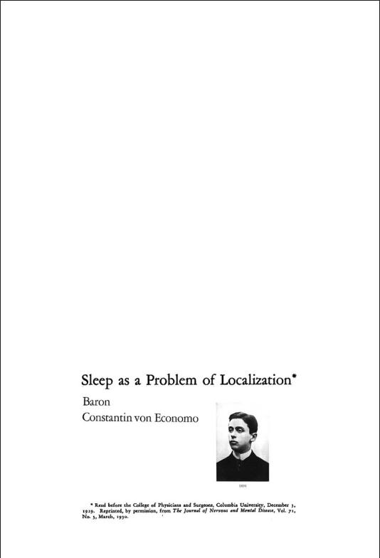
Box 1–1 Constantin von Economo and the Discovery of Intrinsic Wake and Sleep Systems in the Brain
Baron Constantin von Economo von San Serff was born in 1876, the son of Greek parentage. He was brought up in Austrian Trieste, studied medicine in Vienna, and in 1906 took a post in the Psychiatric Clinic under Professor Julius von Wagner-Jauregg. In 1916 during World War I, he began seeing cases of a new and previously unrecorded type of encephalitis and published his first report of this illness in 1917. Although subsequent accounts have often confused this illness with the epidemic of influenza that swept through Europe and then the rest of the world during World War I, von Economo was quite clear that encephalitis lethargica was not associated with respiratory symptoms, and that its appearance preceded the onset of the latter epidemic. Von Economo continued to write and lecture about this experience for the remainder of his life, until his premature death in 1931 from heart disease.
Based on his clinical observations, von Economo proposed a dual center theory for regulation of sleep and wakefulness: a waking influence arising from the upper brainstem and passing through the gray matter surrounding the cerebral aqueduct and the posterior third ventricle; and a rostral hypothalamic sleeppromoting area. These observations became the basis for lesion studies done by Ranson in 1939,20 by Nauta in 1946,21 and by Swett and Hobson in 1968,22 in which they showed that the posterior lateral hypothalamic lesions in monkeys, rats, and cats could reproduce the prolonged sleepiness that von Economo had observed. The rostral hypothalamic sleep-promoting area was confirmed experimentally in rats by Nauta in 194621 and in cats by Sterman and Clemente in the 1960s.23
Interestingly, von Economo also identified a third clinical syndrome, which appeared some months after the acute encephalitis in some patients who had
Figure B1–1A. A photograph of Baron Constantin von Economo, and excerpts from the title page of his lecture on the localization of sleep and wake promoting systems in the brain. (From von Economo,19 with permission.)
(continued)
10
Pathophysiology of Signs and Symptoms of Coma |
11 |
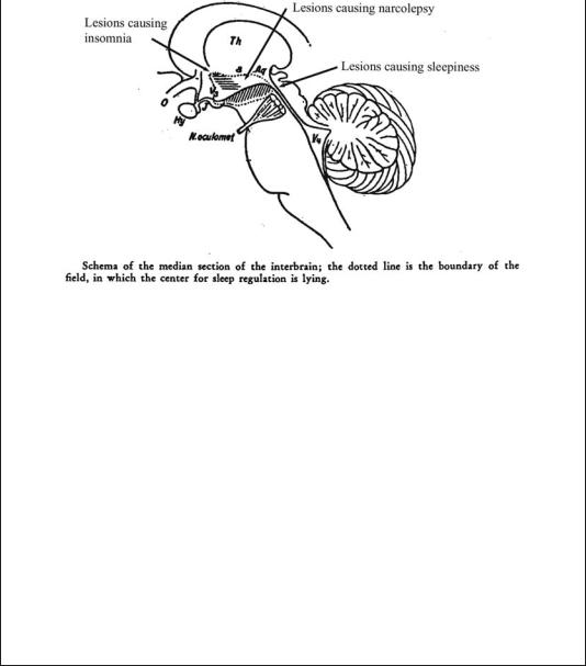
Figure B1–1B. Von Economo’s original drawing of the localization of the lesions in the brain that caused excessive sleepiness and insomnia. (Modified from von Economo,19 with permission.)
posterior hypothalamic lesions, as they were beginning to recover. These individuals would develop episodes of sleep attacks during which they had an overwhelming need to sleep. He noted that they also had attacks of cataplexy in which they lost all muscle tone, often when excited emotionally. Von Economo noted accurately that these symptoms were similar to the rare condition previously identified by Gelinaux as narcolepsy. S.A. Kinnier Wilson described a cohort of similar patients in London in 1928.24 He also noted that they had developed symptoms of narcolepsy after recovering from encephalitis lethargica with posterior hypothalamic lesions. Wilson even described examining a patient in his office, with the young house officer McDonald Critchley, and that the patient indeed had atonic paralysis, with loss of tendon reflexes and an extensor plantar response during the attack.
Von Economo’s theory was highly influential during this period, and a great deal of what was subsequently learned about the organization of brain systems controlling sleep and wakefulness owes its origins to his careful clinicopathologic observations and his imaginative and far-reaching vision about brain organization.
PHYSIOLOGY AND PATHOPHYSIOLOGY OF CONSCIOUSNESS AND COMA
The Ascending Arousal System
In the late 19th century, the great British neurologist John Hughlings-Jackson17 proposed that
consciousness was the sum total of the activity in human cerebral hemispheres. A corollary was that consciousness could only be eliminated by lesions that simultaneously damaged both cerebral hemispheres. However, several clinical observations challenged this view. As early as 1890, Mauthner18 reported that stupor in patients with Wernicke’s encephalopathy was associated with lesions involving the gray matter surrounding the cerebral aqueduct and the caudal part of
12 Plum and Posner’s Diagnosis of Stupor and Coma
the third ventricle. The nascent field of neurosurgery also began to contribute cases in which loss of consciousness was associated with lesions confined to the upper brainstem or caudal diencephalon. However, the most convincing body of evidence was assembled by Baron Constantin von Economo,19 a Viennese neurologist who recorded his observations during an epidemic of a unique disorder, encephalitis lethargica, that occurred in the years surrounding World War I. Most victims of encephalitis lethargica were very sleepy, spending 20 or more hours per day asleep, and awakening only briefly to eat. When awakened, they could interact in a relatively unimpaired fashion with the examiner, but soon fell asleep if not continuously stimulated. Many of these patients suffered from oculomotor abnormalities, and when they died, they were found to have lesions involving the paramedian reticular formation of the midbrain at the junction with the diencephalon. Other patients during the same epidemic developed prolonged wakefulness, sleeping at most a few hours per day. Movement disorders were also common. Von Economo identified the causative lesion in the gray matter surrounding the anterior part of the third ventricle in the hypothalamus and extending laterally into the basal ganglia at that level.
Von Economo suggested that there was specific brainstem circuitry that causes arousal or wakefulness of the forebrain, and that the hypothalamus contains circuitry for inhibiting this system to induce sleep. However, it was difficult to test these deductions because naturally occurring lesions in patients, or experimental lesions in animals that damaged the brainstem, almost invariably destroyed important sensory and motor pathways that complicated the interpretation of the results. As long as the only tool for assessing activity of the cerebral hemispheres remained the clinical examination, this problem could not be resolved.
In 1929, Hans Berger, a Swiss psychiatrist, reported a technologic innovation, the electroencephalogram (EEG), which he developed to assess the cortical function of his psychiatric patients with various types of functional impairment of responsiveness.25 He noted that the waveform pattern that he recorded from the scalps of his patients was generally sinusoidal, and that the amplitude and frequency of the waves in the EEG correlated closely with the level of consciousness of the patient.
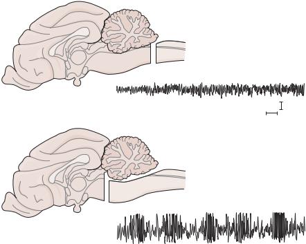
Shortly afterward, in 1935, the Belgian neurophysiologist Frederic Bremer28 (see also29) examined the EEG waveforms in cats into which he had placed lesions of the brainstem. He found that after a transection between the medulla and the spinal cord, a preparation that he called the encephale isole, or isolated brain, animals showed a desynchronized (low voltage, fast, i.e., waking) EEG pattern and appeared to be fully awake. However, when he transected the neuraxis at the level between the superior and inferior colliculus, a preparation he called the cerveau isole, or isolated cerebrum, the EEG showed a synchronized, or high-voltage, slowwave pattern indicative of deep sleep and the animals werebehaviorally unresponsive. Bremer concluded that the forebrain fell asleep due to the lack of somatosensory and auditory sensory inputs. He did not address why the animals failed to respond to visual inputs either with EEG desynchronization or by making vertical eye movements (as do patients who are locked in).
This issue was addressed after World War II by Moruzzi and Magoun,30 who placed more selective lesions in the lateral part of the midbrain tegmentum in cats, interrupting the ascending somatosensory and auditory lemniscal pathways, but leaving the paramedian reticular core of the midbrain intact. Such animals were deaf and did not appear to appreciate somatosensory stimuli, but were fully awake, as indicated both by EEG desynchronization and motor responses to visual stimuli. Conversely, when they placed lesions in the paramedian reticular formation of the midbrain, the animals still showed cortical-evoked responses to somatosensory or auditory stimuli, but the background EEG was synchronized and the animals were behaviorally unresponsive. Later studies showed that electrical stimulation of the midbrain reticular core could excite forebrain desynchronization.31 These observations emphasized the midbrain reticular core as relaying important arousing influences to the cerebral cortex, and this pathway was labeled the ascending reticular activating system. The origin of the pathway was not established in this early work.
Subsequent studies, in which transecting lesions were placed sequentially at different levels of the brainstem in cats, demonstrated that transections at the midpontine level or caudally down to the lower medulla resulted in animals that acutely spent most of their time in

Box 1–2 The Thalamus, Basal Forebrain, and Generation of EEG Waves
The origin of the sinusoidal appearance of the waveforms in the EEG remained a mystery until the 1980s. Although it was understood that the EEG voltages are due to the summated excitatory postsynaptic potentials in dendrites of cortical neurons, the reason for the synchronous waves of dendritic potentials remained elusive. The waves of postsynaptic potentials in the cerebral cortex are now understood to be due to the intrinsic burst firing of neurons in the thalamus, basal forebrain, and the cortex itself, which produce waves of postsynaptic potentials in cortical neurons.
When the membrane potential of burst neurons is close to their firing threshold, they fire single action potentials that transmit sensory and other information. However, when burst neurons have been hyperpolarized to membrane potentials far below their usual threshold for firing sodium action potentials, a low-threshold calcium channel is deinactivated. When the low-threshold calcium channel is triggered, calcium entry brings the membrane potential to a plateau that is above the threshold for firing sodium action potentials. As a result, a series of sodium spikes are fired, until sufficient calcium has entered the cell to activate a calcium-activated potassium current. This potassium current then brings the cell back to a hyperpolarized state, terminating the burst of action potentials. The more deeply the resting
A |
Waking |
B |
Slow-wave sleep |
EEG
Thalamic firing extracellular
Thalamic firing intracellular
Single spikes
0.5 s Single spikes
50 mV
100 ms
Ca2+
Bursts
Bursts
Na+
Figure B1–2. Thalamic relay neurons have transmission and burst modes of firing. (A) During transmission mode, which operates mainly during wakefulness, individual neurons in the thalamus fire single spikes in patterns that reflect their incoming afferent inputs. This correlates with a desynchronized electroencephalogram. (B) During burst mode, the thalamic neurons are hyperpolarized by gamma-aminobutyric acid (GABA)-ergic afferents, deinactivating a low-threshold calcium current with a long plateau. This brings the cell above the threshold for firing sodium action potentials, which are fired in a burst, until this is terminated by a calcium-activated potassium current that hyperpolarizes and silences the cell. These bursts tend to fire rhythmically, in correspondence with high-voltage slow waves in the EEG, which reflect large volleys of synchronized excitatory inputs reaching cortical dendrites. (From Saper, C. Brain stem modulation of sensation, movement, and consciousness. Chapter 45 in: Kandel, ER, Schwartz, JH, Jessel, TM. Principles of Neural Science. 4th ed. McGraw-Hill, New York, 2000, pp. 871–909. By permission of McGraw-Hill.)
(continued)
13

14 Plum and Posner’s Diagnosis of Stupor and Coma
Box 1–2 The Thalamus, Basal Forebrain, and Generation of EEG Waves (cont.)
membrane potential of the cells is hyperpolarized, the less frequent but longer the bursts become.
The bursting behavior of neurons in the thalamic relay nuclei, which are a major source of cortical inputs, is often thought to be a major source of cortical EEG. The synchrony is credited to the thalamic reticular nucleus, which is a thin sheet of GABAergic neurons that covers the thalamus like a shroud. Thalamic axons on their way to the cerebral cortex, and cortical projections to the thalamus, give off collaterals to the reticular nucleus as they pass through it. Neurons in the reticular nucleus provide GABAergic inputs to the thalamic relay nuclei, which hyperpolarizes them and sets them into bursting mode.
However, there is evidence that the synchrony of EEG rhythms across the cerebral cortex is due in large part to corticocortical connections, and that even isolated slabs of cortex can set up rhythmic slow-wave potentials.26 Recent evidence suggests that the basal forebrain may play a critical role in entraining cortical rhythmic activity. Basal forebrain neurons also fire in bursts that are time-locked to cortical rhythms. In addition, cell-specific lesions of the basal forebrain can eliminate fast cortical rhythms, including those associated with wakefulness and rapid eye movement (REM) sleep, whereas large cell-specific thalamic lesions have surprisingly little effect on the cortical EEG.27
Thus, the waveforms of the cortical EEG appear to be due to complex interactions among the burst neurons in the thalamus, cortex, and basal forebrain, all of which receive substantial inputs from the ascending arousal system.
a wakeful state.32 Thus, the lower brainstem was thought to play a synchronizing, or sleeppromoting, role.33 Transections from the rostral pons forward produced EEG slowing and behavioral unresponsiveness. Periods of forebrain arousal returned after several days if the animals were kept alive. However, it is clear that the slab of tissue from the rostral pons through the caudal midbrain (the mesopontine tegmentum) contains neural structures that are critically important to forebrain arousal, at least in the acute setting.
At the time, little was known about the origins of ascending projections from the mesopontine tegmentum to the forebrain, and the arousal effect was attributed to neurons in the reticular formation. However, more recent studies have shown that projections from the mesopontine tegmentum to the forebrain arise from several well-defined populations of neurons. The major source of mesopontine afferents that span the entire thalamus is a collection of cholinergic neurons that form two large clusters, the pedunculopontine and laterodorsal tegmen-
tal nuclei.34 These neurons project through the paramedian midbrain reticular formation to the relay nuclei of the thalamus (which innervate specific cortical regions), as well as the midline and intralaminar nuclei (which innervate the entire cortex more diffusely), and the reticular nucleus. As noted in Box 1–2, the reticular nucleus plays a critical role in regulating thalamocortical transmission by profoundly hyperpolarizing thalamic relay neurons via GABAB receptors.35 Cholinergic inputs in turn hyperpolarize the reticular nucleus. Other neurons in the cholinergic pedunculopontine and laterodorsal tegmental nuclei send axons into the lateral hypothalamus, where they may contact populations of neurons with diffuse cortical projections (see below). Neurons in the pedunculopontine and laterodorsal tegmental nuclei fire fastest during REM sleep (see Box 1–3) and wakefulness,36 two conditions that are characterized by a low-voltage, fast (desynchronized) EEG. They slow down during non-REM (NREM) sleep, when the EEG is dominated by high-voltage slow waves (Figure B1–3A).
Pathophysiology of Signs and Symptoms of Coma |
15 |
A
0.2 mV
1 s
B
Figure 1–1. Electroencephalogram (EEG) from a cat in which Frederic Bremer transected the cervicomedullary junction (A), showing a normal, desynchronized waking activity. However, after a transection at the midcollicular level (B), the EEG consisted of higher voltage slow waves, more typical of sleep or coma. (From Saper, C. Brain stem modulation of sensation, movement, and consciousness. Chapter 45 in: Kandel, ER, Schwartz, JH, Jessel, TM. Principles of Neural Science. 4th ed. McGraw-Hill, New York, 2000, pp. 871–909. By permission of McGraw-Hill.)
In addition, at the mesopontine level the brainstem contains at least three different monoamine groups whose axons project through the hypothalamus to the cerebral cortex.42 The noradrenergic locus coeruleus projects through the paramedian midbrain reticular formation and the lateral hypothalamus, innervating the entire cerebral cortex diffusely.43 Serotoninergic neurons in the dorsal and median raphe nuclei project through a similar course.44 Mixed in with the serotoninergic neurons are a smaller number of dopaminergic cells, which are an extension of the ventral tegmental dopamine group along the midline of the midbrain, into the area under the cerebral aqueduct.45 These dopaminergic neurons also project through the paramedian midbrain reticular formation. Some of them innervate the midline and intralaminar nuclei of the thalamus, and others pass through the lateral hypothalamus to the basal forebrain and prefrontal cortex. Evidence from singleunit recording studies in behaving animals
indicates that neurons in these monoaminergic nuclei are most active during wakefulness, slow down during slow-wave sleep, and stop almost completely during REM sleep.46–49
Application of monoaminergic neurotrans-
mitters to cortical neurons produces complex responses.35,50–52 In most cases, there is inhi-
bition resulting in a decrease in background firing, although firing induced by the specific stimulus to which the neuron is best tuned may not be reduced to as great a degree as background firing. In an awake and aroused individual, this alteration in firing may result in an improvement in signal-to-noise ratio, which may be critical in sharpening cortical information processing to avoid misperception of stimuli, such as occurs during a delirious state.
Although the cholinergic and monoaminergic neurons in the mesopontine tegmentum have traditionally been thought to play a major role in regulating wake-sleep states, lesions of these cell groups have relatively little effect on wakesleep states or cortical EEG.53 Recent studies