

Книги по МРТ КТ на английском языке / PLUM AND POSNER S DIAGNOSIS OF STUPOR AND COM-1
.pdf26 Plum and Posner’s Diagnosis of Stupor and Coma
Figure 1–6. A summary drawing of the laminar organization of the neurons and inputs to the cerebral cortex. The neuronal layers of the cerebral cortex are shown at the left, as seen in a Nissl stain, and in the middle of the drawing as seen in Golgi stains. Layer I has few if any neurons. Layers II and III are composed of small pyramidal cells, and layer V of larger pyramidal cells. Layer IV contains very small granular cells, and layer VI, the polymorph layer, cells of multiple types. Axons from the thalamic relay nuclei (a, b) provide intense ramifications mainly in layer IV. Inputs from the ‘‘nonspecific system,’’ which includes the ascending arousal system, ramify more diffusely, predominantly in layers II, III, and V (c, d). Axons from other cortical areas ramify mainly in layers II, III, and V (e, f). (From Lorente de No R. Cerebral cortex: architecture, intracortical connections, motor projections. In Fulton, JF. Physiology of the Nervous System. Oxford University Press, New York, 1938, pp. 291–340. By permission of Oxford University Press.)
respond only to a complex shape, such as a hand or a brush.
The organization of the cortical column does not vary much from mammals with the most simple cortex, such as rodents, to primates with much larger and more complex cortical development. The depth or width of a column, for example, is only marginally larger in a primate brain than in a rat brain. What has changed most across evolution has been the number of columns. The hugely enlarged sheet of cortical columns in a human brain provides the massively parallel processing power needed to perform a sonata on the piano, solve a differential equation, or send a rocket to another planet.
An important principle of cortical organization is that neurons in different areas of the cerebral cortex specialize in certain types of operations. In a young brain, before school age, it is possible for cortical functions to reorga-
nize themselves to an astonishing degree if one area of cortex is damaged. However, the organization of cortical information processing goes through a series of critical stages during development, in which the maturing cortex gives up a degree of plasticity but demonstrates improved efficiency of processing.95,96 In adults, the ability to perform a specific cognitive process may be irretrievably assigned to a region of cortex, and when that area is damaged, the individual not only loses the ability to perform that operation, but also loses the very concept that the information of that type exists. Hence, the individual with a large right parietal infarct not only loses the ability to appreciate stimuli from the left side of space, but also loses the concept that there is a left side of space. We have witnessed a patient with a large right parietal lobe tumor who ate only the food on the right side of her plate; when done, she would
Pathophysiology of Signs and Symptoms of Coma |
27 |
get up and turn around to the right, until the remaining food appeared on her right side, as she was entirely unable to conceive that the plate or space itself had a left side. Similarly, a patient with aphasia due to damage to Wernicke’s area in the dominant temporal lobe not only cannot appreciate the language symbol content of speech, but also can no longer comprehend that language symbols are an operative component of speech. Such a patient continues to speak meaningless babble and is surprised that others no longer understand his speech because the very concept that language symbols are embedded in speech eludes him.
This concept of fractional loss of consciousness is critical because it explains confusional states caused by focal cortical lesions. It is also a common observation by clinicians that, if the cerebral cortex is damaged in multiple locations by a multifocal disorder, it can eventually cease to function as a whole, producing a state of such severe cognitive impairment as to give the appearance of a global loss of consciousness. During a Wada test, a patient receives an injection of a short-acting barbiturate into the carotid artery to anesthetize one hemisphere so that its role in language can be assessed prior to cortical surgery. When the left hemisphere is acutely anesthetized, the patient gives the appearance of confusion and is typically placid but difficult to test due to the absence of language skills. When the patient recovers, he or she typically is amnestic for the event, as much of memory is encoded verbally. Following a right hemisphere injection, the patient also typically appears to be confused and is unable to orient to his or her surroundings, but can answer simple questions and perform simple commands. The experience also may not be remembered clearly, perhaps because of the sudden inability to encode visuospatial memory.
However, the patient does not appear to be unconscious when either hemisphere is acutely anesthetized. An important principle of examining patients with impaired consciousness is that the condition is not caused by a lesion whose acute effects are confined to a single hemisphere. A very large space-occupying lesion may simultaneously damage both hemispheres or may compress the diencephalon, causing impairment of consciousness, but an acute infarct of one hemisphere does not. Hence, loss of consciousness is not a typical feature of unilateral carotid disease unless both hemispheres
are supplied by a single carotid artery or the patient has had a subsequent seizure.
The concept of the cerebral cortex as a massively parallel processor introduces the question of how all of these parallel streams of information are eventually integrated into a single con-
sciousness, a conundrum that has been called the binding problem.97,98 Embedded in this
question, however, is a supposition: that it is necessary to reassemble all aspects of our experience into a single whole so that they can be monitored by an internal being, like a small person or homunculus watching a television screen. Although most people believe that they experience consciousness in this way, there is no a priori reason why such a self-experience cannot be the neurophysiologic outcome of the massively parallel processing (i.e., the illusion of reassembly, without the brain actually requiring that to occur in physical space). For example, people experience the visual world as an unbroken scene. However, each of us has a pair of holes in the visual fields where the optic nerves penetrate the retina. This blind spot can be demonstrated by passing a small object along the visual horizon until it disappears. However, the visual field is ‘‘seen’’ by the conscious self as a single unbroken expanse, and this hole is papered over with whatever visual material borders it. If the brain can produce this type of conscious impression in the absence of reality, there is no reason to think that it requires a physiologic reassembly of other stimuli for presentation to a central homunculus. Rather, consciousness may be conceived as a property of the integrated activity of the two cerebral hemispheres and not in need of a separate physical manifestation.
Despite this view of consciousness as an ‘‘emergent’’ property of hemispheric information processing, the hemispheres do require a mechanism for arriving at a singularity of thought and action. If each of the independent information streams in the cortical parallel processor could separately command motor responses, human movement would be a hopeless confusion of mixed activities. A good example is seen in patients in whom the corpus callosum has been transected to prevent spread of epileptic seizures.99 In such ‘‘split-brain’’ patients, the left hand may button a shirt and the right hand follow along right behind it unbuttoning. If independent action of the two hemispheres can be so disconcerting, one could only imagine

28 Plum and Posner’s Diagnosis of Stupor and Coma
the effect of each stream of cortical processing commanding its own plan of action.
The brain requires a funnel to narrow down the choices from all of the possible modes of action to the single plan of motor behavior that will be pursued. The physical substrate of this process is the basal ganglia. All cortical regions provide input to the striatum (caudate, putamen, nucleus accumbens, and olfactory tubercle). The output from the striatum is predominantly to the globus pallidus, which it inhibits by using the neurotransmitter GABA.100,101 The pallidal output pathways, in turn, also are GABAergic and constitutively inhibit the motor thalamus, so that when the striatal inhibitory input to the pallidum is activated, movement is disinhibited. By constricting all motor responses that are not specifically activated by this system, the basal ganglia ensure a smooth and steady, unitary stream of action. Basal ganglia disorders that permit too much striatal disinhibition of movement (hyperkinetic movement disorders) result in the emergence of disconnected movements that are outside this unitary stream (e.g., tics, chorea, athetosis).
Similarly, the brain is capable of following only one line of thought at a time. The conscious self is prohibited even from seeing two equally likely versions of an optical illusion simultaneously (e.g., the classic case of the ugly woman vs. the beautiful woman illusion) (Figure 1–7). Rather, the self is aware of the two alternative visual interpretations alternately. Similarly, if it is necessary to pursue two different tasks at the same time, they are pursued alternately rather than simultaneously, until they become so automatic that they can be performed with little conscious thought. The striatal control of thought processes is implemented by the outflow from the ventral striatum to the ventral pallidum, which in turn inhibits the
mediodorsal thalamic nucleus, the relay nucleus for the prefrontal cortex.100,101 By dis-
inhibiting prefrontal thought processes, the striatum ensures that a single line of thought and a unitary view of self will be expressed from the multipath network of the cerebral cortex.
An interesting philosophic question is raised by the hyperkinetic movement disorders, in which the tics, chorea, and athetosis are thought to represent ‘‘involuntary movements.’’ But the use of the term ‘‘involuntary’’ again presupposes
Figure 1–7. A classic optical illusion, illustrating the inability of the brain to view the same scene simultaneously in two different ways. The image of the ugly, older woman or the pretty younger woman may be seen alternately, but not at the same time, as the same visual elements are used in two different percepts. (From W.E. Hill, ‘‘My Wife and My Mother-in-Law,’’ 1915, for Puck magazine. Used by permission. All rights reserved.)
a homunculus that is in control and making decisions. Instead, the interrelationship of involuntary movements, which the self feels ‘‘compelled’’ to make, with self-willed movements is complex. Patients with movement disorders often can inhibit the unwanted movements for a while, but feel uncomfortable doing so, and often report pleasurable release when they can carry out the action. Again, the conscious state is best considered as an emergent property of brain function, rather than directing it.
Similarly, hyperkinetic movement disorders may be associated with disinhibition of larger scale behaviors and even thought processes. In this view, thought disorders can be conceived as chorea (derailing) and dystonia (fixed delusions) of thought. Release of prefrontal cortex inhibition may even permit it to drive mental imagery, producing hallucinations. Under such conditions, we have a tendency to believe that somehow the conscious self is a homunculus that is being tricked by hallucinatory sensory experiences or is unable to command thought processes. In fact, it may be more accurate to view the sensory experience and the behavior as manifestations of an altered consciousness due to malfunction of the brain’s machinery for maintaining a unitary flow of thought and action.
Neurologists tend to take the mechanistic perspective that all that we observe is due to ac-
Pathophysiology of Signs and Symptoms of Coma |
29 |
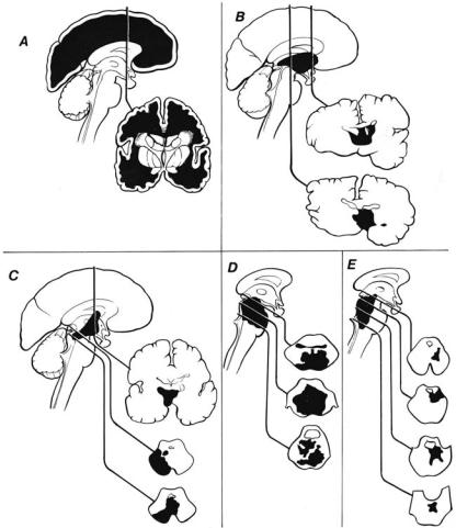
Figure 1–8. Brain lesions that cause coma. (A) Diffuse hemispheric damage, for example, due to hypoxic-ischemic encephalopathy (see Patient 1–1). (B) Diencephalic injury, as in a patient with a tumor destroying the hypothalamus.
(C) Damage to the paramedian portion of the upper midbrain and caudal diencephalon, as in a patient with a tip of the basilar embolus. (D) High pontine and lower midbrain paramedian tegmental injury (e.g., in a case of basilar artery occlusion).
(E) Pontine hemorrhage, because it produces compression of the surrounding brainstem, can cause dysfunction that extends beyond the area of the tissue loss. This case shows the residual area of injury at autopsy 7 months after a pontine hemorrhage. The patient was comatose during the first 2 months.
tion of the nervous system behaving according to fundamental principles. Hence, the evaluation of the comatose patient becomes an exercise in applying those principles to the evaluation of a human with brain failure.
Structural Lesions That Cause
Altered Consciousness in Humans
To produce stupor or coma in humans, a disorder must damage or depress the function of
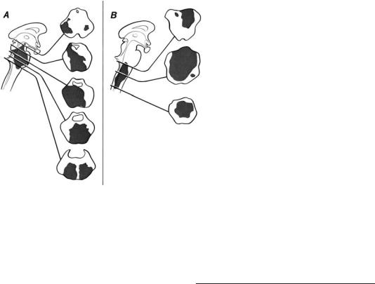
either extensive areas of both cerebral hemispheres or the ascending arousal system, including the paramedian region of the upper brainstem or the diencephalon on both sides of the brain. Figure 1–8 illustrates examples of such lesions that may cause coma. Conversely, unilateral hemispheric lesions, or lesions of the brainstem at the level of the midpons or below, do not cause coma. Figure 1–9 illustrates several such cases that may cause profound sensory and motor deficits but do not impair consciousness.
30 Plum and Posner’s Diagnosis of Stupor and Coma
BILATERAL HEMISPHERIC
DAMAGE
Bilateral and extensive damage to the cerebral cortex occurs most often in the context of hypoxic-ischemic insult. The initial response to loss of cerebral blood flow (CBF) or insufficient oxygenation of the blood includes loss of consciousness. Even if blood flow or oxygenation is restored after 5 or more minutes, there may be widespread cortical injury and neuro-
nal loss even in the absence of frank infarction.102,103 The typical appearance pathologi-
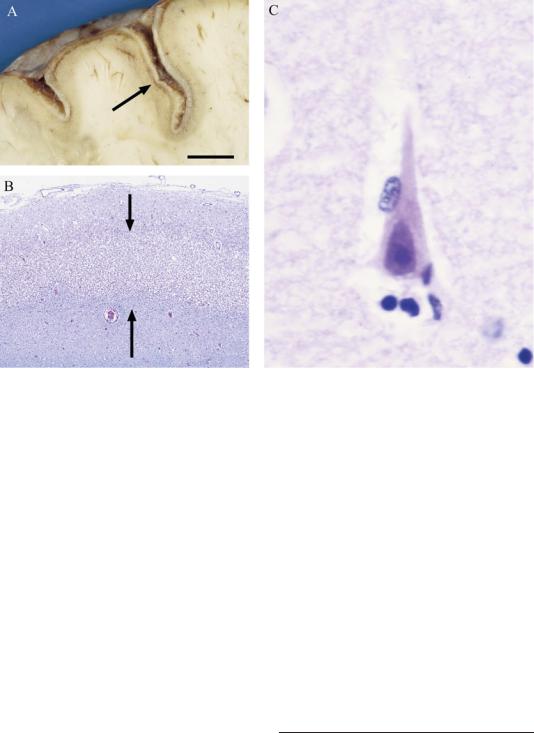
cally is that neurons in layers III and V (which receive the most glutamatergic input from other cortical areas) and in the CA1 region of the hippocampus (which receives extensive glutamatergic input from both the CA3 fields and the entorhinal cortex) demonstrate eosinophilia in the first few days after the injury. Later, the neurons undergo pyknosis and apoptotic cell death (Figure 1–10). The net result is pseudolaminar necrosis, in which the cerebral cortex and the CA1 region both are depopulated of pyramidal cells.
Alternatively, in some patients with less extreme cortical hypoxia, there may be a lucid interval in which the patient appears to recover, followed by a subsequent deterioration. Such a patient is described in the historical vignette on this and the following page. (Throughout this book we will use historical vignettes to describe cases that occurred before the modern era of neurologic diagnosis and treatment, in which
Figure 1–9. Lesions of the brainstem may be very large without causing coma if they do not involve the ascending arousal system bilaterally. (A) Even an extensive infarction at the mesopontine level that does not include the dorsolateral pons on one side and leaves intact the paramedian midbrain can result in preservation of consciousness. (B) Lesions at
a low pontine and medullary level, even if they involve a hemorrhage, do not impair consciousness. (Patient 1–2, p. 33)
the natural history of a disorder unfolded in a way in which it would seldom do today. Fortunately, most such cases included pathologic assessment, which is also all too infrequent in modern cases.)
HISTORICAL VIGNETTE
Patient 1–1
A 59-year-old man was found unconscious in a room filled with natural gas. A companion already had died, apparently the result of an attempted double suicide. On admission the man was unresponsive. His blood pressure was 120/80 mm Hg, pulse 120, and respirations 18 and regular. His rectal temperature was 1028F. His stretch reflexes were hypoactive, and plantar responses were absent. Coarse rhonchi were heard throughout both lung fields.
He was treated with nasal oxygen and began to awaken in 30 hours. On the second hospital day he was alert and oriented. On the fourth day he was afebrile, his chest was clear, and he ambulated. The neurologic examination was normal, and an evaluation by a psychiatrist revealed a clear sensorium with ‘‘no evidence of organic brain damage.’’ He was discharged to his relatives’ care 9 days after the anoxic event.
At home he remained well for 2 days but then became quiet, speaking only when spoken to. The following day he merely shuffled about and res-
Pathophysiology of Signs and Symptoms of Coma |
31 |
Figure 1–10. Hypoxia typically causes more severe damage to large pyramidal cells in the cerebral cortex and hippocampus compared to surrounding structures. (A) shows a low magnification view of the cerebral cortex illustrating pseudolaminar necrosis (arrow), which parallels the pial surface. At higher magnification (B), the area of necrosis involves layers II to V of the cerebral cortex, which contains the large pyramidal cells (region between the two arrows). (C) At high magnification, surviving neurons are pyknotic and eosinophilic, indicating hypoxic injury. Scale in A ¼ 8 mm, B ¼0.6 mm, and C ¼15 micrometers.
ponded in monosyllables. The next day (13 days after the anoxia) he became incontinent and unable to walk, swallow, or chew. He neither spoke to nor recognized his family. He was admitted to a private psychiatric hospital with the diagnosis of depression. Deterioration continued, and 28 days after the initial anoxia he was readmitted to the hospital. His blood pressure was 170/100 mm Hg, pulse 100, respirations 24, and temperature 1018F. There were coarse rales at both lung bases. He perspired profusely and constantly. He did not respond to pain, but would open his eyes momentarily to loud sounds. His extremities were flexed and rigid, his deep tendon reflexes were hyperactive, and his plantar responses extensor. Laboratory studies, including examination of the spinal fluid, were normal. He died 3 days later.
An autopsy examination showed diffuse bronchopneumonia. The brain was grossly normal. There was no cerebral swelling. Coronal sections appeared normal with no evidence of pallidal necrosis. Histologically, neurons in the motor cortex,
hippocampus, cerebellum, and occipital lobes appeared generally well preserved, although a few sections showed minimal cytodegenerative changes and reduction of neurons. There was occasional perivascular lymphocytic infiltration. Pathologic changes were not present in blood vessels, nor was there any interstitial edema. The striking alteration was diffuse demyelination involving all lobes of the cerebral hemispheres and sparing only the arcuate fibers (the immediately subcortical portion of the cerebral white matter). Axons were also reduced in number but were better preserved than was the myelin. Oligodendroglia were preserved in demyelinated areas. Reactive astrocytes were considerably increased. The brainstem and cerebellum were histologically intact. The condition of delayed postanoxic cerebral demyelination observed in this patient is discussed at greater length in Chapter 5.
Another major class of patients with bilateral hemispheric damage causing coma is of those
32 Plum and Posner’s Diagnosis of Stupor and Coma
who suffer from brain trauma.104 These cases usually do not present a diagnostic dilemma, as there is usually history or external evidence of trauma to suggest the cause of the impaired consciousness.
DIENCEPHALIC INJURY
The relay nuclei of the thalamus provide the largest ascending source of input to the cerebral cortex. As a result, it is no exaggeration to
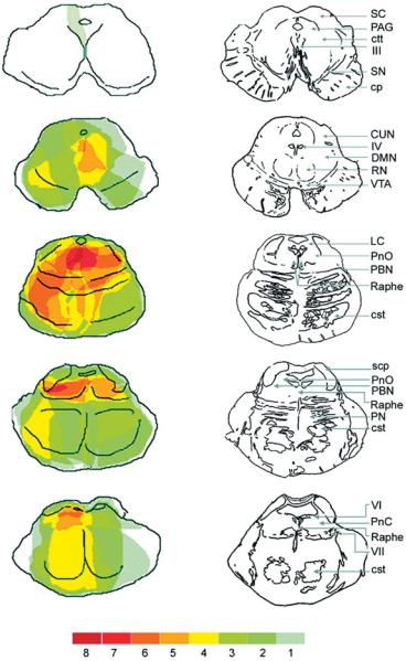
Figure 1–11. A series of drawings illustrating levels through the brainstem at which lesions caused impairment of consciousness. For each case, the extent of the injury at each level was plotted, and the colors indicate the number of cases that involved injury to that area. The overlay illustrates the importance of damage to the dorsolateral pontine tegmentum or the paramedian midbrain in causing coma. (From Parvizi and Damasio,110 with permission.)
say that virtually any deficit due to injury of a discrete cortical area can be mimicked by injury to its thalamic relay nucleus. Hence, thalamic lesions that are sufficiently extensive can produce the same result as bilateral cortical injury. The most common cause of such lesions is the ‘‘tip of the basilar’’ syndrome, in which vascular occlusion of the perforating arteries that arise from the basilar apex or the first segment of the posterior cerebral arteries can produce bilateral thalamic infarction.105

Pathophysiology of Signs and Symptoms of Coma |
33 |
However, careful examination of the MRI scans of such patients, or their brains postmortem, usually shows some damage as well to the paramedian midbrain reticular formation and often in the posterior hypothalamus. Other causes of primarily thalamic damage include thalamic hemorrhage, local infiltrating tumors, and rare cases of diencephalic inflammatory lesions (e.g., Behc¸et’s syndrome).106,107
Another example of severe thalamic injury causing coma was reported by Kinney and colleagues108 in the brain of Karen Anne Quinlan, a famous medicolegal case of a woman who remained in a persistent vegetative state (Chapter 9) for many years after a hypoxic brain injury. Examination of her brain at the time of death disclosed unexpectedly widespread thalamic neuronal loss. However, there was also extensive damage to other brain areas, including the cerebral cortex, so that the thalamic damage alone may not have caused the clinical loss of consciousness. On the other hand, thalamic injury is frequently found in patients with brain injuries who eventually enter a persistent vegetative state (Chapter 9).104
Ischemic lesions of the hypothalamus are rare, because the hypothalamus is literally encircled by the main vessels of the circle of Willis and is fed by local penetrating vessels from all the major arteries. However, the location of the hypothalamus above the pituitary gland results in localized hypothalamic damage in cases of pituitary tumors.109 The hypothalamus also may harbor primary lymphomas of brain, gliomas, or sarcoid granulomas. Patients with hypothalamic lesions often appear to be hypersomnolent rather than comatose. They may yawn, stretch, or sigh, features that are usually lacking in patients with coma due to brainstem lesions.
UPPER BRAINSTEM INJURY
Evidence from clinicopathologic analyses firmly establishes that the midbrain and pontine area critical to consciousness in humans includes the paramedian tegmental zone immediately ventral to the periaqueductal gray matter, from the caudal diencephalon through the rostral pons.110 Numerous cases are on record of small lesions involving this territory bilaterally in which there was profound loss of consciousness (see Figure 1–11). On the other hand, we have not seen loss of consciousness with lesions confined to the medulla or the caudal pons. This principle is
illustrated by the historical vignettes on pages 30 and below.
HISTORICAL VIGNETTES
Patient 1–2
A 62-year-old woman was examined through the courtesy of Dr. Walter Camp. Twenty-five years earlier she had developed weakness and severely impaired position and vibration sense of the right arm and leg. Two years before we saw her, she developed paralysis of the right vocal cord and wasting of the right side of the tongue, followed by insidiously progressing disability with an unsteady gait and more weakness of the right limbs. Four days before coming to the hospital, she became much weaker on the right side, and 2 days later she lost the ability to swallow.
When she entered the hospital she was alert and in full possession of her faculties. She had no difficulty breathing and her blood pressure was 162/ 110 mm Hg. She had upbeat nystagmus on upward gaze and decreased appreciation of pinprick on the left side of the face. The right sides of the pharynx, palate, and tongue were paralyzed. The right arm and leg were weak and atrophic, consistent with disuse. Stretch reflexes below the neck were bilaterally brisk, and the right plantar response was extensor. Position and vibratory sensations were reduced on the right side of the body and the appreciation of pinprick was reduced on the left.
The next day she was still alert and responsive, but she developed difficulty in coughing and speaking and finally she ceased breathing. An endotracheal tube was placed and mechanical ventilation was begun. Later, on that third hospital day, she was still bright and alert and quickly and accurately answered questions by nodding or shaking her head. The opening pressure of cerebrospinal fluid (CSF) at lumbar puncture was 180 mm of water, and the xanthochromic fluid contained 8,500 red blood cells/mm3 and 14 white blood cells/mm3.
She lived for 23 more days. During that time she developed complete somatic motor paralysis below the face. Several hypotensive crises were treated promptly with infusions of pressor agents, but no pressor drugs were needed during the last 2 weeks of life. Intermittently during those final days, she had brief periods of unresponsiveness, but then awakened and signaled quickly and appropriately to questions

34 Plum and Posner’s Diagnosis of Stupor and Coma
demanding a yes or no answer and opened or closed her eyes and moved them laterally when commanded to do so. There was no other voluntary movement. Four days before she died, she developed ocular bobbing when commanded to look laterally, but although she consistently responded to commands by moving her eyes, it was difficult to know whether or not her responses were appropriate. During the ensuing 3 days, evidence of wakefulness decreased. She died of gastrointestinal hemorrhage 26 days after entering the hospital.
The brain at autopsy contained a moderate amount of dark, old blood overlying the right lateral medulla adjacent to the fourth ventricle. A raspberry-appearing arteriovenous malformation, 1.4 cm in greatest diameter, protruded from the right lateral medulla, beginning with its lower border 2.5 cm caudal to the obex. On section, the vascular malformation was seen to originate in the central medulla and to extend rostrally to approximately 2 mm above the obex. From this point, a large hemorrhage extended forward to destroy the central medulla all the way to the pontine junction (Figure 1–9B). Microscopic study demonstrated that, at its most cranial end, the hemorrhage destroyed the caudal part of the right vestibular nuclei and most of the adjacent lower pontine tegmentum on the right. Caudal to this, the hemorrhage widened and destroyed the entire dorsal center of the medulla from approximately the plane of the nucleus of the glossopharyngeal nerve down to just below the plane of the nucleus ambiguus. From this latter point caudally, the hemorrhage was more restricted to the reticular formation of the medulla. The margins of this lesion contained an organizing clot with phagocytosis and reticulum formation indicating a process at least 2 weeks old. The center of the hemorrhage contained a degenerating clot estimated to be at least 72 hours old; at several places along the lateral margin of the lesion were small fresh hemorrhages estimated to have occurred within a few hours of death. It was considered unlikely that the lesion had changed substantially in size or extent of destruction in the few days before death.
Patient 1–3
A 65-year-old woman was admitted to the neurology service for ‘‘coma’’ after an anesthetic procedure. She had rheumatoid arthritis with subluxa-
tion of C1 on C2, and compression of the C2 root causing occipital neuralgia. An anesthesiologist attempted to inject the root with ethanol to eliminate the pain. Almost immediately after the injection, the patient became flaccid and experienced a respiratory arrest. On arrival in the neurology intensive care unit she was hypotensive and apneic. Mechanical ventilation was instituted and blood pressure was supported with pressors.
On examination she had spontaneous eye movements in the vertical direction only and her eyelids fluttered open and closed. There was complete flaccid paralysis of the hypoglossal, vagal, and accessory nerves, as well as all spinal motor function. Twitches of facial and jaw movement persisted. There was no response to pinprick over the face or body. CT scan showed hypodensity of the medulla and lower pons.
The patient responded to commands to open and close her eyes and learned to communicate in this way. She lived another 12 weeks in this setting, without regaining function, and rarely was observed to sleep. No postmortem examination was permitted. However, the injection of ethanol had apparently entered the C2 root sleeve and fixed the lower brainstem up through the facial and abducens nuclei without clouding the state of consciousness of the patient.
Comment: Both of these cases demonstrated the preservation of consciousness in patients with a locked-in state due to destruction of motor pathways below the critical level of the rostral pons. Chapter 2 will explore the ways in which the neurologic examination of a comatose patient can be used to differentiate these different causes of loss of consciousness.
REFERENCES
1.Scheid R, Voltz R, Guthke T, et al. Neuropsychiatric findings in anti-Ma2-positive paraneoplastic limbic encephalitis. Neurology 61, 1159–1160, 2003.
2.Mesulam MM, Waxman SG, Geschwind N, et al. Acute confusional states with right middle cerebral artery infarctions. J Neurol Neurosurg Psychiatry 39, 84–89, 1976.
3.Posner JB, Plum F. The toxic effects of carbon dioxide and acetazolamide in hepatic encephalopathy. J Clin Invest 39, 1246–1258, 1960.
4.Shimojyo S, Scheinberg P, Reinmuth O. Cerebral blood flow and metabolism in the Wernicke-Korsakoff syndrome. J Clin Invest 46, 849–854, 1967.
5.Trzepacz PT, Tarter RE, Shah A, et al. SPECT scan and cognitive findings in subclinical hepatic
Pathophysiology of Signs and Symptoms of Coma |
35 |
encephalopathy. J Neuropsychiatry Clin Neurosci 6, 170–175, 1994.
6.Nilsson K, Warkentin S, Hultberg B, et al. Treatment of cobalamin deficiency in dementia, evaluated clinically and with cerebral blood flow measurements.
Aging (Milano) 12, 199–207, 2000.
7. Van der Mast RC. Pathophysiology of delirium. J Geriatr Psychiatry Neurol 11, 138–145, 1998.
8.Frances A, Pincus HA, First MB. Diagnostic and Statistical Manuel of Mental Disorders: DSM-IV. 4th ed., 1994.
9.Peroutka SJ, Sohmer BH, Kumar AJ, et al. Hallucinations and delusions following a right temporoparietooccipital infarction. Johns Hopkins Med J 151, 181– 185, 1982.
10.Markand ON. Eectroencephalogram in ‘‘locked-in’’ syndrome. Electroencephalogr Clin Neurophysiol 40, 529–534, 1976.
11.Markand ON, Dyken ML. Sleep abnormalities in patients with brain stem lesions. Neurology 26, 769–776, 1976.
12.Billiard M, Dauvilliers Y. Idiopathic hypersomnia. Sleep Med Rev 5, 349–358, 2001.
13.Giacino JT. The vegetative and minimally conscious states: consensus-based criteria for establishing diagnosis and prognosis. NeuroRehabilitation 19, 293–298, 2004.
14.Schlotzhauer AV, Liang BA. Definitions and implications of death. Hematol Oncol Clin North Am 16, 1397–1413, 2002.
15.Towbin A. The respirator brain death syndrome. Hum Pathol 4, 583–594, 1973.
16.Criteria for the diagnosis of brain stem death. Review by a working group convened by the Royal College of Physicians and endorsed by the Conference of Medical Royal Colleges and their Faculties in the United Kingdom. J R Coll Physicians Lond 29, 381–382, 1995.
17.Jackson JH. Selected writings of John Hughlings Jackson. London: Hode and Stoughton, 1931.
18.Mauthner L. Zur Pathologie und Physiologie des Schlafes nebst Bermerkungen ueber die ‘‘Nona.’’ Wien Klin Wochenschr 40, 961–1185, 1890.
19.Von Economo C. Sleep as a problem of localization. J Nerv Ment Dis 71, 249–259, 1930.
20.Ranson SW. Somnolence caused by hypothalamic lesions in monkeys. Arch Neurol Psychiat 41, 1–23, 1939.
21.Nauta WJH. Hypothalamic regulation of sleep in rats. An experimental study. J Neurophysiol 9, 285–314, 1946.
22.Swett CP, Hobson JA. The effects of posterior hypothalamic lesions on behavioral and electrographic manifestations of sleep and waking in cat. Arch Ital Biol 106, 270–282, 1968.
23.Sterman, MB, Clemente, CD. Forebrain inhibitory mechanisms: sleep patterns induced by basal forebrain stimulation in the behaving cat. Exp Neurol 6, 103–117, 1962.
24.Wilson SAK. The Narcolepsies. In: Problems in Neurology. London: Edward Arnold, pp 76–119, 1928.
25.Berger H. Ueber das electroenkephalogramm des menschen. Arch Psychiatr Nervenkr 87, 527–570, 1929.
26.Steriade M. The corticothalamic system in sleep. Front Biosci 8, d878-d899, 2003.
27.Buzsaki G, Bickford RG, Ponomareff G, et al. Nucleus basalis and thalamic control of neocortical activity in the freely moving rat. J Neurosci 8, 4007–4026, 1988.
28.Bremer F. CR. Soc Biol 118, 1235–1241, 1935.
29.Bremer F. Cerebral hypnogenic centers. Ann Neurol 2, 1–6, 1977.
30.Moruzzi G, Magoun HW. Brain stem reticular formation and activation of the EEG. 1949. J Neuropsychiatry Clin Neurosci 7, 251–267, 1995.
31.Starzl TE, Taylor CW, Magoun HW. Ascending conduction in reticular activating system, with special reference to the diencephalon. J Neurophysiol 14, 461– 477, 1951.
32.Zernicki B, Gandolfo G, Glin L, et al. Cerveau isole and pretrigeminal rat preparations. Physiol Bohemoslov 34 Suppl, 183–185, 1985.
33.Magni F, Moruzzi G, Rossi GF, et al. EEG arousal following inactivation of the lower brainstem by selective injection of barbiturate into the vertebral circulation. Arch Ital Biol 97, 33–46, 1959.
34.Hallanger AE, Levey AI, Lee HJ, et al. The origins of cholinergic and other subcortical afferents to the thalamus in the rat. J Comp Neurol 262, 105–124, 1987.
35.McCormick DA, Bal T. Sleep and arousal: thalamocortical mechanisms. Annu Rev Neurosci 20, 185–215, 1997.
36.Strecker RE, Morairty S, Thakkar MM, et al. Adenosinergic modulation of basal forebrain and preoptic/ anterior hypothalamic neuronal activity in the control of behavioral state. Behav Brain Res 115, 183–204, 2000.
37.Aserinsky E, Kleitman N. Regularly occurring periods of eye motility, and concomitant phenomena, during sleep. Science 118, 273–274, 1953.
38.Scammell TE. The neurobiology, diagnosis, and treatment of narcolepsy. Ann Neurol 53, 154–166, 2003.
39.Kales A, Vela-Bueno A, Kales JD. Sleep disorders: sleep apnea and narcolepsy. Ann Intern Med 106, 434–443, 1987.
40.Szymusiak R, Alam N, Steininger TL, et al. Sleepwaking discharge patterns of ventrolateral preoptic/ anterior hypothalamic neurons in rats. Brain Res 803, 178–188, 1998.
41.Vgontzas AN, Kales A. Sleep and its disorders. Annu Rev Med 50, 387–400, 1999.
42.Saper CB. Diffuse cortical projection systems: anatomical organization and role in cortical function. In: F. Plum, ed. Handbook of Physiology. The Nervous System. V. Bethesda, Md.: American Physiological Society, pp 169–210, 1987.
43.Loughlin SE, Foote SL, Fallon JH. Locus coeruleus projections to cortex: topography, morphology and collateralization. Brain Res Bull 19, 287–294, 1982.
44.Bobillier P, Seguin S, Petitjean F, et al. The raphe nuclei of the cat brain stem: a topographical atlas of their efferent projections as revealed by autoradiography. Brain Res 113, 449–486, 1976.
45.Lu J, Jhou TC, Saper CB. Identification of wake-active dopaminergic neurons in the ventral periaqueductal gray matter. J Neurosci 26, 193–202, 2006.
46.Heym J, Steinfels GF, Jacobs BL. Activity of serotonincontaining neurons in the nucleus raphe pallidus of freely moving cats. Brain Res 251, 259–276, 1982.