

Anal Canal |
6 |
|
|
Sofia Gourtsoyianni and Vicky Goh |
|
6.1\ Introduction
A dedicated magnetic resonance imaging (MRI) examination of the lowest part of the gastrointestinal tract, the anus, is performed primarily in two clinical scenarios:
\(a)\ Locoregional staging of anal cancer before and after treatment (b)\\ Detection/road mapping of perianal fistula disease
The MRI protocols for these two clinical scenarios are different; however, diffusion-weighted imaging (DWI) has been shown to be a very helpful adjunctive sequence providing useful functional information in both.
6.2\ Locoregional Staging of Anal Cancer (Baseline)
Squamous cell carcinoma of the anus (SCCA) is a rare malignancy in the general population; however, it is increasingly diagnosed in patients with human immunodeficiency virus (HIV) and chronic inflammatory bowel disease and those having received immunosuppression after transplantation.
Accurate staging of SCCA at presentation, performed according to the 7th UICC/ AJCC tumor-node-metastasis (TNM) system [1], provides prognostic information
S. Gourtsoyianni (*)
Department of Radiology, Guy’s and St Thomas’ Hospitals NHS Foundation Trust, St Thomas’ Hospital, London, UK
V. Goh
Department of Cancer Imaging, School of Biomedical Engineering and Imaging Sciences, King’s College London, London, UK
e-mail: vicky.goh@kcl.ac.uk
© Springer International Publishing AG, part of Springer Nature 2019 |
77 |
S. Gourtsoyianni, N. Papanikolaou (eds.), Diffusion Weighted Imaging of the Gastrointestinal Tract, https://doi.org/10.1007/978-3-319-92819-7_6

78 |
S. Gourtsoyianni and V. Goh |
|
|
and allows for correct therapeutic planning. In particular, the longest diameter of the primary tumor rather than the extramural extension and the site of involved lymph nodes rather than their actual number are important prognosticators and also comprise major differences from rectal adenocarcinoma staging that might also be encountered extending within the anal canal.
According to the most recent practice guidelines of the European Society for Medical Oncology (ESMO) in collaboration with ESSO and ESTRO [2], MRI is the primary imaging modality of choice for accurate locoregional staging of anal cancer.
Multiplanar, high-resolution T2-weighted sequences, covering the anorectal region, using external phased array body coils, performed on ≥1.5 T MRI scanners, allow detailed imaging of the anal sphincter and the surrounding pelvic structures, providing information of local disease extent and nodal involvement, which further assist radiotherapy planning and contouring [2, 3]. In addition short-tau inversion recovery (STIR) sequences are very helpful for identifying fistula tracts [4] present at initial staging or that develop during treatment. The detailed protocol used in our institution for this purpose can be seen in Table 6.1 [5].
The sensitivity of MRI using morphological sequences for the identification of anal SCCs has been reported to approach 90–100%, with high concordance regarding tumor size [6]. On morphological T2W images, SCCA present as intermediate T2W signal intensity mass lesions. MRI unfortunately cannot distinguish low-lying rectal adenocarcinomas from SCCA that may extend upward involving the rectum as the two cancer types share similar morphological imaging characteristics. Qualitative assessment of DWI images of the two entities is also not helpful as both entities are hypercellular and demonstrate a degree of restricted diffusion (Fig. 6.1).
The incidence of involved regional lymph nodes increases with primary tumor size. Lymph node metastases may be present in 25% of cases even with superficial, ≤T2 stage tumors, at initial staging [7]. Nodal staging relies on the distance of
Table 6.1 MRI protocol for locoregional staging of SCCA
MRI |
Sequence |
Acquisition parameters |
Pelvis |
T2 axial TSE |
TR/TE, 6500/133 ms; NEX 1, ST 6 mm |
|
T1 axial SE |
TR/TE, 495/20 ms; NEX 1, ST 6 mm |
|
T2 sagittal TSE |
TR, 6500/135 ms; NEX 1, ST 5 mm |
|
Diffusion axial: |
TR, 3500/97 ms; NEX 6, ST 6 mm |
|
T2-W SS-EPI; three directions; |
|
|
b = 0, 1400 s/mm2 |
|
Tumor |
T2 axiala |
TR/TE, 6500/137 ms; NEX 4, ST 4 mm |
|
T2 coronalb |
|
|
TSE |
|
|
STIR axiala |
TR/TE/TI, 5750/25/150 ms; NEX 1, |
|
STIR coronalb |
ST 4 mm |
|
Diffusion axiala: |
TR/TE, 3000/83; NEX 6, ST 6 mm |
|
T2-W SS-EPI; three directions; |
|
|
b = 0, 50, 100, 400, 800 s/mm2 |
|
NEX number of excitations, ST slice thickness aPerpendicular and bparallel to long axis of anal canal
6 Anal Canal |
79 |
|
|
a |
b |
c |
d |
e
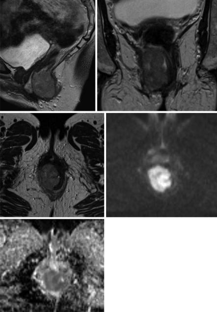
Fig. 6.1 A 63-year-old female patient presented with a SCCA and underwent baseline MRI scan for locoregional staging purposes. High-resolution T2-TSE images on sagittal (a), coronal (b), and axial (c) plane demonstrated a 4.5 cm in longest diameter mass involving the right anal sphincter complex with no visible locoregional lymph nodes rendering this a T2N0. In addition T2-W SS-EPI DWI sequence with three directions, b = 0, 50, 100, 400, 800 s/mm2, was obtained. The mass demonstrates high signal intensity on b800 image (d) and low signal intensity on corresponding ADC map (e) in keeping with restricted diffusion
80 |
S. Gourtsoyianni and V. Goh |
|
|
nodes from the primary tumor site rather than on the number of involved nodes. MRI, especially with the implementation of DWI, is helpful in identifying pelvic lymph nodes. As with rectal adenocarcinoma, it has been found that almost half of all involved lymph nodes have a diameter <5 mm. Short-axis threshold values of 8 mm, 5 mm, and 10 mm have been suggested for pelvic, mesorectal, and inguinal lymph nodes, respectively [8]. Involved lymph nodes have been reported as having signal intensity similar to or the same as that of the primary tumor [3].
A recently published retrospective study investigated in a cohort of 58 patients both qualitative and quantitative DWI parameters, namely, signal intensity and ADC values of primary lesion and lymph nodes [9], and found that neoplastic nodes showed similar signal intensity and ADC value to the anal cancer. The signal of both primary anal cancer and neoplastic lymph nodes was hyperintense on b 800 s/mm2, highest b value obtained, and hypointense on corresponding ADC map in keeping with restricted diffusion and were found to have overlapping ADC values (Fig. 6.2).
a |
b |
c
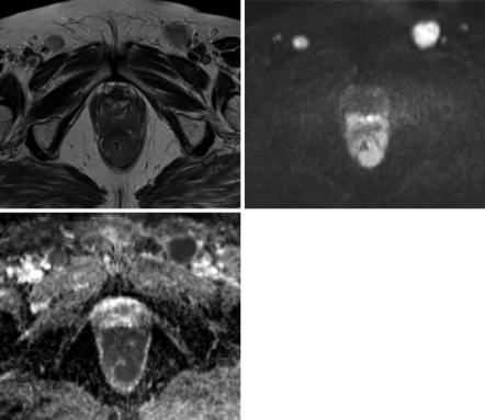
Fig. 6.2 A 77-year-old female patient presented with SCCA extending in the lower rectum (a). b800 image (b) and corresponding ADC map (c) certainly add to the radiologist’s confidence in delineating the primary tumor which demonstrates restricted diffusion, same as would have been in the case of a low-lying rectal adenocarcinoma, in addition to providing an excellent road map of disease extent with prominent bilateral inguinal lymph nodes also depicted