

3 Small Bowel |
43 |
|
|
a |
b |
c |
d |
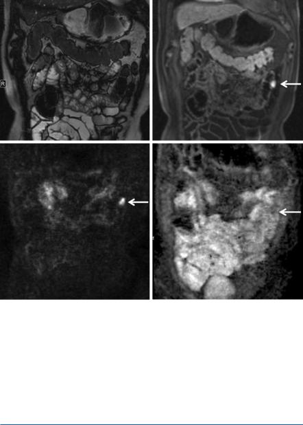
Fig. 3.7 A 72-year-old male patient with a small gastrointestinal stromal tumour (GIST) in the proximal ileum. High signal on TrueFISP imaging (a) and typical homogenous enhancement on arterial dynamic T1-weighted imaging after contrast injection (b). DWI (c) and ADC map (d) show the restricted diffusion, but less low ADC values
1.24 ± 0.34 × 10⁻³ mm2/s [35]. Figure 3.7 shows a 72-year-old patient with a small GIST of the proximal ileum with classical homogenous arterial enhancement. Compared to the lymphoma shown in Fig. 3.6, ADC value is not as low as in the lymphoma, but diffusion restriction is also clearly shown.
3.5\ Other Small Bowel Pathologies
Amongst the wide spectrum of small bowel pathologies, three very important and barely rare entities are presented in this chapter part. For all mentioned small bowel pathologies, patient’s clinical presentation and laboratory markers are essential to help differentiate based on their MR imaging changes including diffusion characteristics.
44 |
S. Kinner |
|
|
3.5.1\ Gluten-Sensitive Enteropathy
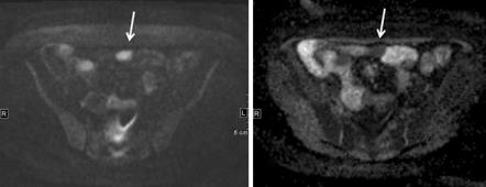
Gluten-sensitive enteropathy also known as celiac disease or celiac sprue is a T-cell- mediated disease occurring in genetically susceptible individuals induced by the ingestion of one of several proteins found in wheat (gliadins), barley (hordeins) and rye (secalinin) [40]. With a prevalence of up to 2%, it is the most frequent enteropathy in Western countries. This food can induce inflammatory processes, which can then destroy the bowel epithelial lining. Symptoms and severity of the disease can be different, and therefore MR imaging not always shows the pathology. If an inflammatory process is present, DWI can help to distinguish non-diseased from diseased bowel parts. Figure 3.8 shows a patient with known gluten-sensitive enteropathy and an inflammatory process in the small bowel.
3.5.2\ Vasculitis
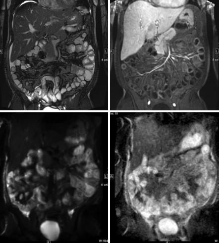
Vasculitis occurs as a systemic disease, e.g. in lupus erythematosus, polyarteritis nodosa and Henoch-Schönlein purpura. Vasculitis affects the distal small vessels, which are usually occluded or narrowed resulting in local ischemia. Usually, a relatively long bowel part is involved and the distribution is non-segmental. Involvement of the duodenum by ischemic changes is nearly always indicative of vasculitis [41]. Figure 3.9 shows a patient with ANCA (anti-neutrophil cytoplasmic autoantibody) vasculitis who was initially diagnosed on computed tomography. The follow-up examination under therapy was performed with MR imaging where changes of the small bowel caused by vasculitis can still be seen. Diffusionweighted imaging shows the restricted diffusion due to inflammatory changes in the bowel wall.
a |
b |
Fig. 3.8 A 43-year-old female patient with known gluten-sensitive enteropathy and an inflammatory process in the small bowel presenting as restricted diffusion on DWI (a) and low signal on ADC map (b)
3 Small Bowel |
45 |
|
|
a |
b |
c |
d |
Fig. 3.9 Patient with ANCA vasculitis, already under therapy. (a) balanced SSFP; (b) arterial dynamic phase; (c) DWI with a b-value of 1000; (d) ADC map still show multiple affected small bowel loops in the left upper and right lower abdomen
3.5.3\ Therapy-Induced Changes of the Small Bowel
Chemotherapy and radiation can alter the small bowel, and these changes can be determined on MR enterography. Subacute changes after radiation therapy (5–12 months after completion) can be ascribed to severe endarteritis obliterans with resultant inflammatory bowel changes which can be seen on DWI as well. Chemotherapy can lead to focal or diffuse small bowel changes.