

3 Small Bowel |
41 |
|
|
3.4\ Small Bowel Neoplasms
Small bowel neoplasms only account for 1–6% of all gastrointestinal tract tumours [34]. Until now only limited experience on DWI is distributed concerning small bowel tumours [35]. Larger studies have to be performed to establish DWI in small bowel tumour imaging.
3.4.1\ Adenocarcinoma
Adenocarcinomas are the most frequent malignancies of the small bowel, accounting for about 40%. They usually appear in the proximal small bowel and present as an annular lesion showing T2 hypointensity frequently and a heterogenous moderate enhancement on gadolinium-enhanced T1-weighted images [36]. On diffusion-weighted imaging, tumours show a diffusion restriction. By now, there is no study reporting explicitly on ADC values for adenocarcinoma. Amzallag-Bellenger et al. have reported on three patients with adenocarcinoma of the jejunum or ileum, who presented with miscellaneous ADC values measuring between 0.76 and 1.28 × 10⁻³ mm2/s.
3.4.2\ Lymphoma
Lymphomas account for about 20% of all small bowel tumours. They can be found mostly in the distal ileum due to the large amount of lymphoid tissue at this site. Patients suffering from Crohn’s disease, celiac disease and extraintestinal lymphoma and those who are immunocompromised or have had chemotherapy have a higher risk to develop small bowel lymphoma. Typically, lymphoma presents as a thickened mass of the wall without complete obstruction showing considerable dilatation. Polypoid lesions with protrusion into the bowel lumen or eccentric masses, which can also show mural ulcerations and fistulation, are much less common. Infiltration of the mesenteric fat in the absence of discrete lymphadenopathy seems to be associated with high-grade non-Hodgkin lymphoma [37].
On diffusion-weighted imaging, lymphomas have been reported to show a mean ADC value of 0.66 ± 0.19 × 10⁻³ mm2/s [35].
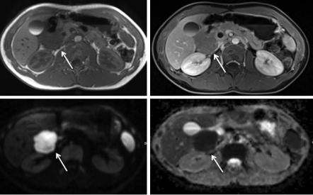
Figure 3.6 shows a 13-year-old boy diagnosed with high-grade lymphoma of the duodenum. Diffusion restriction is obvious in the lesion and ADC value clearly low.
3.4.3\ Carcinoids
Carcinoids account for about 2% of GI tumours. In the small bowel, carcinoids are the second most common tumours (31%). The most common localization for carcinoids is the appendix; second most commonly they can be found in the distal ileum.
42 |
S. Kinner |
|
|
a |
b |
c |
d |
Fig. 3.6 (a–d) A 14-year-old boy with diagnosis of a high-grade lymphoma in the duodenum. Diffusion-weighted imaging (lower row) shows the diffusion restriction, resulting in a low signal on ADC map
In about 30% of cases, carcinoids present in multiple locations. Metastases to the lymph nodes and liver occur depending on the size of the bowel lesion. On MR imaging carcinoids present as mostly isointense lesions on unenhanced T1and T2-weighted images. Especially the tumours that occur in the distal small bowel often show mesenteric masses with similar imaging features, which are 2–4 cm in size. Ileal carcinoids often present as a mesenteric mass with radiating strands of tissue [36, 38]. Annular narrowing is rarely seen with carcinoids, but the involvement of the adjacent mesentery stimulates desmoplastic reactions and fibrosis and can result in angulation and kinking of the bowel leading to obstruction and ischemia.
On diffusion-weighted imaging, carcinoids have been reported to show a mean ADC value of 0.83 ± 0.29 × 10⁻³ mm2/s [35].
3.4.4\ Gastrointestinal Stromal Tumours (GISTs)
GISTs are the most common mesenchymal tumours of the GI tract and can be accounted for 9% of all tumours of the GI tract. Most frequently they can be found in the stomach (up to 60%), followed by the jejunum and ileum (30%) and less so in the duodenum (5%). They can be small or large lesions. In the small bowel, small GISTs usually are round and present with a strong homogeneous arterial enhancement. Larger GISTs tend to present as lobulated lesions with less enhancement and cystic changes [39]. Yu et al. were able to show that the mean ADC value measured in the GISTs and the malignancy risk of the tumour correlated negatively. Diffusion- weighted imaging in GISTs has been reported to show a mean ADC value of