

3 Small Bowel |
37 |
|
|
compared to greyscale, which can help to differentiate pathologies and allocate them to bowel wall, lumen or extraluminal structures.
Quantitative analysis depends on the acquisition of diffusion-weighted images with different b-values. Apparent diffusion coefficient (ADC) values can be calculated. Regions of interests (ROIs) can be drawn in the bowel wall or other structures to record the specific ADC value of this tissue. Areas of restricted diffusion represent with a low ADC value. On DW images this restricted diffusion represents with a hyperintense signal. ADC value changes under therapy can be used to assess therapy response.
3.3\ Inflammatory Bowel Disease
Inflammatory bowel diseases (IBD) represent as recurrent or continuous chronic inflammatory conditions of the bowel due to an autoimmune disease. The two typical and most frequent entities are Crohn’s disease (CD) and ulcerative colitis (UC). Other forms of IBD are less frequent. Inflammation in UC is restricted to the mucosa, while CD shows a transmural inflammation. This is why in CD patients extraintestinal abscesses and fistulae can be found. CD can manifest throughout the whole gastrointestinal tract from the mouth to the anus with normal areas in between, whereas UC shows continuous inflammatory lesions in the colon and rectum. The terminal ileum can be involved in both entities but is much more frequently involved in CD: In the majority of CD, the pathology starts in the terminal ileum for which reason the disease is also often called ileitis terminalis. Figures 3.3 and 3.4 show patients with terminal ileitis. In UC the colonic inflammation can sometimes be “washed back” in the upstream terminal ileum and can represent as a so-called backwash ileitis. To focus on small bowel pathologies, the value of diffusion- weighted imaging in CD will be highlighted in the next section.
3.3.1\ Crohn’s Disease (CD)
Multiple studies have focused on the evaluation of diffusion-weighted imaging in detecting active inflammatory lesions in children and adults. Kiryu et al. and Oto et al. were amongst the first groups reporting on the ability of discriminating inflamed and non-inflamed bowel segments with DWI [17, 22].
Few studies reported higher diagnostic accuracy of DWI compared to gadolinium- enhanced T1-weighted MR imaging [23] and suggested to replace contrast-enhanced MR imaging with DWI. Multiple studies compared standard sequences to protocols with and without DWI: Qi et al. reported that DWI combined with standard sequences has a higher diagnostic accuracy (92%) than standard sequences alone (79%) to detect disease activity [24]. Hahnemann et al. evaluated DWI compared to standard MR imaging in the assessment of inflammatory lesions of the small bowel [25]. They used capsule endoscopy as standard of reference. They found that DWI of the small bowel not only allowed for the detection of inflammatory lesions with
38 |
S. Kinner |
|
|
a |
b |
c |
d |
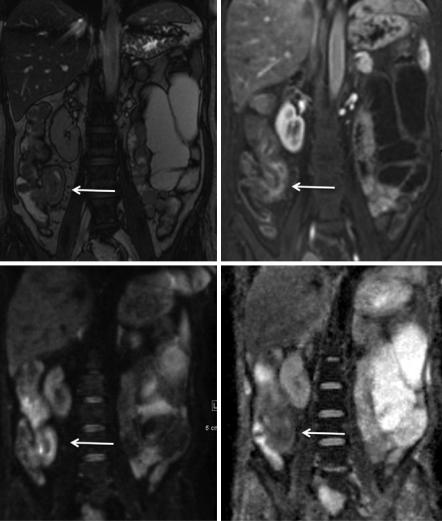
Fig. 3.3 A 15-year-old patient with known Crohn’s disease. MR imaging shows terminal ileitis in (a) TrueFISP and (b) dynamic T1-weighted imaging as well as DWI (c, b-value 1000; d, ADC map)
high accuracy but also enabled the identification of additional lesions that were not found on standard MR imaging alone. Li et al. found similar results when looking at DWI in a paediatric patient population [13]: They found that DWI detected additional inflammatory lesions and reported that in a comprehensive imaging protocol with T2w HASTE and post-contrast scans and DWI, children can be sufficiently examined in <10 min.
Recent studies of Kim et al. and Pendse et al. reported only a small improvement in sensitivity for bowel inflammation by adding DWI to conventional sequences at
3 Small Bowel |
39 |
|
|
a |
b |
c |
d |
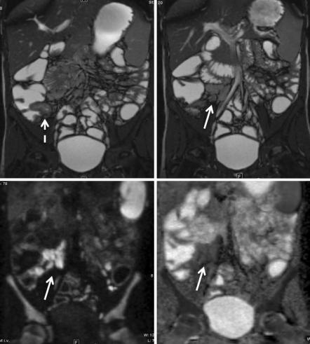
Fig. 3.4 Patient with CD and terminal ileitis (a, balanced SSFP with wall thickening of the terminal ileum) showing reactive lymphadenopathy (b, TrueFISP; c, DWI with b-value 500; d, ADC map)
the expense of reduced specificity [16, 26]. These study data suggest that DWI cannot replace conventional imaging sequences but can be an adjunct to maintain specificity.
For detection of CD, the initial studies relied on qualitative evaluations of DWI sequences alone. Quantitative evaluations using ADC values have been studied to evaluate response to treatment or to discriminate active disease and chronic fibrosing disease.
Li et al. found a strong correlation between ADC values and a simplified endoscopic score when looking at ileocolonic inflammation [27]. Conventional imaging parameters had a less strong correlation.
40 |
S. Kinner |
|
|
Stanescu-Siegmund et al. found an excellent correlation between ADC values and the Harvey Bradshaw Index as clinical parameter [28]. They found an ADC threshold of 1.56 × 10⁻³ mm2/s to be able to differentiate between normal and inflamed bowel wall. Similar cut-off values have been reported by Buisson et al. (1.6 × 10⁻³ mm2/s) [29], while Zhu et al. reported a cut-off value of 1.11 × 10⁻³ mm2/s to have highest sensitivity (100%) and specificity (68.8%).
Kovanlikaya et al. analysed ADC values in paediatric CD patients as a biomarker for fibrosis [30]. They found the ADC values of transmural fibrosis to be lower compared to the reported values of inflammation in Crohn’s disease. Tielbeek et al. showed a significant correlation between ADC values and fibrosis in an adult population [31].
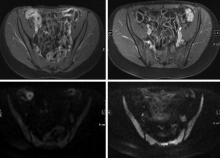
Bhatnagar et al. and Buisson et al. examined ADC values (amongst other parameters) and found them useful in detecting and assessing inflammatory activity but also to predict efficacy of anti-TNF induction therapy in CD [32, 33]. Figure 3.5 shows a young female patient with CD before and after anti-inflammatory therapy. Diffusion-weighted imaging depicts the therapy response in a similar way as contrast-enhanced T1-weighted imaging.
a |
b |
c |
d |
Fig. 3.5 (a–d) CD patient before (left) and after (right) therapy: Upper row, contrast-enhanced T1-weighted imaging; lower row, DWI with b-value 1000. Before therapy inflammatory lesions in the terminal ileum as well as proximal ileum can be found. These changes are nearly gone in the posttherapeutic control