

4 Large Bowel |
57 |
|
|
Solak et al. retrospectively evaluated 26 patients with malignant disease and 15 patients with benign conditions of the colorectum by DW-MRI, visually assessing high b-value (b = 800 s/mm2) DWI images and ADC maps, and also quantifying ADC values, having defined endoscopic biopsy as the gold standard [4]. Results from this study demonstrated that the difference between the mean ADC values of benign and malignant conditions was statistically significant, with ADC values of benign lesions being significantly higher than those of malignant lesions. By applying a cutoff value of 1.21 × 10⁻³ mm2/s, ADC yielded a sensitivity of 100%, a specificity of 87.3%, and an accuracy of 89.3% in the discrimination of malignant colorectal pathology. With the combined visual assessment of the high b-value images and the measurement of ADC values, malignant and benign lesions could be differentiated with 100% sensitivity, 89.2% specificity, and 90.4% accuracy. Importantly also, although some benign lesions were interpreted as malignant, no malignant lesion was judged to be benign on the visual assessment [4].
Other authors directed their attention to the differentiation between a particular inflammatory condition (acute diverticulitis) and cancer. Both clinical conditions may show overlapping signs, symptoms, and imaging features—particularly on CT—and the coexistence of acute diverticulitis superimposed on a colon cancer may obscure the latter on imaging. Therefore, DW-MRI was tested as an alternative to CT in establishing the diagnosis of diverticulitis [5]. Öistämö et al. retrospectively examined patients presenting with either diverticulitis or sigmoid cancer with DW-MRI. This study reported a sensitivity and specificity for the diagnosis of colon cancer and diverticulitis of 100% when using DW-MRI, whereas the sensitivity and specificity for the diagnosis of colon cancer and diverticulitis were 67% and 93%, respectively, using CT [5]. However this study included only two groups of 15 patients, and its results should be confirmed and further validated in larger studies.
In these works, the differentiation between malignant and benign lesions of the colonic wall relied on the identification of hyperintense areas within the colonic wall on high b-value images and lower ADC values, in the former group of diseases.
4.5\ Assessment of Inflammatory Bowel Disease
One major application of DW-MRI consists of the evaluation of inflammatory bowel diseases. In fact, the largest body of evidence on the role of DW-MRI in the colon comes from the assessment of ulcerative colitis and Crohn’s disease. Several works have been published in the field, regarding detection of colonic inflammation, assessment of disease activity, and evaluation of response to therapy.
4.5.1\ Detection of Inflammatory Changes in the Colon
A feasibility study by Oto et al. was designed to determine the possibility of a role for DWI in the detection of bowel inflammation and to investigate the changes in ADC values of the inflamed bowel in patients with Crohn’s disease, with pathologic
58 |
L. C. Semedo |
|
|
a |
b |
c |
d |
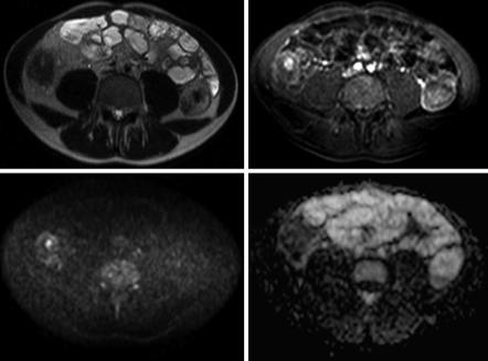
Fig. 4.4 31-year-old male with Crohn’s disease. On the T2-weighted image, there is thickening of the wall of the right colon (a) with correspondent hyperenhancement on the fat-suppressed, gadolinium-enhanced, T1-weighted image (b). On the DWI sequence (b1000), there is restriction to diffusion with hyperintensity of the colonic wall (c) and low signal intensity on the ADC map (d), compatible with inflammatory changes at that level
features as the gold standard [6]. Inflammation of the bowel wall causes restricted diffusion, and as such DWI yields both qualitative (increased signal intensity) and quantitative (decreased ADC values) information that can be helpful in the evaluation of bowel inflammation (Fig. 4.4). In addition to the increased number of inflammatory cells, dilated lymphatic channels, hypertrophied neuronal tissue, and the development of granulomas in the bowel wall can further narrow the extracellular space and therefore contribute to the restricted diffusion of water molecules.
Diffusion-weighted magnetic resonance colonography (DW-MRC) without oral or rectal preparation proved to be a reliable tool for detecting colonic inflammation in several studies. The technique does not need fasting, is noninvasive, and does not require any bowel preparation. Oussalah et al. studied 96 patients with both ulcerative colitis and Crohn’s disease (68 had concomitant endoscopy) with DW-MRC without fasting or oral or rectal preparation [7]. On DW-MRC, six radiological signs were studied: (1) DWI hyperintensity, (2) rapid gadolinium enhancement after intravenous contrast medium administration, (3) differentiation between the mucosa-submucosa complex and the muscularis propria, (4) bowel wall thickening, (5) parietal edema, and
(6) the presence of ulceration(s). In the ulcerative colitis group, the presence of a DWI hyperintensity demonstrated a sensitivity and a specificity of 90.79% and 80%, respectively, for the detection of endoscopic inflammation, with an area under the ROC curve
4 Large Bowel |
59 |
|
|
of 0.854. DWI hyperintensity was statistically more effective for the detection of endoscopic colonic inflammation in ulcerative colitis than in Crohn’s disease. Comparatively, in the ulcerative colitis group, rapid gadolinium enhancement correlated with endoscopic inflammation in the colon with a sensitivity and specificity of 72.37% and 96.67%, respectively, with an area under the ROC curve of 0.845. Rapid gadolinium enhancement was significantly more effective for the detection of endoscopic inflammation in ulcerative colitis than in Crohn’s disease. Of note, there was no statistically significant difference in accuracy between DWI hyperintensity and rapid gadolinium enhancement areas under the ROC curves in ulcerative colitis and Crohn’s disease. In ulcerative colitis, ROC analyses for the four remaining parameters of the MR score for the detection of endoscopic inflammation demonstrated a good sensitivity (88.16%) and specificity (83.33%) for the “differentiation between the mucosa-submucosa complex and the muscularis propria.” For the three other items, the sensitivity was low, ranging from 38.16% to 67.11%, with excellent specificities ranging from 93.33% to 96.67%. In Crohn’s disease, ROC analyses for the same four parameters revealed low sensitivities ranging from 36.11% to 62.5% and good to excellent specificities ranging from 75% to 100%. Among these four parameters, the “differentiation between the mucosa-submucosa complex and the muscularis propria” and “ulcerations” exhibited better accuracy for the detection of endoscopic inflammation in ulcerative colitis than in Crohn’s disease. The accuracy was similar for the other two items in both ulcerative colitis and Crohn’s disease. Logistic regression analysis showed that DWI hyperintensity was predictive of the presence of endoscopic inflammation in both the ulcerative colitis and the Crohn’s disease groups (odds ratio = 13.26 and 2.67, respectively) [7].
Similarly, Sirin et al. used DW-MRC to assess whether intravenous contrast was needed to depict inflammatory lesions in the bowel when DWI was also available, in a pediatric population [8]. In this retrospective study, patients received bowel preparation, and optical colonoscopy was the gold standard for the 37 individuals studied. Mean sensitivity and specificity for two readers for the depiction of inflammatory lesions were, respectively, 78.4% and 100% using gadolinium-enhanced T1-weighted MRC, 95.2% and 100% using DWI, and 93.5% and 100% combining both imaging techniques compared with colonoscopy (including results of the histopathological samples). In six patients, inflammatory lesions were only detected by DWI; in another six patients, DWI detected additional lesions. The preferred b-value with the best detectability of the lesions was b = 1000 s/mm2 in 28 of the 30 patients (93.3%) (8).
A recent study also assessed the role of DW-MRI without bowel preparation in the detection of ulcerative colitis in 20 patients with optical colonoscopy as the gold standard [9]. The authors assessed the following imaging signs: (1) DWI hyperintensity (b = 800 s/mm2), (2) rapid gadolinium enhancement after intravenous contrast medium administration (20–25 s after gadolinium infusion), (3) differentiation between the mucosa-submucosa complex and the muscularis, (4) bowel wall thickening (exceeding 5 mm), (5) parietal edema, (6) the presence of ulceration(s), and (7) comb sign of engorged vasa recta that perpendicularly penetrated the bowel wall. The results showed that DWI provided qualitative and quantitative information when this technique was combined with conventional magnetic resonance imaging without bowel preparation; the combined technique demonstrated a good diagnostic performance to detect colonic inflammation in ulcerative colitis. DWI hyperintensity at b = 800 s/mm2 detected
60 |
L. C. Semedo |
|
|
endoscopic colonic inflammation with a sensitivity of 93.0% and a specificity of 79.3% with an area under the ROC curve of 0.867. With rapid gadolinium enhancement, endoscopic colonic inflammation was detected with a sensitivity of 73.2%, a specificity of 93.1%, and an area under the ROC curve of 0.853. The accuracy between DWI hyperintensity and rapid gadolinium enhancement was not significantly different. Differentiation between the mucosa-submucosa complex and the muscles revealed a good sensitivity (80.3%) and specificity (86.2%). The four other signs demonstrated low sensitivities (range, 43.7–66.2%) and excellent specificities (range, 89.7–93.1%) [9].
From the analysis of the published works, it seems reasonable to recognize that DW-MRC, which combines morphological MRI and DWI, even without oral or rectal preparation might be used in clinical practice to evaluate colonic inflammation, particularly in ulcerative colitis. DWI has the potential to replace gadolinium- enhanced sequences in order to detect inflammatory changes in the colon, therefore reducing the likelihood of adverse effects from the use of gadolinium-based contrast media and, at the same time, reducing examination costs.
4.5.2\ Assessment of Disease Activity
With the aim of assessing disease activity in patients with ulcerative colitis, Kılıçkesmez et al. prospectively studied 28 patients in different stages of the disease by means of DW-MRI without preparation, measuring ADC of the bowel wall in sigmoid colon and rectum and comparing the findings with endoscopy [10]. Results disclosed no statistically significant difference in the ADC of the sigmoid colon in patients with active, subacute, and remissive ulcerative colitis. On the contrary, ADC values of the rectum were statistically different between patients in the active (1.08 ± 0.14 × 10⁻³ mm2/s) and subacute phases (1.13 ± 0.23 × 10⁻³ mm2/s) of disease and those in remission (1.29 ± 0.17 × 10⁻³ mm2/s). Therefore, an increased activity of the disease was correlated with lower ADC values [10].
Similarly, Kiryu et al. investigated the application of free-breathing DW-MRI to assess active Crohn’s disease [11]. The findings of a conventional barium study or surgery were regarded as the gold standard. The ADC was significantly lower in the disease-active segments than in the disease-inactive segments in the large bowel (1.52 ± 0.43 × 10⁻³ mm2/s versus 2.31 ± 0.59 × 10⁻³ mm2/s, respectively). The sensitivity, specificity, and accuracy were 85.7%, 75.7%, and 77.3%, respectively, in the large bowel. The accuracy was 82.6% in the ascending colon, 85.0% in the transverse colon, 80.8% in the descending colon, 72.4% in the sigmoid colon, and 70.0% in the rectum [11].
Diffusion-weighted magnetic resonance enterocolonography (DW-MREC) with no bowel cleansing and no rectal enema was performed by Buisson et al. to prospectively evaluate patients with Crohn’s disease, specifically for the indirect detection of ulcerations in this setting [12]. Forty-four patients were studied and results were compared to the gold standard (ileocolonoscopy): a total of 158 colorectal segments were assessed. The authors showed that not only the segmental ADC measured on the bowel wall in these segments was correlated with endoscopic scores but also that MRI accuracy to detect endoscopic ulcerations, using a ADC < 1.88 for colon/
4 Large Bowel |
61 |
|
|
rectum, ranged from 63.2% (cecum/right colon) to 84.6% (left/sigmoid colon). This work also disclosed a relationship between ulcer size and ADC: the segmental ADC values decreased when the ulceration size increased [12].
Sato et al. also aimed to compare the findings of DW-MREC with endoscopically identified lesions according to inflammatory grades and assess the diagnostic accuracy of DW-MREC for sensitivity and grading severity in Crohn’s disease [13]. A total of 27 patients were evaluated. A positive lesion was defined as having at least one of the following: wall thickness, edema, high intensity on DWI images, and relative contrast enhancement on MREC. The sensitivities were 100% for ulcer, 84.6% for erosion, and 52.9% for redness, suggesting an ability to detect milder lesions such as erosion or redness in MREC. For DWI in specific, the sensitivities for endoscopic ulcer, erosion, and redness were 80.9%, 69.2%, and 33.3%, respectively. The specificities for endoscopically identified lesions were high (92.1%). When the lesion was defined as having two or three positive MREC findings, sensitivity increased: sensitivities for either wall thickness or DWI high intensity, either edema or DWI high intensity, and either wall thickness or edema or DWI high intensity were 80.9%, 80.9%, and 80.9% for ulcer, respectively; 76.9%, 69.2%, and 76.9% for erosion, respectively; and 41.2%, 35.3%, and 41.2% for redness, respectively. Specificities were 92%, 92%, and 92%, respectively [13].
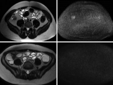
Therefore, DWI could be an adjunct to differentiate between active inflammation and quiescent disease (Fig. 4.5).
a |
b |
c |
d |
Fig. 4.5 Two different patients (a, b, 19-year-old male; c, d, 16-year-old female) with Crohn’s disease showing areas of wall thickening at the level of the right colon on the T2-weighted images (a, c). On the DWI sequence (b1000), there is restriction of diffusion with hyperintensity of the colonic wall in the first patient (b) indicating active disease, whereas in the second patient, no areas of restricted diffusion are seen, corresponding to quiescent disease