


34 |
S. Kinner |
|
|
therefore on tissue microarchitecture, it provides functional quantitative information and helps to distinguish diseased and normal tissue and therefore to assess therapy response [3]. DWI is a non-contrast technique, which is of extreme importance when it comes to imaging patients in whom the injection of a gadoliniumbased contrast agent should be omitted. This is the case in pregnant patients, patients with renal impairment and also at times for children. In general, with the recent discussion of gadolinium deposition in the brain [4], each contrast agent injection should be carefully considered, and if DWI proves to be able to replace contrastenhanced imaging, it should be used as a first-line technique.
3.2\ Prerequisites
3.2.1\ Patient Preparation
For small bowel MR imaging, patients should fast for 4–6 h at least. If only the small bowel is of interest, a thorough cleansing protocol does not have to be followed. Generally, there are two ways to achieve distension of the small bowel. Patients can drink a hyperosmolar solution about 45–60 min prior to the examination (MR enterography). For the alternate technique, MR enteroclysis, the solution is given via a nasoenteric tube, which is placed under fluoroscopic guidance before MR imaging [5]. This technique requires for the patient to change location, is more time-consuming and is burdened with associated tube placement radiation exposure. Some studies show that MR enteroclysis is able to distend the small bowel loops a little better than MR enterography [6–8]; others did not find a difference [9].
In our institution, we perform MR enterography and use an in-house pharmacy- made solution of locust bean gum and mannitol (LBG-mannitol). The patient is asked to drink at least 1 L of the solution before imaging [10]. Other oral solutions (containing polyethylene glycol, barium sulphate or alike) have been tested and perform similarly [11].
Recent studies suggest that DWI can be performed without bowel preparation reliably if looking at the large bowel [12]. For small bowel imaging, we still believe that bowel distension is of extreme importance as collapsed bowel loops mostly show higher signal in DWI and can therefore mimic pathologies, which have also been discussed by Li et al. in their paper. In their opinion poor loop distension results in apparent thickening and signal increase of the collapsed bowel segment mimicking inflammation on standard sequences and DWI [13].
To suppress peristalsis and concomitant artefacts, intravascular or intramuscular agents have to be injected. We use hyoscine butylbromide, also known as scopolamine butylbromide (Buscopan, Boehringer Ingelheim, Ingelheim, Germany), which is injected intravenously and has a half-life of 5 h. If contraindications for scopolamine are present (glaucoma, benign prostate hyperplasia) or in countries where butylscopolamine is not approved, glucagon (GlucaGen, Novo Nordisk Inc., Princeton, New Jersey) can be used [14]. The half-life is much shorter with 8–18 min but sufficient for imaging. We usually administer the spasmolytic agent shortly
3 Small Bowel |
35 |
|
|
before dynamic imaging and DWI intravenously as balanced steady state free precession images are less prone to motion artefacts and the onset of the effect lies around 1 min. If an intramuscular administration is chosen, patients have to be injected before imaging, as the onset of effect takes about 10 min.
Patients in our department are placed in prone position on the MR table. We use a 50 cm field of view for imaging to cover the whole abdomen from the diaphragm to the symphysis. The prone position reduces the diameter of the abdomen that has to be imaged and thus shortens scan time and also reduces motion artefacts.
3.2.2\ Imaging Protocol
Diffusion-weighted imaging can be performed in axial and coronal views. Most authors include one view, but from our own experience, we would always add both orientations, as short pathologies can be missed in one or the other view. Furthermore, coronal views are often more hampered by artefacts [15].
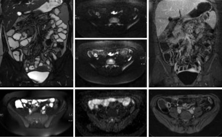
Figure 3.1 shows a typical imaging protocol used in our institution.
The typical slice thickness for DWI lies between 5 and 9 mm [16, 17]. Our protocol includes a coronal DWI sequence with a slice thickness of 7 mm and an axial DWI sequence with a slice thickness of 5 mm.
Diffusion-weighted imaging can be acquired during free breathing, but also breath-hold techniques and respiratory gated sequences are available. We use a free- breathing technique in our study as Kwee et al. showed that DWI data in liver
Fig. 3.1 Protocol for small bowel MR imaging used in our clinical routine: left column T2-weighted imaging and TrueFISP; middle column DWI with b-values of 50 and 1000 and corresponding ADC map; right column dynamic and post-contrast T1-weighted imaging
36 |
S. Kinner |
|
|
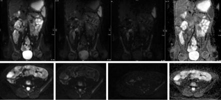
a |
b |
c |
d |
Fig. 3.2 Coronal (upper row) and transverse (lower row) diffusion-weighted imaging (a–c) and ADC map (d) showing terminal ileitis in a 17-year-old patient. B-values of 50 (a) show the lesion less good as b-values of 500 (b) and 1000 (c)
imaging were less reproducible when using a breath-hold DWI technique [18]. In a study by Muro et al. in a moving phantom, the authors were able to show that stable motion such as calm respiration does not cause signal loss on DWI [19]. Therefore, DWI using a free-breathing technique is well suited for DWI of the small bowel as patients in prone position mostly do not significantly move on the MR table and bowel movements can be sufficiently suppressed using spasmolytic agents.
Until now, there is no general agreement on which b-values should be used for DWI of the small bowel. Most groups perform DWI with two or three b-values. The lower b-value is chosen as 0 or 50 and reflects a T2-weighted image, where all water-containing structures (especially liquid bowel content) are displayed with high signal intensity. If two b-values are chosen, the second one is between 500 and 1000. The higher the b-value, the more “normal” fluids are suppressed, and only the tissues with restricted diffusion represent with a higher signal on diffusion-weighted imaging (Fig. 3.2). However, this comes at the cost of lower SNR performance.
We typically use three b-values (b = 50, 500 and 1000), and from our experience the highest b-value shows pathologies in the bowel best most of the time [20].
3.2.3\ DWI Analysis
Diffusion-weighted images can be viewed in a qualitative and/or quantitative way. For qualitative viewing images can be analysed as they are or can be colour coded and also superimposed with anatomic T1or T2-weighted images. The latter resembles hybrid imaging techniques like positron emission tomography (PET)/CT or PET/MR and can help to allocate areas with restricted diffusion to the right anatomic localization more easily than viewing anatomic and DWI images side by side [21]. Furthermore, viewing software provides a wider range of colour contrast