

66 |
D. M. J. Lambregts and R. G. H. Beets-Tan |
|
|
primary staging setting, as well as for restaging of tumours after neoadjuvant treatment. Finally, the use of DWI for follow-up after treatment, its potential value as a prognostic marker and main interpretation pitfalls for rectal DWI will be discussed.
5.2\ DWI for Primary Rectal Cancer Staging
5.2.1\ DWI for Rectal Tumour Detection
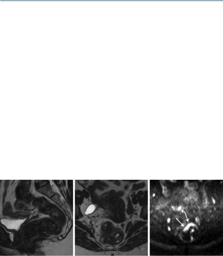
Only a few reports have focussed on the primary detection of rectal tumours using DWI. This makes sense, since the primary detection of rectal tumours is not so much a task for the radiologist. Once a patient is referred for imaging, the diagnosis is typically already clear from clinical examination and endoscopy with biopsy. The main role of imaging is therefore not to detect, but rather to stage the tumour. The studies that did focus on rectal tumour detection reported very high detection rates for DWI between 93% and 100% [2, 3]. DWI may be helpful to improve tumour conspicuity in specific cases, particularly for less experienced readers, for example, when the tumour is small and/or when there is a lot of faeces present in the rectal lumen, as a result of which the tumour may be more difficult to discern (Fig. 5.1). DWI is less useful for the assessment of mucinous-type rectal adenocarcinoma since—as opposed to tubular-type adenocarcinomas that have a dense cellular structure leading to diffusion restriction with high signal on DWI—mucinous tumours typically do not show restricted diffusion but have a relatively low signal intensity on high b-value DWI with corresponding high ADC signal (Fig. 5.2) [4].
a |
b |
c |
Fig. 5.1 Example of a female patient with a small tumour in the mid-rectum. On the sagittal (a) and transverse (b) T2-weighted images, the tumour is hardly visible and difficult to discern within the stool present in the rectal lumen. On b1000 DWI (c), the small tumour is easily recognised (arrows)
5 Rectum |
|
67 |
|
|
|
a |
b |
c |
Fig. 5.2 Transverse T2-weighted image (a), b1000 DWI (b) and ADC map (c) of a female patient with a largely mucinous tumour in the distal rectum. The mucinous components of the tumour show high signal on T2-weighted MRI with corresponding high signal on the ADC map and only slightly increased signal on b1000 DWI (caused by T2 shine through effects). On the anterior side, the tumour has a small solid component (arrows) with restricted diffusion resulting in high signal on b1000 DWI and corresponding low signal in the ADC map
5.2.2\ DWI for Rectal Tumour Staging
Key principles of rectal tumour staging are to determine the depth of tumour invasion into and beyond the different layers of the bowel wall and the minimum distance between the tumour and the mesorectal fascia (MRF) and to assess whether the tumour invades any nearby organs or structures. These factors are mainly assessed using T2-weighted MRI sequences in multiple planes parallel and perpendicular to the tumour axis as these provide good anatomical detail to visualise the morphology of the rectal wall, the mesorectal compartment and the mesorectal fascia. There appears to be little role for DWI in primary tumour staging. The few studies that investigated the use of DWI for local tumour staging reported similar sensitivities ranging between 64% and 90% for T1-T2 tumours and 88% and 95% for T3-T4 tumours and specificities of 88–95% for T1-T2 tumours and 82–100% for T3-T4 tumours, results similar to those found for T2-weighted MRI with no significant contributing value for DWI [5, 6]. The value of DWI to assess mesorectal fascia involvement has so far only been reported in the restaging setting (see Sect. 5.3.2).
5.2.3\ DWI for Lymph Node Staging
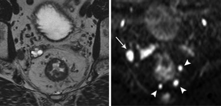
Staging of rectal cancer lymph nodes remains one the most difficult tasks for the radiologist. Theoretically, DWI could be a suitable technique to assess lymph nodes, since lymphoid tissue has a dense cellular structure which makes it easily detectable on DWI (Fig. 5.3). Different reports have indeed shown that DWI is a highly sensitive technique to detect rectal cancer lymph nodes and that it can help identify over
68 |
D. M. J. Lambregts and R. G. H. Beets-Tan |
|
|
a |
b |
Fig. 5.3 T2-weighted (a) and b1000 diffusion-weighted (b) images that illustrate how DWI can be used to help detect lymph nodes. The mesorectal lymph nodes (arrowheads) show markedly bright signal on DWI making them easier to detect than on the corresponding T2-weighted image. Also note the enlarged extramesorectal lymph node in the right obturator area (arrow)
25% more nodes compared to routine morphological T2-weighted MR sequences [7–11]. There is however no consensus on the value of DWI to discriminate between benign/reactive and metastatic lymph nodes. Heijnen et al. reported that benign and metastatic rectal cancer lymph nodes show equally high signal intensities on DWI. When using the signal intensity of nodes on DWI as a criterion to discriminate malignant nodes, results were disappointing with areas under the receiver-operator characteristics (ROC) curve (AUC) of only 0.45–50 [7]. Similarly, in a study by Mizukami and colleagues, positive predictive value was only 52% when using high signal on DWI as a criterion for malignancy, indicating that almost half of the nodes that show high signal on DWI are in fact benign [8]. Two studies quantitatively studied nodal ADC values to differentiate between benign and metastatic nodes at the time of primary staging. Significant differences in nodal ADC between benign and metastatic nodes were found, but sensitivity (67–78%) and specificity (60–67%) for nodal ADC were suboptimal [7, 10] and do not offer a considerable improvement compared to the use of the more widely used MRI criterion nodal size, with reported sensitivities and specificities of 55–78% [12, 13]. Moreover, in many of the typically small rectal cancer nodes, it can be very difficult to reproducibly measure ADC [11]. Altogether, the main benefit of DWI for primary nodal staging thus appears to help detect rather than help characterise the nodes. One report showed that studying morphologic characteristics of nodes on DWI may be helpful to characterise them and that metastatic nodes typically show a more heterogeneous signal and ill-defined border on DWI [14]. Similar results have however previously also been reported for the assessment of nodal morphology using routine T2-weighted sequences [15, 16].