


Large Bowel |
4 |
|
|
Luís Curvo Semedo |
|
4.1\ Introduction
Magnetic resonance imaging (MRI) is being increasingly performed for the assessment of lesions of the large bowel, whereby mainly morphometric macroscopic tissue information is usually obtained. Yielding insights at a cellular level, diffusion-weighted imaging (DWI) provides images whose signal intensity is sensitized to the random motion of free water molecules. The mobility of water molecules within a given voxel is determined by the microscopic cellular structure, i.e., the presence of barriers, such as cell membranes and macromolecules. Thus, DWI offers a theoretical possibility for the assessment of colonic diseases, both inflammatory and neoplastic, on a more “functional” level. DW-MR images may be evaluated both qualitatively and quantitatively: the former results from a visual assessment of the DW-MR sequences, in which areas of restricted diffusion will appear hyperintense against a hypointense background on highest b-values obtained and hypointense on corresponding ADC map; quantitative evaluation of the water diffusion characteristics is performed by expressing them as an apparent diffusion coefficient (ADC) value. Both qualitative and quantitative information have been the subject of several works on colonic diseases.
A critical review of colonic polyp and cancer detection, characterization of colonic wall thickening, and assessment of inflammatory bowel disease of the colon will be performed in this chapter. A perspective on future applications and trends will also be discussed.
L. C. Semedo
Medical Imaging Department, Coimbra Hospital and University Centre, Faculty of Medicine, University of Coimbra, Coimbra, Portugal
© Springer International Publishing AG, part of Springer Nature 2019 |
53 |
S. Gourtsoyianni, N. Papanikolaou (eds.), Diffusion Weighted Imaging of the Gastrointestinal Tract, https://doi.org/10.1007/978-3-319-92819-7_4

54 |
L. C. Semedo |
|
|
4.2\ Technical Considerations
Imaging of the large bowel with DWI is challenging. In fact, relatively long acquisition times causing an increased sensitivity to bowel motion and the presence of T2 shine-through effect, which is frequently encountered in the bowel lumen, may hamper the diagnostic information conveyed by MR images [1].
A combination of high magnetic field MR scanners, decreased acquisition times, multichannel coils, and parallel imaging techniques have been helpful to reduce these technical limitations. New pulse sequences such as echo-planar imaging have contributed to a reduction in acquisition time, therefore also decreasing sensitivity to bowel and respiratory movements [1]. At present, no clear benefit from the use of antiperistaltic agents has been clearly shown, although theoretically they may reduce bowel motion artifacts.
The T2 shine-through effect may be reduced with the use of high b-values and short echo times.
For imaging of the bowel, both a low b-value ranging from 0 to 50 s/mm2 and at least one high b-value (800–1000 s/mm2) are useful. In our daily practice, usually three b-values (b = 50, b = 500, and b = 1000 s/mm2) are routinely acquired in a high-field magnet. This allows for the calculation of ADC values minimizing the effects of microperfusion allowing at the same time the acquisition of images with high contrast and signal-to-noise ratios.
Axial images should be preferably acquired, since they are less prone to motion artifacts than those acquired in other planes, namely in the coronal plane [1].
For image acquisition, different approaches may be used: e.g., a navigator- triggered technique, which helps reduce motion artifact and increases signal-to- noise ratio, the main drawback being the increased acquisition time. Sequences acquired with the patient breathing freely or in several breath holds are generally faster but suffer from motion artifacts and less detail [1].
The use of oral and rectal cleansing before DW-MRI is questionable nowadays. If it seems reasonable to think that colonic distention would improve detection and evaluation of lesions, it is also true that there is a growing body of evidence showing that colonic assessment without bowel preparation is feasible, yields satisfactory results, and improves patient compliance by reducing discomfort.
4.3\ Detection of Polyps and Cancer
The rationale behind the use of DW-MRI for the detection of colonic polypoid lesions and cancer is that those lesions will exhibit restriction to diffusion and therefore will show high signal intensity at high b-value DW images. As such, the high lesion-to-background contrast that can be provided by DW-MRI will theoretically
4 Large Bowel |
55 |
|
|
a |
b |
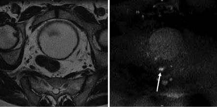
Fig. 4.1 64-year-old male with a history of occult blood loss in stool. On the T2-weighted image (a), no abnormalities are found, whereas on the DWI sequence (b1000), a small focus of high signal intensity is apparent on the topography of the upper rectum (white arrow on b), which corresponded to a sessile polyp on colonoscopy. Both sequences were obtained without previous bowel preparation
provide an advantage compared to conventional sequences (Fig. 4.1). Furthermore, DWI does not require the administration of any contrast agents and can be performed without any bowel preparation [2].
A feasibility study published by Dutch authors on the detection of polyps included 26 patients and achieved a lesion-based sensitivity of 80.0% for clinically relevant lesions (polyps ≥6 mm and cancer) [2]. Nevertheless, the authors suggested that further technical developments are required in order to increase the diagnostic yield of DW-MRI in the detection of polypoid lesions of the colon.
Some authors have also investigated the value of DW-MRI in the detection of colorectal cancer. Ichikawa et al. retrospectively assessed the diagnostic value of DWI for the detection of colorectal cancer in 33 patients with neoplasms and 15 controls and reported a sensitivity of 90.9% and specificity of 100% for the diagnosis of colonic adenocarcinoma [3]. Neoplastic lesions, due to its high cellularity, presented with high signal intensity on the high b-value images, by contrast to a dark background (Fig. 4.2). On the ADC maps, these lesions characteristically appeared with low signal intensity. Again, the detection of neoplastic lesions could be achieved on non-gadolinium-enhanced sequences, and previous bowel cleaning was not performed.
In polypoid cancers, DWI can clearly demarcate the areas within the polyp that show a high cellular content, helping to distinguish them from the low-cellular areas (Fig. 4.3).
56 |
L. C. Semedo |
|
|
a |
b |
Fig. 4.2 77-year-old female with an endoluminal lesion on colonoscopy. On the T2-weighted image, an irregular wall thickening is disclosed at the level of the distal sigmoid (white arrow on a) with some perilesional adenopathies (white arrowheads on a). On the DWI sequence (b1000), both the primary lesion (white arrow on b) and the adenopathies (white arrowheads on b) reveal predominant hyperintensity against a dark background
a |
b |
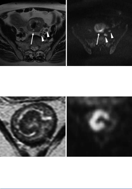
Fig. 4.3 62-year-old male with a polyp on colonoscopy. On the T2-weighted image, a large pedunculated polypoid cancer is seen at the level of the rectum (a). On the DWI sequence (b1000), the head of the polyp demonstrates high signal intensity, corresponding to the more cellular areas, in contrast to the stalk, which, because of the relatively low cellularity, appears dark on DWI (b)
4.4\ Characterization ofWall Thickening
DW-MRI has been used as a tool to help characterize diffuse bowel wall thickening, namely to distinguish between malignancy and various benign conditions, including inflammatory, ischemic, or infectious bowel diseases.