


46 |
S. Kinner |
|
|
Another entity that occurs frequently after therapy is graft-versus-host disease (GvHD) of the bowel in patients after stem cell transplantation (SCT). GvHD is one of the major causes of mortality and morbidity after allogenic SCT. It develops frequently after SCT (typically 3–11 weeks after transplantation), and 30–50% of patients are affected. Intestinal manifestation is one of the most frequent. The findings are similar to those in CD, but the extent is usually greater [42]. Diffusion- weighted imaging can help to show diseased bowel parts. Figure 3.10 shows a patient with graft-versus-host disease after stem cell transplantation for acute myeloic leucemia.
With the knowledge of previous therapy in the future, it might be possible to omit contrast injection in this patient as DWI is able to show the changes just quite as well as contrast-enhanced T1-weighted sequences.
3.6\ Appendicitis
Acute appendicitis is a common medical emergency condition that can affect children and adults. The incidence of appendicitis has been shown to increase, and the lifetime risk lies around 9%. Clinical symptoms and laboratory parameters alone often render the diagnosis of appendicitis. Ultrasound has been the diagnostic method of choice to evaluate for appendicitis but is dependent on the system used, the operator as well as the patient, especially if patients are larger [43] or if the appendix lies in a not well-visible area, e.g. retrocaecal. CT can visualize the location and condition of the appendix well but is burdened by use of ionizing radiation, which is especially a problem in paediatric and pregnant patients. The increasing prevalence and accessibility of MR have led many institutions to choose MR as the primary cross-sectional imaging tool for appendicitis. MR imaging combines the advantages of ultrasound (noninvasive, lack of ionizing radiation) with the high- resolution 3D cross-sectional information of CT [44].
Standard imaging sequences alone have shown high specificity and sensitivity in diagnosing acute appendicitis [45, 46]. Diffusion-weighted imaging has been proven to add important information [47]. Acute appendicitis appears bright on fat- suppressed DWI due to the combination of restricted diffusion and oedema that appears bright with T2 weighting. As with small bowel imaging, we typically use two to three b-values for appendicitis visualization, most often b = 0 and b = 500. The b = 0 image is effectively a fat-suppressed T2-weighted image and is ideal for visualizing periappendiceal fluids. We have found that b = 500 is a good compromise for imaging of acute appendicitis, balancing adequate diffusion weighting and good SNR performance [47]. In general, we have not found the quantitative ADC maps particularly useful for the diagnosis of acute appendicitis, although we have found the high b-value images to be very helpful for qualitative detection of oedema and inflammatory changes. Finally, we routinely use externally calibrated parallel imaging with all DWI acquisitions to reduce distortion in the phase encoding direction due to the sensitivity of echo-planar methods to magnetic field inhomogeneities.
3 Small Bowel |
47 |
|
|
a |
b |
c |
d |
Fig. 3.10 Patient with graft-versus-host disease after stem cell transplantation for acute myeloic leucemia. Small bowel and stomach are affected as shown by bowel wall thickening in TrueFISP (a), contrast enhancement in T1-weighted imaging after contrast injection (b) and DWI (c, b-value 500; d, ADC map)
48 |
S. Kinner |
|
|
|
Studies have shown the value of adding DWI to a routine imaging protocol to |
diagnose acute appendicitis: Bayraktutan et al. studied 45 consecutive children suspected of having appendicitis and compared the diagnostic performance of DWI with standard sequences and surgical findings [48]: A combination of DWI and conventional MR imaging showed highest sensitivity and specificity compared to standard sequences and DWI alone. They found mean ADC values for inflamed appendices to be 1.12 ± 0.17 × 10⁻³ mm2/s, while normal appendices showed a mean ADC value of 2.17 × 0.11 × 10−3 mm2/s. This is in accordance with results found in adults: Inci et al. examined 119 patients with a suspicion of acute appendicitis and 50 control patients [49]. They found mean ADC values in healthy appendices to be 2.02 ± 0.19 × 10⁻³ mm2/s, and in inflamed appendices the mean ADC value was 1.22 ± 0.18 × 10⁻³ mm2/s. Like us, they found a b-value of 500 to be of highest value for visualizing appendiceal inflammation, even if a higher b-value of 1000 was also used.
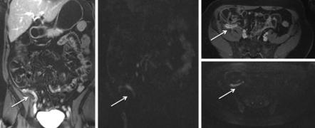
Figure 3.11 shows an image example of a patient diagnosed with appendicitis who went straight to appendectomy after the examination: diffusion-weighted imaging is able to show the inflammation as well as contrast-enhanced MR imaging. As not all patients undergo surgery for inflammatory appendiceal masses (IAM), DWI was studied as a follow-up tool in appendicitis by Özdemir et al. [50]: they concluded that DWI may be used with a significant success for follow-up of patients with IAM. As a monitoring imaging method, DWI may also aid in determining the most appropriate timing for interval appendectomy as well as help in diagnosing alternative diagnoses (e.g. malignancy and inflammatory bowel disease) that can mimic IAM.
As DWI is a quite new tool to diagnose appendicitis, most radiologists have limited or no experience in the evaluation and most likely will need training to achieve the diagnostic accuracy that has been reported in the literature [51].
a b c
d
Fig. 3.11 A patient presenting to the emergency room with a high suspicion of appendicitis and an unequivocal ultrasound. Contrast-enhanced T1-weighted images (a, coronal; c, axial) show contrast enhancement as correlate for acute inflammation. Diffusion-weighted imaging (b, coronal; d, axial) shows high signal as result of restricted diffusion due to inflammation

3 Small Bowel |
49 |
|
|
3.7\ Summary
Diffusion-weighted imaging has proven to be of help in the imaging of the small bowel and appendix. While conventional imaging sequences still are the “backbone” of small bowel imaging, DWI can add quantitative and qualitative information. DWI therefore should be part of a routine small bowel protocol to diagnose appendicitis in MR imaging. Standard MR imaging sequences including T2-weighted images with and without fat suppression as well as contrast-enhanced imaging will continue to be the reference standards until larger studies have been performed, especially in patients with small bowel tumours.
References
\1.\Rondonotti E, Herrerias JM, Pennazio M, Caunedo A, Mascarenhas-Saraiva M, de Franchis R. Complications, limitations, and failures of capsule endoscopy: a review of 733 cases. Gastrointest Endosc. 2005;62(5):712–6; . quiz 52, 54. https://doi.org/10.1016/j. gie.2005.05.002.
\2.\Sinha R, Rajiah P, Ramachandran I, Sanders S, Murphy PD. Diffusion-weighted MR imaging of the gastrointestinal tract: technique, indications, and imaging findings. Radiographics. 2013;33(3):655–76; . discussion 76–80. https://doi.org/10.1148/rg.333125042.
\3.\Dillman JR, Smith EA, Sanchez R, Adler J, Fazeli S, Zhang B, et al. DWI in pediatric small- bowel Crohn disease: are apparent diffusion coefficients surrogates for disease activity in patients receiving infliximab therapy? AJR Am J Roentgenol. 2016;207(5):1002–8. https:// doi.org/10.2214/AJR.16.16477.
\4.\Kanda T, Ishii K, Kawaguchi H, Kitajima K, Takenaka D. High signal intensity in the dentate nucleus and globus pallidus on unenhanced T1-weighted MR images: relationship with increasing cumulative dose of a gadolinium-based contrast material. Radiology. 2014;270(3):834–41. https://doi.org/10.1148/radiol.13131669.
\5.\Umschaden HW, Gasser J. MR enteroclysis. Radiol Clin North Am. 2003;41(2):231–48.
\6.\Masselli G, Casciani E, Polettini E, Gualdi G. Comparison of MR enteroclysis with MR enterography and conventional enteroclysis in patients with Crohn’s disease. Eur Radiol. 2008;18(3):438–47. https://doi.org/10.1007/s00330-007-0763-2.
\7.\Arrivé L, El Mouhadi S. MR enterography versus MR enteroclysis. Radiology. 2013;266(2):688. https://doi.org/10.1148/radiol.12121840.
\8.\Negaard A, Paulsen V, Sandvik L, Berstad A, Borthne A, Try K, et al. A prospective randomized comparison between two MRI studies of the small bowel in Crohn’s disease, the oral contrast method and MR enteroclysis. Eur Radiol. 2007;17(9):2294–301. https://doi.org/10.1007/ s00330-007-0648-4.
\9.\Schreyer AG, Geissler A, Albrich H, Schölmerich J, Feuerbach S, Rogler G, et al. Abdominal MRI after enteroclysis or with oral contrast in patients with suspected or proven Crohn’s disease. Clin Gastroenterol Hepatol. 2(6):491–7. https://doi.org/10.1016/S1542-3565(04)00168-5.
10\ .\Lauenstein TC, Umutlu L, Kloeters C, Aschoff AJ, Ladd ME, Kinner S. Small bowel imaging with MRI. Acad Radiol. 2012;19(11):1424–33. https://doi.org/10.1016/j.acra.2012.05.019.
11\ .\Young BM, Fletcher JG, Booya F, Paulsen S, Fidler J, Johnson CD, et al. Head-to-head comparison of oral contrast agents for cross-sectional enterography: small bowel distention, timing, and side effects. J Comput Assist Tomogr. 2008;32(1):32–8. https://doi.org/10.1097/ RCT.0b013e318061961d.
\12.\Oussalah A, Laurent V, Bruot O, Bressenot A, Bigard M-A, Régent D, et al. Diffusion-weighted magnetic resonance without bowel preparation for detecting colonic inflammation in inflammatory bowel disease. Gut. 2010;59(8):1056–65. https://doi.org/10.1136/gut.2009.197665.
50 |
S. Kinner |
|
|
13\ .\Li M, Dick A, Hassold N, Pabst T, Bley T, Kostler H, et al. CAIPIRINHA-accelerated T1w 3D-FLASH for small-bowel MR imaging in pediatric patients with Crohn’s disease: assessment of image quality and diagnostic performance. World J Pediatr. 2016;12(4):455–62. https://doi.org/10.1007/s12519-016-0047-5.
14\ .\Froehlich JM, Daenzer M, von Weymarn C, Erturk SM, Zollikofer CL, Patak MA. Aperistaltic effect of hyoscine N-butylbromide versus glucagon on the small bowel assessed by magnetic resonance imaging. Eur Radiol. 2009;19(6):1387–93. https://doi.org/10.1007/ s00330-008-1293-2.
\15.\Sirin S, Kathemann S, Schweiger B, Hahnemann ML, Forsting M, Lauenstein TC, et al. Magnetic resonance colonography including diffusion-weighted imaging in children and adolescents with inflammatory bowel disease: do we really need intravenous contrast? Invest Radiol. 2015;50(1):32–9. https://doi.org/10.1097/RLI.0000000000000092.
16\ .\Pendse DA, Makanyanga JC, Plumb AA, Bhatnagar G, Atkinson D, Rodriguez-Justo M, et al. Diffusion-weighted imaging for evaluating inflammatory activity in Crohn’s disease: comparison with histopathology, conventional MRI activity scores, and faecal calprotectin. Abdom Radiol. 2017;42(1):115–23. https://doi.org/10.1007/s00261-016-0863-z.
\17.\Oto A, Kayhan A, Williams JTB, Fan X, Yun L, Arkani S, et al. Active Crohn’s Disease in the small bowel: evaluation by diffusion weighted imaging and quantitative dynamic contrast enhanced MR imaging. J Magn Reson Imaging. 2011;33(3):615–24. https://doi.org/10.1002/ jmri.22435.
18\ .\Kwee TC, Takahara T, Koh D-M, Nievelstein RAJ, Luijten PR. Comparison and reproducibility of ADC measurements in breathhold, respiratory triggered, and free-breathing diffusion- weighted MR imaging of the liver. J Magn Reson Imaging. 2008;28(5):1141–8. https://doi. org/10.1002/jmri.21569.
\19.\Muro I, Takahara T, Horie T, Honda M, Kamiya A, Okumura Y et al. [Influence of respiratory motion in body diffusion weighted imaging under free breathing (examination of a moving phantom)]. Nihon Hoshasen Gijutsu Gakkai Zasshi 2005;61(11):1551–1558.
20\ .\van Rijswijck C, Lauenstein T, Kinner S. Detectability of inflammatory bowel disease in Diffusion-weighted MR imaging (DWI): which imaging plane and b-values should be preferred? Insights Imaging. 2017:262.
\21.\Ehman EC, Phelps AS, Ohliger MA, Rhee SJ, MacKenzie JD, Courtier JL. Detection of bowel inflammation with fused DWI/T2 images versus contrast-enhanced images in pediatric MR enterography with histopathologic correlation. Clin Imaging. 2016;40(6):1135–9. https://doi. org/10.1016/j.clinimag.2016.07.006.
22\ .\Kiryu S, Dodanuki K, Takao H, Watanabe M, Inoue Y, Takazoe M, et al. Free-breathing diffusion-weighted imaging for the assessment of inflammatory activity in Crohn’s disease. J Magn Reson Imaging. 2009;29(4):880–6. https://doi.org/10.1002/jmri.21725.
\23.\Dubron C, Avni F, Boutry N, Turck D, Duhamel A, Amzallag-Bellenger E. Prospective evaluation of free-breathing diffusion-weighted imaging for the detection of inflammatory bowel disease with MR enterography in childhood population. Br J Radiol. 2016;89(1060):20150840. https://doi.org/10.1259/bjr.20150840.
24\ .\Qi F, Jun S, Qi QY, Chen PJ, Chuan GX, Jiong Z, et al. Utility of the diffusion-weighted imaging for activity evaluation in Crohn’s disease patients underwent magnetic resonance enterography. BMC Gastroenterol. 2015;15:12. https://doi.org/10.1186/s12876-015-0235-0.
\25.\Hahnemann ML, Dechene A, Kathemann S, Sirin S, Gerken G, Lauenstein TC, et al. Diagnostic value of diffusion-weighted imaging (DWI) for the assessment of the small bowel in patients with inflammatory bowel disease. Clin Radiol. 2017;72(1):95.e1–8. https://doi.org/10.1016/j. crad.2016.08.007.
26\ .\Kim KJ, Lee Y, Park SH, Kang BK, Seo N, Yang SK, et al. Diffusion-weighted MR enterography for evaluating Crohn’s disease: how does it add diagnostically to conventional MR enterography? Inflamm Bowel Dis. 2015;21(1):101–9. https://doi.org/10.1097/ MIB.0000000000000222.
\27.\Li XH, Sun CH, Mao R, Huang SY, Zhang ZW, Yang XF, et al. Diffusion-weighted MRI enables to accurately grade inflammatory activity in patients of ileocolonic Crohn’s disease:
3 Small Bowel |
51 |
|
|
results from an observational study. Inflamm Bowel Dis. 2017;23(2):244–53. https://doi. org/10.1097/MIB.0000000000001001.
\28.\Stanescu-Siegmund N, Nimsch Y, Wunderlich AP, Wagner M, Meier R, Juchems MS, et al. Quantification of inflammatory activity in patients with Crohn’s disease using diffusion weighted imaging (DWI) in MR enteroclysis and MR enterography. Acta Radiol. 2017;58(3):264–71. https://doi.org/10.1177/0284185116648503.
29\ .\Buisson A, Joubert A, Montoriol PF, Ines DD, Hordonneau C, Pereira B, et al. Diffusion- weighted magnetic resonance imaging for detecting and assessing ileal inflammation in Crohn’s disease. Aliment Pharmacol Ther. 2013;37(5):537–45. https://doi.org/10.1111/ apt.12201.
\30.\Kovanlikaya A, Beneck D, Rose M, Renjen P, Dunning A, Solomon A, et al. Quantitative apparent diffusion coefficient (ADC) values as an imaging biomarker for fibrosis in pediatric Crohn’s disease: preliminary experience. Abdom Imaging. 2015;40(5):1068–74. https://doi. org/10.1007/s00261-014-0247-1.
31\ .\Tielbeek JA, Ziech ML, Li Z, Lavini C, Bipat S, Bemelman WA, et al. Evaluation of conventional, dynamic contrast enhanced and diffusion weighted MRI for quantitative Crohn’s disease assessment with histopathology of surgical specimens. Eur Radiol. 2014;24(3):619–29. https://doi.org/10.1007/s00330-013-3015-7.
\32.\Bhatnagar G, Dikaios N, Prezzi D, Vega R, Halligan S, Taylor SA. Changes in dynamic contrast-enhanced pharmacokinetic and diffusion-weighted imaging parameters reflect response to anti-TNF therapy in Crohn’s disease. Br J Radiol. 2015;88(1055):20150547. https://doi.org/10.1259/bjr.20150547.
33\ .\Buisson A, Hordonneau C, Goutte M, Scanzi J, Goutorbe F, Klotz T, et al. Diffusion-weighted magnetic resonance enterocolonography in predicting remission after anti-TNF induction therapy in Crohn’s disease. Dig Liver Dis. 2016;48(3):260–6. https://doi.org/10.1016/j. dld.2015.10.019.
\34.\Gourtsoyiannis N, Papanikolaou N. MR enteroclysis. In: Gore R, Levine M, editors. Textbook of gastrointestinal radiology. Philadelphia, PA: Saunders Elsevier; 2008. p. 765–74.
35\ .\Amzallag-Bellenger E, Soyer P, Barbe C, Nguyen TL, Amara N, Hoeffel C. Diffusion-weighted imaging for the detection of mesenteric small bowel tumours with Magnetic Resonance-- enterography. Eur Radiol. 2014;24(11):2916–26. https://doi.org/10.1007/s00330-014-3303-x.
\36.\Semelka RC, John G, Kelekis NL, Burdeny DA, Ascher SM. Small bowel neoplastic disease: demonstration by MRI. J Magn Reson Imaging. 1996;6(6):855–60.
37\ .\Lohan DG, Alhajeri AN, Cronin CG, Roche CJ, Murphy JM. MR enterography of small-bowel lymphoma: potential for suggestion of histologic subtype and the presence of underlying celiac disease. AJR Am J Roentgenol. 2008;190(2):287–93. https://doi.org/10.2214/AJR.07.2721.
\38.\Masselli G, Gualdi G. Evaluation of small bowel tumors: MR enteroclysis. Abdom Imaging. 2010;35(1):23–30. https://doi.org/10.1007/s00261-008-9490-7.
39\ .\Yu MH, Lee JM, Baek JH, Han JK, Choi BI. MRI features of gastrointestinal stromal tumors. AJR Am J Roentgenol. 2014;203(5):980–91. https://doi.org/10.2214/AJR.13.11667.
\40.\Gheller-Rigoni AI, Yale SH, Abdulkarim AS. Celiac Disease: celiac sprue, gluten-sensitive enteropathy. Clin Med Res. 2004;2(1):71–2.
41\ .\Rha SE, Ha HK, Lee SH, Kim JH, Kim JK, Kim JH, et al. CT and MR imaging findings of bowel ischemia from various primary causes. Radiographics. 2000;20(1):29–42. https://doi. org/10.1148/radiographics.20.1.g00ja0629.
\42.\Derlin T, Laqmani A, Veldhoen S, Apostolova I, Ayuk F, Adam G, et al. Magnetic resonance enterography for assessment of intestinal graft-versus-host disease after allogeneic stem cell transplantation. Eur Radiol. 2015;25(5):1229–37. https://doi.org/10.1007/s00330-014-3503-4.
43\ .\Pickuth D, Heywang-Kobrunner SH, Spielmann RP. Suspected acute appendicitis: is ultrasonography or computed tomography the preferred imaging technique? Eur J Surg. 2000;166(4):315–9. https://doi.org/10.1080/110241500750009177.
\44.\Liu B, Ramalho M, AlObaidy M, Busireddy KK, Altun E, Kalubowila J, et al. Gastrointestinal imaging-practical magnetic resonance imaging approach. World J Radiol. 2014;6(8):544–66. https://doi.org/10.4329/wjr.v6.i8.544.
52 |
S. Kinner |
|
|
45\ .\Repplinger MD, Levy JF, Peethumnongsin E, Gussick ME, Svenson JE, Golden SK, et al. Systematic review and meta-analysis of the accuracy of MRI to diagnose appendicitis in the general population. J Magn Reson Imaging. 2016;43(6):1346–54. https://doi.org/10.1002/ jmri.25115.
46\ .\Heverhagen JT, Pfestroff K, Heverhagen AE, Klose KJ, Kessler K, Sitter H. Diagnostic accuracy of magnetic resonance imaging: a prospective evaluation of patients with suspected appendicitis (diamond). J Magn Reson Imaging. 2012;35(3):617–23. https://doi.org/10.1002/ jmri.22854.
\47.\Kinner S, Repplinger MD, Pickhardt PJ, Reeder SB. Contrast-enhanced abdominal MRI for suspected appendicitis: how we do it. AJR Am J Roentgenol. 2016;207(1):49–57. https://doi. org/10.2214/AJR.15.15948.
48\ .\Bayraktutan U, Oral A, Kantarci M, Demir M, Ogul H, Yalcin A, et al. Diagnostic performance of diffusion-weighted MR imaging in detecting acute appendicitis in children: comparison with conventional MRI and surgical findings. J Magn Reson Imaging. 2014;39(6):1518–24. https://doi.org/10.1002/jmri.24316.
\49.\Inci E, Kilickesmez O, Hocaoglu E, Aydin S, Bayramoglu S, Cimilli T. Utility of diffusion- weighted imaging in the diagnosis of acute appendicitis. Eur Radiol. 2011;21(4):768–75. https://doi.org/10.1007/s00330-010-1981-6.
50\ .\Özdemir O, Metin Y, Metin NO, Küpeli A, Kalcan S, Taşçı F. Contribution of diffusion- weighted MR imaging in follow-up of inflammatory appendiceal mass: preliminary results and review of the literature. Eur J Radiol Open. 2016;3:207–15. https://doi.org/10.1016/j. ejro.2016.08.005.
\51.\Leeuwenburgh MM, Wiarda BM, Bipat S, Nio CY, Bollen TL, Kardux JJ, et al. Acute appendicitis on abdominal MR images: training readers to improve diagnostic accuracy. Radiology. 2012;264(2):455–63. https://doi.org/10.1148/radiol.12111896.