

- •Preface to the First Edition
- •Preface to the Second Edition
- •Contents
- •Diagnostic Challenges
- •Expert Centers
- •Patient Organizations
- •Clinical Trials
- •Research in Orphan Lung Diseases
- •Orphan Drugs
- •Orphanet
- •Empowerment of Patients
- •Conclusions
- •References
- •Introduction
- •Challenges to Overcome in Order to Undertake Quality Clinical Research
- •Lack of Reliable Data on Prevalence
- •Small Number of Patients
- •Identifying Causation/Disease Pathogenesis
- •Disease Complexity
- •Lack of Access to a Correct Diagnosis
- •Delay in Diagnosis
- •Challenges But Not Negativity
- •Some Success Stories
- •The Means to Overcome the Challenges of Clinical Research: Get Bigger Numbers of Well-Characterized Patients
- •The Importance of Patient Organizations
- •National and International Networks
- •End Points for Trials: Getting Them Right When Numbers Are Small and Change Is Modest
- •Orphan Drug Development
- •Importance of Referral Centers
- •Looking at the Future
- •The Arguments for Progress
- •Concluding Remarks
- •References
- •3: Chronic Bronchiolitis in Adults
- •Introduction
- •Cellular Bronchiolitis
- •Follicular Bronchiolitis
- •Respiratory Bronchiolitis
- •Airway-Centered Interstitial Fibrosis
- •Proliferative Bronchiolitis
- •Diagnosis
- •Chest Imaging Studies
- •Pulmonary Function Testing
- •Lung Biopsy
- •Mineral Dusts
- •Organic Dusts
- •Volatile Flavoring Agents
- •Infectious Causes of Bronchiolitis
- •Idiopathic Forms of Bronchiolitis
- •Connective Tissue Diseases
- •Organ Transplantation
- •Hematopoietic Stem Cell Transplantation
- •Drug-Induced Bronchiolitis
- •Treatment
- •Constrictive Bronchiolitis
- •Follicular Bronchiolitis
- •Airway-Centered Interstitial Fibrosis
- •Proliferative Bronchiolitis
- •References
- •Background and Epidemiology
- •Pathophysiology
- •Host Characteristics
- •Clinical Manifestations
- •Symptoms
- •Laboratory Evaluation
- •Skin Testing
- •Serum Precipitins
- •Eosinophil Count
- •Total Serum Immunoglobulin E Levels
- •Recombinant Antigens
- •Radiographic Imaging
- •Pulmonary Function Testing
- •Histology
- •Diagnostic Criteria
- •Historical Diagnostic Criteria
- •Rosenberg and Patterson Diagnostic Criteria
- •ISHAM Diagnostic Criteria
- •Cystic Fibrosis Foundation Diagnostic Criteria
- •General Diagnostic Recommendations
- •Allergic Aspergillus Sinusitis (AAS)
- •Natural History
- •Treatment
- •Corticosteroids
- •Antifungal Therapy
- •Monoclonal Antibodies
- •Monitoring for Treatment Response
- •Conclusions
- •References
- •5: Orphan Tracheopathies
- •Introduction
- •Anatomical Considerations
- •Clinical Presentation
- •Etiological Considerations
- •Idiopathic Subglottic Stenosis
- •Introduction
- •Clinical Features
- •Pulmonary Function Studies
- •Imaging Studies
- •Bronchoscopy
- •Treatment
- •Introduction and Clinical Presentation
- •Clinical Features
- •Pulmonary Function Studies
- •Imaging Studies
- •Bronchoscopy
- •Treatment
- •Tracheomalacia
- •Introduction
- •Clinical Features
- •Pulmonary Function Studies
- •Imaging Studies
- •Bronchoscopy
- •Treatment
- •Tracheobronchomegaly
- •Introduction
- •Clinical Features
- •Pathophysiology
- •Pulmonary Function Studies
- •Imaging Studies
- •Treatment
- •Tracheopathies Associated with Systemic Diseases
- •Relapsing Polychondritis
- •Introduction
- •Clinical Features
- •Laboratory Findings
- •Pulmonary Function and Imaging Studies
- •Treatment
- •Introduction
- •Clinical Features
- •Pulmonary Function Studies
- •Imaging Studies
- •Bronchoscopy
- •Treatment
- •Tracheobronchial Amyloidosis
- •Introduction
- •Clinical Features
- •Pulmonary Function Studies
- •Imaging Studies
- •Bronchoscopy
- •Treatment
- •Sarcoidosis
- •Introduction
- •Pulmonary Function Studies
- •Imaging Studies
- •Bronchoscopy
- •Treatment
- •Orphan Tracheopathies: Conclusions
- •References
- •6: Amyloidosis and the Lungs and Airways
- •Introduction
- •Diagnosis and Evaluation of Amyloidosis
- •Systemic AA Amyloidosis
- •Systemic AL Amyloidosis
- •Amyloidosis Localised to the Respiratory Tract
- •Laryngeal Amyloidosis
- •Tracheobronchial Amyloidosis
- •Parenchymal Pulmonary Amyloidosis
- •Pulmonary Amyloidosis Associated with Sjögren’s Disease
- •Conclusions
- •References
- •Introduction
- •Pathophysiology
- •Genetic Predisposition
- •Immune Dysregulation
- •Epidemiology
- •Incidence and Prevalence
- •Triggering Factors
- •Clinical Manifestations
- •General Symptoms
- •Pulmonary Manifestations
- •Ear, Nose, and Throat (ENT) Manifestations
- •Neurological Manifestations
- •Skin Manifestations
- •Cardiac Manifestations
- •Gastrointestinal Involvement
- •Renal Manifestations
- •Ophthalmological Manifestations
- •Complementary Investigations
- •Diagnosis
- •Diagnostic Criteria
- •Prognosis and Outcomes
- •Phenotypes According to the ANCA Status
- •Treatment
- •Therapeutic Strategies
- •Remission Induction
- •Maintenance Therapy
- •Other Treatments
- •Prevention of AEs
- •Conclusions
- •References
- •8: Granulomatosis with Polyangiitis
- •A Brief Historical Overview
- •Epidemiology
- •Pathogenesis
- •Clinical Manifestations
- •Constitutional Symptoms
- •Ear, Nose, and Throat (ENT) Manifestations
- •Pulmonary Manifestations
- •Kidney and Urological Manifestations
- •Kidney Manifestations
- •Urological Manifestations
- •Neurological Manifestations
- •Peripheral Nervous System (PNS) Manifestations
- •Central Nervous System (CNS) Manifestations
- •Spinal Cord and Cranial Nerve Involvement
- •Skin and Oral Mucosal Manifestations
- •Eye Manifestations
- •Cardiac Involvement
- •Gastrointestinal Manifestations
- •Gynecological and Obstetric Manifestations
- •Venous Thrombosis and Other Vascular Events
- •Other Manifestations
- •Pediatric GPA
- •Diagnosis
- •Diagnostic Approach
- •Laboratory Investigations
- •Biology
- •Immunology
- •Pathology
- •Treatment
- •Glucocorticoids
- •Cyclophosphamide
- •Rituximab
- •Other Current Induction Approaches
- •Other Treatments in GPA
- •Intravenous Immunoglobulins
- •Plasma Exchange
- •CTLA4-Ig (Abatacept)
- •Cotrimoxazole
- •Other Agents
- •Principles of Treatment for Relapsing and Refractory GPA
- •Outcomes and Prognostic Factors
- •Survival and Causes of Deaths
- •Relapse
- •Damage and Disease Burden on Quality of Life
- •Conclusions
- •References
- •9: Alveolar Hemorrhage
- •Introduction
- •Clinical Presentation
- •Diagnosis (Table 9.1, Fig. 9.3)
- •Pulmonary Capillaritis
- •Histology (Fig. 9.4)
- •Etiologies
- •ANCA-Associated Small Vessel Vasculitis: Granulomatosis with Polyangiitis (GPA)
- •ANCA-Associated Small Vessel Vasculitis: Microscopic Polyangiitis
- •Isolated Pulmonary Capillaritis
- •Systemic Lupus Erythematosus
- •Antiphospholipid Antibody Syndrome
- •Anti-Basement Membrane Antibody Disease (Goodpasture Syndrome)
- •Lung Allograft Rejection
- •Others
- •Bland Pulmonary Hemorrhage (Fig. 9.5)
- •Histology
- •Etiologies
- •Idiopathic Pulmonary Hemosiderosis
- •Drugs and Medications
- •Coagulopathy
- •Valvular Heart Disease and Left Ventricular Dysfunction
- •Other
- •Histology
- •Etiologies
- •Hematopoietic Stem Cell Transplantation (HSCT)
- •Cocaine Inhalation
- •Acute Exacerbation of Interstitial Lung Disease
- •Acute Interstitial Pneumonia
- •Acute Respiratory Distress Syndrome
- •Miscellaneous Causes
- •Etiologies
- •Pulmonary Capillary Hemangiomatosis
- •Treatment
- •Conclusions
- •References
- •Takayasu Arteritis
- •Epidemiology
- •Pathologic Features
- •Pathogenesis
- •Clinical Features
- •Laboratory Findings
- •Imaging Studies
- •Therapeutic Management
- •Prognosis
- •Behçet’s Disease
- •Epidemiology
- •Pathologic Features
- •Pathogenesis
- •Diagnostic Criteria
- •Clinical Features
- •Pulmonary Artery Aneurysm
- •Pulmonary Artery Thrombosis
- •Pulmonary Parenchymal Involvement
- •Laboratory Findings
- •Imaging Studies
- •Therapeutic Management
- •Treatment of PAA
- •Treatment of PAT
- •Prognosis
- •References
- •Introduction
- •Portopulmonary Hypertension (PoPH)
- •Epidemiology and Risk Factors
- •Molecular Pathogenesis
- •PoPH Treatment
- •Hepatopulmonary Syndrome (HPS)
- •Epidemiology and Risk Factors
- •Molecular Pathogenesis
- •HPS Treatment
- •Conclusion
- •References
- •12: Systemic Sclerosis and the Lung
- •Introduction
- •Risk factors for SSc-ILD
- •Genetic Associations
- •Clinical Presentation of SSc-ILD
- •Pulmonary Function Tests (PFTs)
- •Imaging
- •Management
- •References
- •13: Rheumatoid Arthritis and the Lungs
- •Introduction
- •Epidemiology
- •Risk Factors for ILD (Table 13.3)
- •Pathogenesis
- •Clinical Features and Diagnosis
- •Treatments
- •Prognosis
- •Epidemiology
- •Risk Factors
- •Clinical Features, Diagnosis, and Outcome
- •Subtypes or RA-AD
- •Obliterative Bronchiolitis
- •Bronchiectasis
- •COPD
- •Cricoarytenoid Involvement
- •Pleural Disease
- •Conclusion
- •References
- •Introduction
- •Systemic Lupus Erythematosus
- •Epidemiology
- •Pathophysiology
- •Pulmonary Manifestations
- •Pleural Disease
- •Shrinking Lung Syndrome
- •Thrombotic Manifestations
- •Interstitial Lung Disease
- •Other Pulmonary Manifestations
- •Prognosis
- •Sjögren’s Syndrome
- •Epidemiology
- •Pathophysiology
- •Pulmonary Manifestations
- •Airway Disorders
- •Lymphoproliferative Disease
- •Interstitial Lung Disease
- •Prognosis
- •Mixed Connective Tissue Disease
- •Epidemiology
- •Pathophysiology
- •Pulmonary Manifestations
- •Pulmonary Hypertension
- •Interstitial Lung Disease
- •Prognosis
- •Myositis
- •Epidemiology
- •Pathophysiology
- •Pulmonary Manifestations and Treatments
- •Interstitial Lung Disease
- •Respiratory Muscle Weakness
- •Other Pulmonary Manifestations
- •Prognosis
- •Other Therapeutic Options in CTD-ILD
- •Lung Transplantation
- •Conclusion
- •References
- •Introduction
- •Diagnostic Criteria
- •Controversies in the Diagnostic Criteria
- •Typical Clinical Features
- •Disease Progression and Prognosis
- •Summary
- •References
- •Introduction
- •Histiocytes and Dendritic Cells
- •Introduction
- •Cellular and Molecular Pathogenesis
- •Pathology
- •Clinical Presentation
- •Treatment and Prognosis
- •Erdheim-Chester Disease
- •Epidemiology
- •Cellular and Molecular Pathogenesis
- •Histopathology and Immunohistochemistry
- •Clinical Presentation
- •Investigation/Diagnosis
- •Chest Studies
- •Cardiovascular Imaging
- •CNS Imaging
- •Bone Radiography
- •Other Imaging Findings and Considerations
- •Disease Monitoring
- •Pathology
- •Management/Treatment
- •Prognosis
- •Rosai-Dorfman Destombes Disease
- •Epidemiology
- •Etiology/Pathophysiology
- •Histopathology and Immunohistochemistry
- •Clinical Presentation
- •Investigation/Diagnosis
- •Management/Treatment
- •Prognosis
- •Conclusions
- •Diagnostic Criteria for Primary Histiocytic Disorders of the Lung
- •References
- •17: Eosinophilic Pneumonia
- •Introduction
- •Eosinophil Biology
- •Physiologic and Immunologic Role of Eosinophils
- •Release of Mediators
- •Targeting the Eosinophil Cell Lineage
- •Historical Perspective
- •Clinical Presentation
- •Pathology
- •Diagnosis
- •Eosinophilic Lung Disease of Undetermined Cause
- •Idiopathic Chronic Eosinophilic Pneumonia
- •Clinical Features
- •Imaging
- •Laboratory Studies
- •Bronchoalveolar Lavage
- •Lung Function Tests
- •Treatment
- •Outcome and Perspectives
- •Clinical Features
- •Imaging
- •Laboratory Studies
- •Bronchoalveolar Lavage
- •Lung Function Tests
- •Lung Biopsy
- •Treatment and Prognosis
- •Eosinophilic Granulomatosis with Polyangiitis
- •History and Nomenclature
- •Pathology
- •Clinical Features
- •Imaging
- •Laboratory Studies
- •Pathogenesis
- •Diagnosis
- •Treatment and Prognosis
- •Long-Term Outcome
- •Hypereosinophilic Syndrome
- •Pathogenesis
- •Clinical and Imaging Features
- •Laboratory Studies
- •Treatment and Prognosis
- •Eosinophilic Pneumonias of Parasitic Origin
- •Tropical Eosinophilia [191]
- •Ascaris Pneumonia
- •Eosinophilic Pneumonia in Larva Migrans Syndrome
- •Strongyloides Stercoralis Infection
- •Eosinophilic Pneumonias in Other Infections
- •Allergic Bronchopulmonary Aspergillosis
- •Pathogenesis
- •Diagnostic Criteria
- •Biology
- •Imaging
- •Treatment
- •Bronchocentric Granulomatosis
- •Miscellaneous Lung Diseases with Associated Eosinophilia
- •References
- •Introduction
- •Pulmonary Langerhans’ Cell Histiocytosis
- •Epidemiology
- •Pathogenesis
- •Diagnosis
- •Clinical Features
- •Extrathoracic Lesions
- •Pulmonary Function Tests
- •Chest Radiography
- •High-Resolution Computed Tomography (HRCT)
- •Bronchoscopy and Bronchoalveolar Lavage (BAL)
- •Lung Biopsy
- •Pathology
- •Treatment
- •Course and Prognosis
- •Case Report I
- •Introduction
- •Epidemiology
- •Clinical Features
- •Histopathological Findings
- •Radiologic Findings
- •Prognosis and Therapy
- •Desquamative Interstitial Pneumonia
- •Epidemiologic and Clinical Features
- •Histopathological Findings
- •Radiological Findings
- •Prognosis and Therapy
- •Conclusion
- •References
- •19: Lymphangioleiomyomatosis
- •Introduction
- •Pathogenesis
- •Presentation
- •Prognosis
- •Management
- •General Measures
- •Parenchymal Lung Disease
- •Pleural Disease
- •Renal Angiomyolipoma
- •Abdominopelvic Lymphatic Disease
- •Pregnancy
- •Tuberous Sclerosis
- •Drug Treatment
- •Bronchodilators
- •mTOR Inhibitors
- •Anti-Oestrogen Therapy
- •Experimental Therapies
- •Interventions for Advanced Disease
- •Oxygen Therapy
- •Pulmonary Hypertension
- •References
- •20: Diffuse Cystic Lung Disease
- •Introduction
- •Lymphangioleiomyomatosis
- •Pathogenesis
- •Pathologic and Radiographic Characteristics
- •Diagnostic Approach
- •Pulmonary Langerhans Cell Histiocytosis (PLCH)
- •Pathogenesis
- •Pathological and Radiographic Characteristics
- •Diagnostic Approach
- •Birt-Hogg-Dubé Syndrome (BHD)
- •Pathogenesis
- •Pathological and Radiographic Characteristics
- •Diagnostic Approach
- •Lymphoproliferative Disorders
- •Pathogenesis
- •Pathological and Radiographic Characteristics
- •Diagnostic Approach
- •Amyloidosis
- •Light Chain Deposition Disease (LCDD)
- •Conclusion
- •References
- •Introduction
- •Lymphatic Development
- •Clinical Presentation of Lymphatic Disorders
- •Approaches to Diagnosis and Management of Congenital Lymphatic Anomalies
- •Generalized Lymphatic Anomaly
- •Etiopathogenesis
- •Clinical Presentation and Diagnosis
- •Course/Prognosis
- •Management
- •Kaposiform Lymphangiomatosis
- •Etiopathogenesis
- •Clinical Presentation and Diagnosis
- •Management
- •Course/Prognosis
- •Gorham Stout Disease
- •Etiopathogenesis
- •Clinical Presentation and Diagnosis
- •Management
- •Course/Prognosis
- •Channel-Type LM/Central Conducting LM
- •Etiopathogenesis
- •Clinical Presentation and Diagnosis
- •Management
- •Course/Prognosis
- •Yellow Nail Syndrome
- •Etiopathogenesis
- •Clinical Presentation and Diagnosis
- •Management
- •Course/Prognosis
- •Summary
- •References
- •Introduction
- •Historical Note
- •Epidemiology
- •Pathogenesis
- •Surfactant Homeostasis in PAP
- •GM-CSF Signaling Disruption
- •Myeloid Cell Dysfunction
- •GM-CSF Autoantibodies
- •Lymphocytosis
- •Clinical Manifestations
- •Clinical Presentation
- •Secondary Infections
- •Pulmonary Fibrosis
- •Diagnosis
- •Pulmonary Function Testing
- •Radiographic Assessment
- •Bronchoscopy and Bronchoalveolar Lavage
- •Laboratory Studies and Biomarkers
- •GM-CSF Autoantibodies
- •Genetic Testing
- •Lung Pathology
- •Diagnostic Approach to the Patient with PAP
- •Natural History and Prognosis
- •Treatment
- •Whole-Lung Lavage
- •Subcutaneous GM-CSF
- •Inhaled GM-CSF
- •Other Approaches
- •Conclusions and Future Directions
- •References
- •Introduction
- •Epidemiology
- •Gastric Contents
- •Pathobiology of GER/Microaspirate in the Lungs of Patients with IPF
- •GER and the Microbiome
- •Diagnosis
- •Clinical History/Physical Exam
- •Investigations
- •Esophageal Physiology
- •Upper Esophageal Sphincter
- •Esophagus and Peristalsis
- •Lower Esophageal Sphincter and Diaphragm
- •Esophageal pH and Impedance Testing
- •High Resolution Esophageal Manometry
- •Esophagram/Barium Swallow
- •Bronchoalveolar Lavage/Sputum: Biomarkers
- •Treatment
- •Anti-Acid Therapy (PPI/H2 Blocker)
- •GER and Acute Exacerbations of IPF
- •Suggested Approach
- •Summary and Future Directions
- •References
- •Introduction
- •Familial Interstitial Pneumonia
- •Telomere Related Genes
- •Genetic
- •Telomere Length
- •Pulmonary Involvement
- •Interstitial Lung Disease
- •Other Lung Disease
- •Hepatopulmonary Syndrome
- •Emphysema
- •Extrapulmonary Manifestations
- •Mucocutaneous Involvement
- •Hematological Involvement
- •Liver Involvement
- •Other Manifestations
- •Treatment
- •Telomerase Complex Agonists
- •Lung Transplantation
- •Surfactant Pathway
- •Surfactant Protein Genes
- •Pulmonary Involvement
- •Treatment
- •Heritable Forms of Pulmonary Fibrosis with Autoimmune Features
- •TMEM173
- •COPA
- •Pulmonary Alveolar Proteinosis
- •GMCSF Receptor Mutations
- •GATA2
- •MARS
- •Lysinuric Protein Intolerance
- •Lysosomal Diseases
- •Hermansky-Pudlak Syndrome
- •Lysosomal Storage Disorders
- •FAM111B, NDUFAF6, PEPD
- •Conclusion
- •References
- •Introduction
- •Pathophysiology
- •Clinical Presentation
- •Epidemiology
- •Genetic Causes of Bronchiectasis
- •Disorders of Mucociliary Clearance
- •Cystic Fibrosis
- •Primary Ciliary Dyskinesia
- •Other Ciliopathies
- •X-Linked Agammaglobulinemia
- •Chronic Granulomatous Disease and Other Disorders of Neutrophil Function
- •Other Genetic Disorders Predisposing to Bronchiectasis
- •Idiopathic Bronchiectasis
- •Diagnosis of Bronchiectasis
- •Management of Patients with Bronchiectasis
- •Airway Clearance Therapy (ACT)
- •Management of Infections
- •Immune Therapy
- •Surgery
- •Novel Therapies for Managing Cystic Fibrosis
- •Summary
- •References
- •Pulmonary Arteriovenous Malformations
- •Background Pulmonary AVMs
- •Anatomy Pulmonary AVMs
- •Clinical Presentation of Pulmonary AVMs
- •Screening Pulmonary AVMs
- •Treatment Pulmonary AVMs
- •Children with Hereditary Hemorrhagic Telangiectasia
- •Pulmonary Hypertension
- •Pulmonary Hypertension Secondary to Liver Vascular Malformations
- •Pulmonary Arterial Hypertension
- •Background HHT
- •Pathogenesis
- •References
- •27: Pulmonary Alveolar Microlithiasis
- •Introduction
- •Epidemiology
- •Pathogenesis
- •Clinical Features
- •Diagnosis
- •Management
- •Summary
- •References
- •Introduction
- •Hermansky-Pudlak Syndrome
- •Telomerase-Associated Pulmonary Fibrosis
- •Lysosomal Storage Diseases
- •Lysinuric Protein Intolerance
- •Familial Hypocalciuric Hypercalcemia
- •Surfactant Dysfunction Disorders
- •Concluding Remarks
- •References
- •Introduction
- •Background
- •Image Acquisition
- •Key Features of Fibrosis
- •Ancillary Features of Fibrosis
- •Other Imaging Findings in FLD
- •Probable UIP-IPF
- •Indeterminate
- •Alternative Diagnosis
- •UIP in Other Fibrosing Lung Diseases
- •Pleuroparenchymal Fibroelastosis (PPFE)
- •Combined Pulmonary Fibrosis and Emphysema
- •Chronic Hypersensitivity Pneumonitis
- •Other Fibrosing Lung Diseases
- •Fibrosing Sarcoidosis
- •CTD-ILD and Drug-Induced FLD
- •Complications
- •Prognosis
- •Computer Analysis of CT Imaging
- •The Progressive Fibrotic Phenotype
- •Other Imaging Techniques
- •Conclusion
- •References
- •Introduction
- •Bronchoalveolar Lavage (BAL)
- •Technique
- •Interpretation
- •Transbronchial Biopsy (TBB)
- •Transbronchial Lung Cryobiopsy (TLCB)
- •References
- •Introduction
- •Overview of ILD Diagnosis
- •Clinical Assessment
- •Radiological Assessment
- •Laboratory Assessment
- •Integration of Individual Features
- •Multidisciplinary Discussion
- •Diagnostic Ontology
- •Conclusions
- •References
- •Introduction
- •Idiopathic Pulmonary Fibrosis
- •Chronic Hypersensitivity Pneumonitis
- •Connective Tissue Disease
- •Drug-Induced Lung Diseases
- •Radiation Pneumonitis
- •Asbestosis
- •Hermansky-Pudlak Syndrome
- •Risk Factors for Progression
- •Diagnosis
- •Pharmacological Management
- •Conclusions
- •References
- •Historical Perspective
- •Epidemiology and Etiologies
- •Tobacco Smoking and Male Sex
- •Genetic Predisposition
- •Systemic Diseases
- •Other Etiological Contexts
- •Clinical Manifestations
- •Pulmonary Function and Physiology
- •Imaging
- •Computed Tomography Characteristics and Patterns
- •Thick-Walled Large Cysts
- •Imaging Phenotypes
- •Pitfalls
- •Pathology
- •Diagnosis
- •CPFE Is a Syndrome
- •Biology
- •Complications and Outcome
- •Mortality
- •Pulmonary Hypertension
- •Lung Cancer
- •Acute Exacerbation of Pulmonary Fibrosis
- •Other Comorbidities and Complications
- •Management
- •General Measures and Treatment of Emphysema
- •Treatment of Pulmonary Fibrosis
- •Management of Pulmonary Hypertension
- •References
- •Acute Interstitial Pneumonia (AIP)
- •Epidemiology
- •Presentation
- •Diagnostic Evaluation
- •Radiology
- •Histopathology
- •Clinical Course
- •Treatment
- •Epidemiology
- •Presentation
- •Diagnostic Evaluation
- •Radiology
- •Histopathology
- •Clinical Course
- •Desquamative Interstitial Pneumonia (DIP)
- •Presentation
- •Diagnostic Evaluation
- •Radiology
- •Histopathology
- •Clinical Course
- •Treatment
- •Epidemiology
- •Presentation
- •Diagnostic Evaluation
- •Radiology
- •Histopathology
- •Clinical Course
- •Treatment
- •References
- •Organizing Pneumonias
- •Epidemiology
- •Pathogenesis
- •Clinical Features
- •Imaging
- •Multifocal Form
- •Isolated Nodular Form
- •Other Imaging Patterns
- •Histopathological Diagnosis of OP Pattern
- •Etiological Diagnosis of OP
- •Treatment
- •Clinical Course and Outcome
- •Severe Forms of OP with Respiratory Failure
- •Acute Fibrinous and Organizing Pneumonia
- •Granulomatous Organizing Pneumonia
- •Acute Interstitial Pneumonia
- •Epidemiology
- •Clinical Picture
- •Imaging
- •Histopathology
- •Diagnosis
- •Treatment
- •Outcome
- •References
- •36: Pleuroparenchymal Fibroelastosis
- •Introduction
- •Epidemiology
- •Clinical Manifestations
- •Laboratory Findings
- •Respiratory Function
- •Radiologic Features
- •Pathologic Features
- •Diagnosis
- •Treatment
- •Prognosis
- •Conclusions
- •References
- •Introduction
- •Acute Berylliosis
- •Chronic Beryllium Disease
- •Exposure
- •Epidemiology
- •Immunopathogenesis and Pathology
- •Genetics
- •Clinical Description and Natural History
- •Treatment and Monitoring
- •Indium–Tin Oxide-Lung Disease
- •Hard Metal Lung
- •Flock Worker’s Disease
- •Asbestosis
- •Nanoparticle Induced ILD
- •Flavoring-Induced Lung Disease
- •Silica-Induced Interstitial Lung Disease
- •Chronic Silicosis
- •Acute and Accelerated Silicosis
- •Chronic Obstructive Disease in CMDLD
- •Simple CMDLD
- •Complicated CMDLD
- •Conclusion
- •References
- •38: Unclassifiable Interstitial Lung Disease
- •Introduction
- •Diagnostic Scenarios
- •Epidemiology
- •Clinical Presentation
- •Diagnosis
- •Clinical Features
- •Radiology
- •Laboratory Investigations
- •Pathology
- •Conclusion
- •References
- •39: Lymphoproliferative Lung Disorders
- •Introduction
- •Nodular Lymphoid Hyperplasia
- •Lymphocytic Interstitial Pneumonia (LIP)
- •Follicular Bronchitis/Bronchiolitis
- •Castleman Disease
- •Primary Pulmonary Lymphomas
- •Primary Pulmonary MALT B Cell Lymphoma
- •Pulmonary Plasmacytoma
- •Follicular Lymphoma
- •Lymphomatoid Granulomatosis
- •Primary Pulmonary Hodgkin Lymphoma (PPHL)
- •Treatment
- •References
- •Introduction
- •Late-Onset Pulmonary Complications
- •Bronchiolitis Obliterans (BO)
- •Pathophysiology
- •Diagnosis
- •Management of BOS
- •Post-HSCT Organizing Pneumonia
- •Other Late-Onset NonInfectious Pulmonary Complications (LONIPCs)
- •Conclusion
- •References
- •Introduction
- •Pulmonary Hypertension Associated with Sarcoidosis (Group 5.2)
- •PH Associated with Pulmonary Langerhans Cell Histiocytosis (Group 5.2)
- •PH in Combined Pulmonary Fibrosis and Emphysema (Group 3.3)
- •PH Associated with Lymphangioleiomyomatosis (Group 3)
- •Hereditary Hemorrhagic Telangiectasia (Group 1.2)
- •Pulmonary Veno-Occlusive Disease (Group 1.5)
- •Small Patella Syndrome (Group 1.2)
- •Conclusion
- •References
- •Introduction
- •Epidemiology
- •Timing, Chronology, Delay Time
- •Route of Administration
- •Patterns of Involvement [3, 4]
- •Drugs and Agents Fallen Out of Favor
- •Drug-Induced Noncardiac Pulmonary Edema
- •Drug-Induced Cardiogenic Pulmonary Edema
- •The “Chemotherapy Lung”
- •Drug-Induced/Iatrogenic Alveolar Hemorrhage
- •Drugs
- •Superwarfarin Rodenticides
- •Transfusion Reactions: TACO–TRALI
- •Acute Eosinophilic Pneumonia
- •Acute Granulomatous Interstitial Lung Disease
- •Acute Organizing Pneumonia (OP), Bronchiolitis Obliterans Organizing Pneumonia (BOOP), or Acute Fibrinous Organizing Pneumonia (AFOP) Patterns
- •Acute Amiodarone-Induced Pulmonary Toxicity (AIPT)
- •Accelerated Pulmonary Fibrosis
- •Acute Exacerbation of Previously Known (Idiopathic) Pulmonary Fibrosis
- •Anaphylaxis
- •Acute Vasculopathy
- •Drug-Induced/Iatrogenic Airway Emergencies
- •Airway Obstruction as a Manifestation of Anaphylaxis
- •Drug-Induced Angioedema
- •Hematoma Around the Upper Airway
- •The “Pill Aspiration Syndrome”
- •Catastrophic Drug-Induced Bronchospasm
- •Peri-operative Emergencies (Table 42.8)
- •Other Rare Presentations
- •Pulmonary Nodules and Masses
- •Pleuroparenchymal Fibroelastosis
- •Late Radiation-Induced Injury
- •Chest Pain
- •Rebound Phenomenon
- •Recall Pneumonitis
- •Thoracic Bezoars: Gossipybomas
- •Respiratory Diseases Considered Idiopathic That May Be Drug-Induced (Table 42.4)
- •Eye Catchers
- •Conclusion
- •References
- •Cancer Mimics of Organizing Pneumonia
- •Lung Adenocarcinoma/Bronchioloalveolar Carcinoma
- •Primary Pulmonary Lymphoma
- •Cancer Mimics of Interstitial Lung Diseases
- •Lymphangitic Carcinomatosis
- •Epithelioid Hemangio-Endothelioma
- •Lymphomatoid Granulomatosis
- •Cystic Tumors
- •Cavitating Tumors
- •Intrathoracic Pseudotumors
- •Respiratory Papillomatosis
- •Pulmonary Langerhans Cell Histiocytosis
- •References
- •Index

Complex Thoracic Lymphatic Disorders |
21 |
of Adults |
Hassan Mujahid, Anita Gupta, Adrienne Hammill,
Christopher T. Towe, and Francis X. McCormack
Introduction
Complex lymphatic anomalies (CLAs) not only comprise a diverse set of congenital disorders that usually come to medical attention early in life, either in utero or during early childhood, but can also manifest in adulthood [1]. The major members of this family include generalized lymphatic anomaly (GLA), Gorham-Stout disease (GSD), kaposiform lymphangiomatosis (KLA), and central collecting lymphatic anomaly (CCLA). Depending on the anomaly, these manifestations can be systemic or isolated to a single site or organ system. Lung involvement with
CLAs is not uncommon, and can be the primary manifestation [2]. Lymphangioleiomyomatosis, which can present with lymphatic masses, cystic lung disease, and chylous effusions, is the subject of another chapter and will not be addressed here. Although not considered a CLA, Yellow Nail Syndrome is an acquired lymphatic disorder that typically presents in adulthood with some combination of discolored nails, lymphedema, sinusitis, bronchiectasis, and pleural effusion that is occasionally chylous [3]. This chapter will focus on these ve lymphatic disorders that may present to an adult pulmonologist (Table 21.1).
H. Mujahid · F. X. McCormack (*)
Division of Pulmonary, Critical Care, and Sleep Medicine, Department of Internal Medicine, University of Cincinnati, Cincinnati, OH, USA
e-mail: mujahihn@ucmail.uc.ede; frank.mccormack@uc.edu
A. Gupta
Division of Pathology and Laboratory Medicine, Ann and Robert H. Lurie Children’s Hospital of Chicago, Chicago, IL, USA e-mail: angupta@luriechildrens.org
A. Hammill
Division of Hematology, Cancer and Blood Diseases Institute, Cincinnati, OH, USA
Department of Pediatrics, University of Cincinnati College of Medicine, Cincinnati, OH, USA
e-mail: adrienne.hammill@cchmc.org
C. T. Towe
Division of Pulmonary Medicine, Cincinnati Children’s Hospital Medical Center, Cincinnati, OH, USA
Department of Pediatrics, University of Cincinnati College of Medicine, Cincinnati, OH, USA
e-mail: christopher.towe@cchmc.org
© Springer Nature Switzerland AG 2023 |
369 |
V. Cottin et al. (eds.), Orphan Lung Diseases, https://doi.org/10.1007/978-3-031-12950-6_21 |
|
Данная книга находится в списке для перевода на русский язык сайта https://meduniver.com/

370 |
|
|
|
|
|
|
|
H. Mujahid et al. |
|
|
|
|
|||||
Table 21.1 Complex lymphatic anomalies (CTA)–clinical manifestations, diagnosis, and treatments |
|
|
|
|||||
|
|
|
|
|
|
|
|
|
|
|
|
|
|
|
Known |
|
|
Disease |
Clinical manifestation |
Diagnosis |
mutations |
Treatment |
||||
Generalized lymphatic |
|
– |
Multifocal lymphatic |
MRI, CT and bone scintigraphy |
PIK3CA |
– |
Sirolimus |
|
anomaly (GLA) |
|
|
malformations can involve liver |
|
|
[27] |
– |
Interferon 2b |
|
|
|
and spleen |
|
|
|
– |
Zoledronic acid |
|
|
– |
Bone involvement medullary, |
|
|
|
– |
Embolization/ |
|
|
|
non-contiguous, and without |
|
|
|
|
sclerosis |
|
|
|
cortical destruction |
|
|
|
– |
Surgical |
|
|
– |
Pleural effusions |
|
|
|
|
|
Kaposiform |
|
– |
All of the above and hematologic |
Spindle cells on biopsy |
NRAS |
All of the above and |
||
Lymphangiomatosis |
|
|
abnormalities |
|
|
[47] |
Steroids |
|
(KLA) |
|
– |
Hemorrhagic effusions (pleural, |
|
|
CBL [48] |
Vincristine |
|
|
|
|
pericardial) |
|
|
|
MEK inhibitora |
|
Gorham stout disease |
|
– |
Multifocal lymphatic |
Cortical destruction on bone |
KRAS |
– |
Sirolimus |
|
(GSD) |
|
|
proliferations with cortical bone |
imaging |
[111] |
– |
Interferon 2b |
|
|
|
|
destruction |
|
|
|
– |
Zoledronic acid |
|
|
– |
Predilection for axial skeleton |
|
|
|
– |
Surgical |
|
|
|
|
|
|
|
|
stabilization |
|
|
|
|
|
|
|
– |
Radiation MEK |
|
|
|
|
|
|
|
|
inhibitora |
Central conducting |
Chylous effusions |
– |
Lymphangiography |
ARAF |
Sirolimus |
|||
lymphatic anomaly |
Protein-losing enteropathy |
– |
Dynamic magnetic |
[73] |
MEK inhibitora |
|||
(CCLA) |
Chylous leaks |
|
resonance |
EPHB4 |
Surgical correction |
|||
|
|
|
|
|
lymphangiography |
[72] |
|
|
Yellow nail syndrome |
Sinusitis |
2/3 symptoms |
|
Vitamin E/antifungal |
||||
(YNS) |
Bronchiectasis |
|
|
|
Symptomatic |
|||
|
Lymphedema |
|
|
|
|
|
||
|
Yellow nails |
|
|
|
|
|
||
|
Pleural effusion |
|
|
|
|
|
||
aWhen underlying genetic cause has been identi ed as RAS/RAF/MAPK pathway
The Pulmonary Lymphatic System: Structure
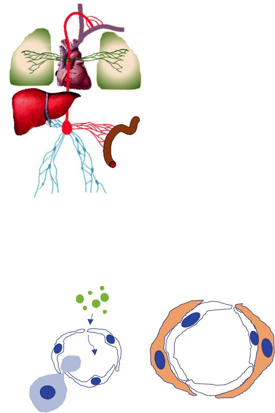
and Function
Lymphatics are a blind-ended organ system of interconnected vessels, lymph nodes and lymphatic tissues that transport 2–4 L per day of a clear fuid called lymph from peripheral tissues toward the heart [4] (Fig. 21.1). They play an essential role in the circulatory system by returning extravasated cells, plasma, macromolecules, and interstitial components to the bloodstream. Chylomicrons are triglyceride rich lipoprotein particles that are generated in the endoplasmic reticulum of enterocytes and secreted into gut lacteals where they enter the lymphatic stream to become chyle. They ultimately fow into the venous circulation to transport lipid to adipose tissue and skeletal and cardiac muscle. As an integral component of the immune system, the lymphatics are responsible for transporting antigen-
loaded dendritic cells and memory/effector T cells to draining lymph nodes, providing a platform for initiation of adaptive immune responses. Lymphatic capillaries are present in all tissues, with the exception of the bone marrow and cartilage. Lymphatic networks were recently discovered in the brain [5], an organ that was thought to be devoid of these structures.
Below the diaphragm, the lymphatic system consists of three major components: the soft tissue lymphatic system, the intestinal lymphatic system, and the hepatic lymphatic system (Fig. 21.1) [4]. These organ systems drain into cisterna chyli and ultimately into the thoracic duct (TD) which courses through the thorax and inserts into the left innominate vein at the junction with the internal jugular vein. The pulmonary lymphatic system primarily drains into the thoracic duct. A smaller lymphatic network comprises the right upper lobe, right head and neck, and right
21 Complex Thoracic Lymphatic Disorders of Adults |
371 |
|
|
Thoracic duct flow 2-4 liters per day
.25L |
.25L |
1.4L
1.4L
.7L
Fig. 21.1 Lymphatic fuid courses through the soft tissue, hepatic and intestinal lymphatic networks, and converges on the cisterna chyli in the abdomen. The approximately daily volume of fuid generated from each source is shown. Chylomicrons generated in the lacteals of the gut impart a milky white appearance to the lymphatic fuid as it fows cephalad via the thoracic duct to the junction between the left innominate and left internal jugular vein [113]
arm drains into right thoracic duct, which inserts into the right subclavian vein [2, 4]. It is important to note that chyle is generated in the abdomen and can only enter the lung or pleural space through abnormal abdominal pulmonary lymphatic communications with the airways or pleural space, or via refux from the thoracic duct into the pulmonary lymphatic network when the pressure gradient for fow is reversed [6].
On microscopy, lymphatics capillaries are characterized by thin walled vessels with a mostly round/irregular lumen lined by a single layer of endothelium resting on a discontinuous basement membrane [7]. Smooth muscle cells or pericytes may be absent or only partially surround the vessel [4, 8] (Fig. 21.2). These lymphatics drain into pre-collecting vessels and then into contractile collecting vessels with a continuous muscular layer made up of intima, media, and adventitia layers that propel lymph forward, augmented by skeletal muscle contraction and arterial pulsations, with one-way valves to prevent backfow. Afferent collecting vessels deliver soluble and antigen presenting cell-associated antigens to lymph nodes, and efferent vessels directly processed lymph to the venous system via the thoracic duct or the right lymphatic duct.
The pulmonary lymphatics transport cells and fluids from the peripheral lung to the venous system via central lymphatic conduits, to regulate tissue pressure, keep the alveolus dry for optimal gas exchange, and to facilitate regional immune responses (Fig. 21.3a) [8, 9]. There are two major lymphatic networks in the lung; the subpleural superficial plexus located within the connective tissues of the visceral pleura and the deep peribronchovascular
a |
b |
|
Fig. 21.2 (a) Lymphatic capillary showing single layer of lymphatic endothelial cells allowing access for cells (blue dumbbell-shaped structure and small molecules (green dots)). (b) Central lymphatic vessel
composed of lymphatic endothelial cells surrounded by smooth muscle cells (orange cells)
Данная книга находится в списке для перевода на русский язык сайта https://meduniver.com/

372 |
H. Mujahid et al. |
|
|
plexus comprised of interand of intra-lobular lymphatics located in the connective tissues lining vascular structures and airways. The subpleural lymphatics are most abundant in the lower lobes and join with the lymphatics of the deep plexus near the hilum, or more rarely drain directly into the mediastinum. The more distal, smaller lymphatics associated with arterioles are involved in absorption and propulsion of lymph with the help of arterial, respiratory, and cardiac movements, with the peribronchovascular plexus providing most of the drainage (Fig. 21.3b).
Lymphatic Development
A comprehensive discussion of lymphatic development is beyond the scope of this review, but a basic awareness of the process of lymphangiogenesis provides a platform for understanding adult lymphatic disorders, including biomarkers and molecular targets. Florence Sabin is credited with recognizing that the lymphatic system arises from the cardinal vein [10]. Endothelial cells differentiate from angioblasts, and undergo arterial or venous speci cation [11–13]. Embryonic venous endothelial cells express high levels of VEGFR3, LYVE-1 (in a lateralizing subpopula-
tion), and SOX18, which in turn upregulates PROX-1, therst step in lymphatic endothelial cell speci cation. As VEGFR3 expression is diminishing in blood vessels, neuropilin 1 expression is induced in LECs making them more responsive to VEGF-C signals arising from the lateral mesenchyme, which promotes sprouting of lymphatic sacs from central veins. LECs express podoplanin (D2–40), which through Clec-2 promotes platelet aggregation to form a barrier between the vein and the budding lymphatic sac, separating the blood and lymphatic vascular systems. Further maturation of the differentiating lymphatic collecting vessels follows, including the formation of intraluminal valves, recruitment of smooth muscle, and assembly of a basement membrane.
Clinical Presentation of Lymphatic Disorders
Patients with pulmonary lymphatic disorders may develop chylous complications, including chyloptysis, chylous pulmonary congestion or lymph collections in the pleural space or pericardium [2, 6]. Chyloptysis presents with expectoration of milky white material and can occur when chylous lymphatic fuid gains access to airways by direct communication through abnormal or stulous tracks, rup-
|
|
|
Vasomotor nerves |
|
|
|
||
|
|
Pulmonary artery |
|
|
|
|
|
|
|
Thoracic duct |
Bronchus |
|
|
|
Lymphatics |
|
|
|
|
|
|
|
|
|
||
Right bronchomedastinal |
Left bronchomediastinal |
Bronchial artery |
|
|
|
|
|
|
trunk |
trunk |
|
|
|
|
|
|
|
Superior |
Aortic arch node |
Bronchomotor nerve |
|
|
|
|
|
|
Trecheobronchial |
|
|
|
|
|
|
|
|
node |
|
|
|
|
|
|
|
|
|
Broncnopulmonary |
|
|
|
|
|
|
|
|
(hilar) nodes |
|
|
|
TB |
|
|
|
|
Pulmonary nodes |
Pulmonary vein |
Lymphatics |
A |
RB |
|
|
|
|
|
|
|
|
|
|
||
|
|
|
|
|
|
|
|
|
|
|
|
|
|
|
|
A |
|
|
|
|
AD |
|
|
A |
|
|
|
|
Vasomotor nerves |
|
AD |
|
A |
AD |
|
|
|
|
|
AD |
A |
|||
|
|
|
|
|
|
|
|
|
Anterior view
Koeppen & Stanton: Berne and Levy Physiology, 6th Edition,
Copyright © 2008 by Mosby, an imprint of Elsevier, Inc. All rights reserved
Fig. 21.3 The pulmonary lymphatics transport cells and fuids from the periphery of the lung via central lymphatic conduits to the venous system, to regulate tissue pressure, facilitate regional immune responses, and to provide a mechanism for antigens and infectious pathogens to
interact with immune cells within lymph nodes. Flow is propelled forward by arterial pulsations and respiratory motion, and backfow is prevented by one way valves. (Used with permission from [112])

21 Complex Thoracic Lymphatic Disorders of Adults |
373 |
|
|
ture of airway lymphatics, or fooding of alveoli with lymphatic fuid due to refux of chyle via retrograde fow through pulmonary lymphatics in a condition known as chylous pulmonary congestion (CPC) or lymphatic pulmonary edema [14]. CPC not only was originally described in lymphangioleiomyomatosis (LAM) but also occurs in other lymphatic disorders (Fig. 21.4). In the case of LAM, CPC is usually accompanied by thickened alveolar septa and often by chylous pleural effusion and tends to respond to sirolimus treatment [15]. Occasionally, chylous material that lls the airway can solidify and be expectorated as branching multi-antennary structures that represent molds of the bronchial tree (Fig. 21.5) [6, 16]. This condition, known as plastic bronchitis, has also been described in nonlymphatic disorders, including Mycobacteria infections, allergic bronchopulmonary disorders, and post-Fontan repair [17]. Chylopericardium is a rare manifestation of thoracic lymphatic disorders [18]. In addition to the CLAs, chylous pleural effusions can occur in patients with LAM, lymphoma, other neoplasms, or infectious diseases that obstruct or violate the thoracic duct [19]. The hallmarks of chylous pleural effusions are a milky white appearance with lymphocyte predominance and excess triglycerides on laboratory evaluation.
Pulmonary lymphatic disorders, such as GLA, may present with malformed lymphatic channels within the pleura, bronchovascular bundles, and interstitium (Fig. 21.6). Similarly, the bronchiectasis that occurs in patients with Yellow Nail Syndrome may be a consequence of lymphatic dysregulation (Fig. 21.7). Finally, in the CLAs, solid or cystic lymphangiomas can occur in the chest or abdomen (Fig. 21.8), as well as hepatic, splenic, or bony involvement.
Fig. 21.4 (a) chylous pulmonary congestion associated with interlobular septa thickening, patchy consolidation, and pleural effusion on a background of cystic parenchymal lung disease due to LAM. (b) Near complete resolution after treatment with sirolimus. (Used with permission from [15])
Fig. 21.5 Branching multi-antennary bronchial casts from a patient with plastic bronchitis due to a lymphatic anomaly
Данная книга находится в списке для перевода на русский язык сайта https://meduniver.com/
374 |
H. Mujahid et al. |
|
|
Fig. 21.7 Basilar mucous plugging and bronchiectasis in a patient with Yellow Nail Syndrome
Fig. 21.6 Dilated pulmonary lymphatics in a patient with GLA
Fig. 21.8 T2-weighted MRI demonstrating a lymphangioma involving the neck and upper thorax