

- •Preface to the First Edition
- •Preface to the Second Edition
- •Contents
- •Diagnostic Challenges
- •Expert Centers
- •Patient Organizations
- •Clinical Trials
- •Research in Orphan Lung Diseases
- •Orphan Drugs
- •Orphanet
- •Empowerment of Patients
- •Conclusions
- •References
- •Introduction
- •Challenges to Overcome in Order to Undertake Quality Clinical Research
- •Lack of Reliable Data on Prevalence
- •Small Number of Patients
- •Identifying Causation/Disease Pathogenesis
- •Disease Complexity
- •Lack of Access to a Correct Diagnosis
- •Delay in Diagnosis
- •Challenges But Not Negativity
- •Some Success Stories
- •The Means to Overcome the Challenges of Clinical Research: Get Bigger Numbers of Well-Characterized Patients
- •The Importance of Patient Organizations
- •National and International Networks
- •End Points for Trials: Getting Them Right When Numbers Are Small and Change Is Modest
- •Orphan Drug Development
- •Importance of Referral Centers
- •Looking at the Future
- •The Arguments for Progress
- •Concluding Remarks
- •References
- •3: Chronic Bronchiolitis in Adults
- •Introduction
- •Cellular Bronchiolitis
- •Follicular Bronchiolitis
- •Respiratory Bronchiolitis
- •Airway-Centered Interstitial Fibrosis
- •Proliferative Bronchiolitis
- •Diagnosis
- •Chest Imaging Studies
- •Pulmonary Function Testing
- •Lung Biopsy
- •Mineral Dusts
- •Organic Dusts
- •Volatile Flavoring Agents
- •Infectious Causes of Bronchiolitis
- •Idiopathic Forms of Bronchiolitis
- •Connective Tissue Diseases
- •Organ Transplantation
- •Hematopoietic Stem Cell Transplantation
- •Drug-Induced Bronchiolitis
- •Treatment
- •Constrictive Bronchiolitis
- •Follicular Bronchiolitis
- •Airway-Centered Interstitial Fibrosis
- •Proliferative Bronchiolitis
- •References
- •Background and Epidemiology
- •Pathophysiology
- •Host Characteristics
- •Clinical Manifestations
- •Symptoms
- •Laboratory Evaluation
- •Skin Testing
- •Serum Precipitins
- •Eosinophil Count
- •Total Serum Immunoglobulin E Levels
- •Recombinant Antigens
- •Radiographic Imaging
- •Pulmonary Function Testing
- •Histology
- •Diagnostic Criteria
- •Historical Diagnostic Criteria
- •Rosenberg and Patterson Diagnostic Criteria
- •ISHAM Diagnostic Criteria
- •Cystic Fibrosis Foundation Diagnostic Criteria
- •General Diagnostic Recommendations
- •Allergic Aspergillus Sinusitis (AAS)
- •Natural History
- •Treatment
- •Corticosteroids
- •Antifungal Therapy
- •Monoclonal Antibodies
- •Monitoring for Treatment Response
- •Conclusions
- •References
- •5: Orphan Tracheopathies
- •Introduction
- •Anatomical Considerations
- •Clinical Presentation
- •Etiological Considerations
- •Idiopathic Subglottic Stenosis
- •Introduction
- •Clinical Features
- •Pulmonary Function Studies
- •Imaging Studies
- •Bronchoscopy
- •Treatment
- •Introduction and Clinical Presentation
- •Clinical Features
- •Pulmonary Function Studies
- •Imaging Studies
- •Bronchoscopy
- •Treatment
- •Tracheomalacia
- •Introduction
- •Clinical Features
- •Pulmonary Function Studies
- •Imaging Studies
- •Bronchoscopy
- •Treatment
- •Tracheobronchomegaly
- •Introduction
- •Clinical Features
- •Pathophysiology
- •Pulmonary Function Studies
- •Imaging Studies
- •Treatment
- •Tracheopathies Associated with Systemic Diseases
- •Relapsing Polychondritis
- •Introduction
- •Clinical Features
- •Laboratory Findings
- •Pulmonary Function and Imaging Studies
- •Treatment
- •Introduction
- •Clinical Features
- •Pulmonary Function Studies
- •Imaging Studies
- •Bronchoscopy
- •Treatment
- •Tracheobronchial Amyloidosis
- •Introduction
- •Clinical Features
- •Pulmonary Function Studies
- •Imaging Studies
- •Bronchoscopy
- •Treatment
- •Sarcoidosis
- •Introduction
- •Pulmonary Function Studies
- •Imaging Studies
- •Bronchoscopy
- •Treatment
- •Orphan Tracheopathies: Conclusions
- •References
- •6: Amyloidosis and the Lungs and Airways
- •Introduction
- •Diagnosis and Evaluation of Amyloidosis
- •Systemic AA Amyloidosis
- •Systemic AL Amyloidosis
- •Amyloidosis Localised to the Respiratory Tract
- •Laryngeal Amyloidosis
- •Tracheobronchial Amyloidosis
- •Parenchymal Pulmonary Amyloidosis
- •Pulmonary Amyloidosis Associated with Sjögren’s Disease
- •Conclusions
- •References
- •Introduction
- •Pathophysiology
- •Genetic Predisposition
- •Immune Dysregulation
- •Epidemiology
- •Incidence and Prevalence
- •Triggering Factors
- •Clinical Manifestations
- •General Symptoms
- •Pulmonary Manifestations
- •Ear, Nose, and Throat (ENT) Manifestations
- •Neurological Manifestations
- •Skin Manifestations
- •Cardiac Manifestations
- •Gastrointestinal Involvement
- •Renal Manifestations
- •Ophthalmological Manifestations
- •Complementary Investigations
- •Diagnosis
- •Diagnostic Criteria
- •Prognosis and Outcomes
- •Phenotypes According to the ANCA Status
- •Treatment
- •Therapeutic Strategies
- •Remission Induction
- •Maintenance Therapy
- •Other Treatments
- •Prevention of AEs
- •Conclusions
- •References
- •8: Granulomatosis with Polyangiitis
- •A Brief Historical Overview
- •Epidemiology
- •Pathogenesis
- •Clinical Manifestations
- •Constitutional Symptoms
- •Ear, Nose, and Throat (ENT) Manifestations
- •Pulmonary Manifestations
- •Kidney and Urological Manifestations
- •Kidney Manifestations
- •Urological Manifestations
- •Neurological Manifestations
- •Peripheral Nervous System (PNS) Manifestations
- •Central Nervous System (CNS) Manifestations
- •Spinal Cord and Cranial Nerve Involvement
- •Skin and Oral Mucosal Manifestations
- •Eye Manifestations
- •Cardiac Involvement
- •Gastrointestinal Manifestations
- •Gynecological and Obstetric Manifestations
- •Venous Thrombosis and Other Vascular Events
- •Other Manifestations
- •Pediatric GPA
- •Diagnosis
- •Diagnostic Approach
- •Laboratory Investigations
- •Biology
- •Immunology
- •Pathology
- •Treatment
- •Glucocorticoids
- •Cyclophosphamide
- •Rituximab
- •Other Current Induction Approaches
- •Other Treatments in GPA
- •Intravenous Immunoglobulins
- •Plasma Exchange
- •CTLA4-Ig (Abatacept)
- •Cotrimoxazole
- •Other Agents
- •Principles of Treatment for Relapsing and Refractory GPA
- •Outcomes and Prognostic Factors
- •Survival and Causes of Deaths
- •Relapse
- •Damage and Disease Burden on Quality of Life
- •Conclusions
- •References
- •9: Alveolar Hemorrhage
- •Introduction
- •Clinical Presentation
- •Diagnosis (Table 9.1, Fig. 9.3)
- •Pulmonary Capillaritis
- •Histology (Fig. 9.4)
- •Etiologies
- •ANCA-Associated Small Vessel Vasculitis: Granulomatosis with Polyangiitis (GPA)
- •ANCA-Associated Small Vessel Vasculitis: Microscopic Polyangiitis
- •Isolated Pulmonary Capillaritis
- •Systemic Lupus Erythematosus
- •Antiphospholipid Antibody Syndrome
- •Anti-Basement Membrane Antibody Disease (Goodpasture Syndrome)
- •Lung Allograft Rejection
- •Others
- •Bland Pulmonary Hemorrhage (Fig. 9.5)
- •Histology
- •Etiologies
- •Idiopathic Pulmonary Hemosiderosis
- •Drugs and Medications
- •Coagulopathy
- •Valvular Heart Disease and Left Ventricular Dysfunction
- •Other
- •Histology
- •Etiologies
- •Hematopoietic Stem Cell Transplantation (HSCT)
- •Cocaine Inhalation
- •Acute Exacerbation of Interstitial Lung Disease
- •Acute Interstitial Pneumonia
- •Acute Respiratory Distress Syndrome
- •Miscellaneous Causes
- •Etiologies
- •Pulmonary Capillary Hemangiomatosis
- •Treatment
- •Conclusions
- •References
- •Takayasu Arteritis
- •Epidemiology
- •Pathologic Features
- •Pathogenesis
- •Clinical Features
- •Laboratory Findings
- •Imaging Studies
- •Therapeutic Management
- •Prognosis
- •Behçet’s Disease
- •Epidemiology
- •Pathologic Features
- •Pathogenesis
- •Diagnostic Criteria
- •Clinical Features
- •Pulmonary Artery Aneurysm
- •Pulmonary Artery Thrombosis
- •Pulmonary Parenchymal Involvement
- •Laboratory Findings
- •Imaging Studies
- •Therapeutic Management
- •Treatment of PAA
- •Treatment of PAT
- •Prognosis
- •References
- •Introduction
- •Portopulmonary Hypertension (PoPH)
- •Epidemiology and Risk Factors
- •Molecular Pathogenesis
- •PoPH Treatment
- •Hepatopulmonary Syndrome (HPS)
- •Epidemiology and Risk Factors
- •Molecular Pathogenesis
- •HPS Treatment
- •Conclusion
- •References
- •12: Systemic Sclerosis and the Lung
- •Introduction
- •Risk factors for SSc-ILD
- •Genetic Associations
- •Clinical Presentation of SSc-ILD
- •Pulmonary Function Tests (PFTs)
- •Imaging
- •Management
- •References
- •13: Rheumatoid Arthritis and the Lungs
- •Introduction
- •Epidemiology
- •Risk Factors for ILD (Table 13.3)
- •Pathogenesis
- •Clinical Features and Diagnosis
- •Treatments
- •Prognosis
- •Epidemiology
- •Risk Factors
- •Clinical Features, Diagnosis, and Outcome
- •Subtypes or RA-AD
- •Obliterative Bronchiolitis
- •Bronchiectasis
- •COPD
- •Cricoarytenoid Involvement
- •Pleural Disease
- •Conclusion
- •References
- •Introduction
- •Systemic Lupus Erythematosus
- •Epidemiology
- •Pathophysiology
- •Pulmonary Manifestations
- •Pleural Disease
- •Shrinking Lung Syndrome
- •Thrombotic Manifestations
- •Interstitial Lung Disease
- •Other Pulmonary Manifestations
- •Prognosis
- •Sjögren’s Syndrome
- •Epidemiology
- •Pathophysiology
- •Pulmonary Manifestations
- •Airway Disorders
- •Lymphoproliferative Disease
- •Interstitial Lung Disease
- •Prognosis
- •Mixed Connective Tissue Disease
- •Epidemiology
- •Pathophysiology
- •Pulmonary Manifestations
- •Pulmonary Hypertension
- •Interstitial Lung Disease
- •Prognosis
- •Myositis
- •Epidemiology
- •Pathophysiology
- •Pulmonary Manifestations and Treatments
- •Interstitial Lung Disease
- •Respiratory Muscle Weakness
- •Other Pulmonary Manifestations
- •Prognosis
- •Other Therapeutic Options in CTD-ILD
- •Lung Transplantation
- •Conclusion
- •References
- •Introduction
- •Diagnostic Criteria
- •Controversies in the Diagnostic Criteria
- •Typical Clinical Features
- •Disease Progression and Prognosis
- •Summary
- •References
- •Introduction
- •Histiocytes and Dendritic Cells
- •Introduction
- •Cellular and Molecular Pathogenesis
- •Pathology
- •Clinical Presentation
- •Treatment and Prognosis
- •Erdheim-Chester Disease
- •Epidemiology
- •Cellular and Molecular Pathogenesis
- •Histopathology and Immunohistochemistry
- •Clinical Presentation
- •Investigation/Diagnosis
- •Chest Studies
- •Cardiovascular Imaging
- •CNS Imaging
- •Bone Radiography
- •Other Imaging Findings and Considerations
- •Disease Monitoring
- •Pathology
- •Management/Treatment
- •Prognosis
- •Rosai-Dorfman Destombes Disease
- •Epidemiology
- •Etiology/Pathophysiology
- •Histopathology and Immunohistochemistry
- •Clinical Presentation
- •Investigation/Diagnosis
- •Management/Treatment
- •Prognosis
- •Conclusions
- •Diagnostic Criteria for Primary Histiocytic Disorders of the Lung
- •References
- •17: Eosinophilic Pneumonia
- •Introduction
- •Eosinophil Biology
- •Physiologic and Immunologic Role of Eosinophils
- •Release of Mediators
- •Targeting the Eosinophil Cell Lineage
- •Historical Perspective
- •Clinical Presentation
- •Pathology
- •Diagnosis
- •Eosinophilic Lung Disease of Undetermined Cause
- •Idiopathic Chronic Eosinophilic Pneumonia
- •Clinical Features
- •Imaging
- •Laboratory Studies
- •Bronchoalveolar Lavage
- •Lung Function Tests
- •Treatment
- •Outcome and Perspectives
- •Clinical Features
- •Imaging
- •Laboratory Studies
- •Bronchoalveolar Lavage
- •Lung Function Tests
- •Lung Biopsy
- •Treatment and Prognosis
- •Eosinophilic Granulomatosis with Polyangiitis
- •History and Nomenclature
- •Pathology
- •Clinical Features
- •Imaging
- •Laboratory Studies
- •Pathogenesis
- •Diagnosis
- •Treatment and Prognosis
- •Long-Term Outcome
- •Hypereosinophilic Syndrome
- •Pathogenesis
- •Clinical and Imaging Features
- •Laboratory Studies
- •Treatment and Prognosis
- •Eosinophilic Pneumonias of Parasitic Origin
- •Tropical Eosinophilia [191]
- •Ascaris Pneumonia
- •Eosinophilic Pneumonia in Larva Migrans Syndrome
- •Strongyloides Stercoralis Infection
- •Eosinophilic Pneumonias in Other Infections
- •Allergic Bronchopulmonary Aspergillosis
- •Pathogenesis
- •Diagnostic Criteria
- •Biology
- •Imaging
- •Treatment
- •Bronchocentric Granulomatosis
- •Miscellaneous Lung Diseases with Associated Eosinophilia
- •References
- •Introduction
- •Pulmonary Langerhans’ Cell Histiocytosis
- •Epidemiology
- •Pathogenesis
- •Diagnosis
- •Clinical Features
- •Extrathoracic Lesions
- •Pulmonary Function Tests
- •Chest Radiography
- •High-Resolution Computed Tomography (HRCT)
- •Bronchoscopy and Bronchoalveolar Lavage (BAL)
- •Lung Biopsy
- •Pathology
- •Treatment
- •Course and Prognosis
- •Case Report I
- •Introduction
- •Epidemiology
- •Clinical Features
- •Histopathological Findings
- •Radiologic Findings
- •Prognosis and Therapy
- •Desquamative Interstitial Pneumonia
- •Epidemiologic and Clinical Features
- •Histopathological Findings
- •Radiological Findings
- •Prognosis and Therapy
- •Conclusion
- •References
- •19: Lymphangioleiomyomatosis
- •Introduction
- •Pathogenesis
- •Presentation
- •Prognosis
- •Management
- •General Measures
- •Parenchymal Lung Disease
- •Pleural Disease
- •Renal Angiomyolipoma
- •Abdominopelvic Lymphatic Disease
- •Pregnancy
- •Tuberous Sclerosis
- •Drug Treatment
- •Bronchodilators
- •mTOR Inhibitors
- •Anti-Oestrogen Therapy
- •Experimental Therapies
- •Interventions for Advanced Disease
- •Oxygen Therapy
- •Pulmonary Hypertension
- •References
- •20: Diffuse Cystic Lung Disease
- •Introduction
- •Lymphangioleiomyomatosis
- •Pathogenesis
- •Pathologic and Radiographic Characteristics
- •Diagnostic Approach
- •Pulmonary Langerhans Cell Histiocytosis (PLCH)
- •Pathogenesis
- •Pathological and Radiographic Characteristics
- •Diagnostic Approach
- •Birt-Hogg-Dubé Syndrome (BHD)
- •Pathogenesis
- •Pathological and Radiographic Characteristics
- •Diagnostic Approach
- •Lymphoproliferative Disorders
- •Pathogenesis
- •Pathological and Radiographic Characteristics
- •Diagnostic Approach
- •Amyloidosis
- •Light Chain Deposition Disease (LCDD)
- •Conclusion
- •References
- •Introduction
- •Lymphatic Development
- •Clinical Presentation of Lymphatic Disorders
- •Approaches to Diagnosis and Management of Congenital Lymphatic Anomalies
- •Generalized Lymphatic Anomaly
- •Etiopathogenesis
- •Clinical Presentation and Diagnosis
- •Course/Prognosis
- •Management
- •Kaposiform Lymphangiomatosis
- •Etiopathogenesis
- •Clinical Presentation and Diagnosis
- •Management
- •Course/Prognosis
- •Gorham Stout Disease
- •Etiopathogenesis
- •Clinical Presentation and Diagnosis
- •Management
- •Course/Prognosis
- •Channel-Type LM/Central Conducting LM
- •Etiopathogenesis
- •Clinical Presentation and Diagnosis
- •Management
- •Course/Prognosis
- •Yellow Nail Syndrome
- •Etiopathogenesis
- •Clinical Presentation and Diagnosis
- •Management
- •Course/Prognosis
- •Summary
- •References
- •Introduction
- •Historical Note
- •Epidemiology
- •Pathogenesis
- •Surfactant Homeostasis in PAP
- •GM-CSF Signaling Disruption
- •Myeloid Cell Dysfunction
- •GM-CSF Autoantibodies
- •Lymphocytosis
- •Clinical Manifestations
- •Clinical Presentation
- •Secondary Infections
- •Pulmonary Fibrosis
- •Diagnosis
- •Pulmonary Function Testing
- •Radiographic Assessment
- •Bronchoscopy and Bronchoalveolar Lavage
- •Laboratory Studies and Biomarkers
- •GM-CSF Autoantibodies
- •Genetic Testing
- •Lung Pathology
- •Diagnostic Approach to the Patient with PAP
- •Natural History and Prognosis
- •Treatment
- •Whole-Lung Lavage
- •Subcutaneous GM-CSF
- •Inhaled GM-CSF
- •Other Approaches
- •Conclusions and Future Directions
- •References
- •Introduction
- •Epidemiology
- •Gastric Contents
- •Pathobiology of GER/Microaspirate in the Lungs of Patients with IPF
- •GER and the Microbiome
- •Diagnosis
- •Clinical History/Physical Exam
- •Investigations
- •Esophageal Physiology
- •Upper Esophageal Sphincter
- •Esophagus and Peristalsis
- •Lower Esophageal Sphincter and Diaphragm
- •Esophageal pH and Impedance Testing
- •High Resolution Esophageal Manometry
- •Esophagram/Barium Swallow
- •Bronchoalveolar Lavage/Sputum: Biomarkers
- •Treatment
- •Anti-Acid Therapy (PPI/H2 Blocker)
- •GER and Acute Exacerbations of IPF
- •Suggested Approach
- •Summary and Future Directions
- •References
- •Introduction
- •Familial Interstitial Pneumonia
- •Telomere Related Genes
- •Genetic
- •Telomere Length
- •Pulmonary Involvement
- •Interstitial Lung Disease
- •Other Lung Disease
- •Hepatopulmonary Syndrome
- •Emphysema
- •Extrapulmonary Manifestations
- •Mucocutaneous Involvement
- •Hematological Involvement
- •Liver Involvement
- •Other Manifestations
- •Treatment
- •Telomerase Complex Agonists
- •Lung Transplantation
- •Surfactant Pathway
- •Surfactant Protein Genes
- •Pulmonary Involvement
- •Treatment
- •Heritable Forms of Pulmonary Fibrosis with Autoimmune Features
- •TMEM173
- •COPA
- •Pulmonary Alveolar Proteinosis
- •GMCSF Receptor Mutations
- •GATA2
- •MARS
- •Lysinuric Protein Intolerance
- •Lysosomal Diseases
- •Hermansky-Pudlak Syndrome
- •Lysosomal Storage Disorders
- •FAM111B, NDUFAF6, PEPD
- •Conclusion
- •References
- •Introduction
- •Pathophysiology
- •Clinical Presentation
- •Epidemiology
- •Genetic Causes of Bronchiectasis
- •Disorders of Mucociliary Clearance
- •Cystic Fibrosis
- •Primary Ciliary Dyskinesia
- •Other Ciliopathies
- •X-Linked Agammaglobulinemia
- •Chronic Granulomatous Disease and Other Disorders of Neutrophil Function
- •Other Genetic Disorders Predisposing to Bronchiectasis
- •Idiopathic Bronchiectasis
- •Diagnosis of Bronchiectasis
- •Management of Patients with Bronchiectasis
- •Airway Clearance Therapy (ACT)
- •Management of Infections
- •Immune Therapy
- •Surgery
- •Novel Therapies for Managing Cystic Fibrosis
- •Summary
- •References
- •Pulmonary Arteriovenous Malformations
- •Background Pulmonary AVMs
- •Anatomy Pulmonary AVMs
- •Clinical Presentation of Pulmonary AVMs
- •Screening Pulmonary AVMs
- •Treatment Pulmonary AVMs
- •Children with Hereditary Hemorrhagic Telangiectasia
- •Pulmonary Hypertension
- •Pulmonary Hypertension Secondary to Liver Vascular Malformations
- •Pulmonary Arterial Hypertension
- •Background HHT
- •Pathogenesis
- •References
- •27: Pulmonary Alveolar Microlithiasis
- •Introduction
- •Epidemiology
- •Pathogenesis
- •Clinical Features
- •Diagnosis
- •Management
- •Summary
- •References
- •Introduction
- •Hermansky-Pudlak Syndrome
- •Telomerase-Associated Pulmonary Fibrosis
- •Lysosomal Storage Diseases
- •Lysinuric Protein Intolerance
- •Familial Hypocalciuric Hypercalcemia
- •Surfactant Dysfunction Disorders
- •Concluding Remarks
- •References
- •Introduction
- •Background
- •Image Acquisition
- •Key Features of Fibrosis
- •Ancillary Features of Fibrosis
- •Other Imaging Findings in FLD
- •Probable UIP-IPF
- •Indeterminate
- •Alternative Diagnosis
- •UIP in Other Fibrosing Lung Diseases
- •Pleuroparenchymal Fibroelastosis (PPFE)
- •Combined Pulmonary Fibrosis and Emphysema
- •Chronic Hypersensitivity Pneumonitis
- •Other Fibrosing Lung Diseases
- •Fibrosing Sarcoidosis
- •CTD-ILD and Drug-Induced FLD
- •Complications
- •Prognosis
- •Computer Analysis of CT Imaging
- •The Progressive Fibrotic Phenotype
- •Other Imaging Techniques
- •Conclusion
- •References
- •Introduction
- •Bronchoalveolar Lavage (BAL)
- •Technique
- •Interpretation
- •Transbronchial Biopsy (TBB)
- •Transbronchial Lung Cryobiopsy (TLCB)
- •References
- •Introduction
- •Overview of ILD Diagnosis
- •Clinical Assessment
- •Radiological Assessment
- •Laboratory Assessment
- •Integration of Individual Features
- •Multidisciplinary Discussion
- •Diagnostic Ontology
- •Conclusions
- •References
- •Introduction
- •Idiopathic Pulmonary Fibrosis
- •Chronic Hypersensitivity Pneumonitis
- •Connective Tissue Disease
- •Drug-Induced Lung Diseases
- •Radiation Pneumonitis
- •Asbestosis
- •Hermansky-Pudlak Syndrome
- •Risk Factors for Progression
- •Diagnosis
- •Pharmacological Management
- •Conclusions
- •References
- •Historical Perspective
- •Epidemiology and Etiologies
- •Tobacco Smoking and Male Sex
- •Genetic Predisposition
- •Systemic Diseases
- •Other Etiological Contexts
- •Clinical Manifestations
- •Pulmonary Function and Physiology
- •Imaging
- •Computed Tomography Characteristics and Patterns
- •Thick-Walled Large Cysts
- •Imaging Phenotypes
- •Pitfalls
- •Pathology
- •Diagnosis
- •CPFE Is a Syndrome
- •Biology
- •Complications and Outcome
- •Mortality
- •Pulmonary Hypertension
- •Lung Cancer
- •Acute Exacerbation of Pulmonary Fibrosis
- •Other Comorbidities and Complications
- •Management
- •General Measures and Treatment of Emphysema
- •Treatment of Pulmonary Fibrosis
- •Management of Pulmonary Hypertension
- •References
- •Acute Interstitial Pneumonia (AIP)
- •Epidemiology
- •Presentation
- •Diagnostic Evaluation
- •Radiology
- •Histopathology
- •Clinical Course
- •Treatment
- •Epidemiology
- •Presentation
- •Diagnostic Evaluation
- •Radiology
- •Histopathology
- •Clinical Course
- •Desquamative Interstitial Pneumonia (DIP)
- •Presentation
- •Diagnostic Evaluation
- •Radiology
- •Histopathology
- •Clinical Course
- •Treatment
- •Epidemiology
- •Presentation
- •Diagnostic Evaluation
- •Radiology
- •Histopathology
- •Clinical Course
- •Treatment
- •References
- •Organizing Pneumonias
- •Epidemiology
- •Pathogenesis
- •Clinical Features
- •Imaging
- •Multifocal Form
- •Isolated Nodular Form
- •Other Imaging Patterns
- •Histopathological Diagnosis of OP Pattern
- •Etiological Diagnosis of OP
- •Treatment
- •Clinical Course and Outcome
- •Severe Forms of OP with Respiratory Failure
- •Acute Fibrinous and Organizing Pneumonia
- •Granulomatous Organizing Pneumonia
- •Acute Interstitial Pneumonia
- •Epidemiology
- •Clinical Picture
- •Imaging
- •Histopathology
- •Diagnosis
- •Treatment
- •Outcome
- •References
- •36: Pleuroparenchymal Fibroelastosis
- •Introduction
- •Epidemiology
- •Clinical Manifestations
- •Laboratory Findings
- •Respiratory Function
- •Radiologic Features
- •Pathologic Features
- •Diagnosis
- •Treatment
- •Prognosis
- •Conclusions
- •References
- •Introduction
- •Acute Berylliosis
- •Chronic Beryllium Disease
- •Exposure
- •Epidemiology
- •Immunopathogenesis and Pathology
- •Genetics
- •Clinical Description and Natural History
- •Treatment and Monitoring
- •Indium–Tin Oxide-Lung Disease
- •Hard Metal Lung
- •Flock Worker’s Disease
- •Asbestosis
- •Nanoparticle Induced ILD
- •Flavoring-Induced Lung Disease
- •Silica-Induced Interstitial Lung Disease
- •Chronic Silicosis
- •Acute and Accelerated Silicosis
- •Chronic Obstructive Disease in CMDLD
- •Simple CMDLD
- •Complicated CMDLD
- •Conclusion
- •References
- •38: Unclassifiable Interstitial Lung Disease
- •Introduction
- •Diagnostic Scenarios
- •Epidemiology
- •Clinical Presentation
- •Diagnosis
- •Clinical Features
- •Radiology
- •Laboratory Investigations
- •Pathology
- •Conclusion
- •References
- •39: Lymphoproliferative Lung Disorders
- •Introduction
- •Nodular Lymphoid Hyperplasia
- •Lymphocytic Interstitial Pneumonia (LIP)
- •Follicular Bronchitis/Bronchiolitis
- •Castleman Disease
- •Primary Pulmonary Lymphomas
- •Primary Pulmonary MALT B Cell Lymphoma
- •Pulmonary Plasmacytoma
- •Follicular Lymphoma
- •Lymphomatoid Granulomatosis
- •Primary Pulmonary Hodgkin Lymphoma (PPHL)
- •Treatment
- •References
- •Introduction
- •Late-Onset Pulmonary Complications
- •Bronchiolitis Obliterans (BO)
- •Pathophysiology
- •Diagnosis
- •Management of BOS
- •Post-HSCT Organizing Pneumonia
- •Other Late-Onset NonInfectious Pulmonary Complications (LONIPCs)
- •Conclusion
- •References
- •Introduction
- •Pulmonary Hypertension Associated with Sarcoidosis (Group 5.2)
- •PH Associated with Pulmonary Langerhans Cell Histiocytosis (Group 5.2)
- •PH in Combined Pulmonary Fibrosis and Emphysema (Group 3.3)
- •PH Associated with Lymphangioleiomyomatosis (Group 3)
- •Hereditary Hemorrhagic Telangiectasia (Group 1.2)
- •Pulmonary Veno-Occlusive Disease (Group 1.5)
- •Small Patella Syndrome (Group 1.2)
- •Conclusion
- •References
- •Introduction
- •Epidemiology
- •Timing, Chronology, Delay Time
- •Route of Administration
- •Patterns of Involvement [3, 4]
- •Drugs and Agents Fallen Out of Favor
- •Drug-Induced Noncardiac Pulmonary Edema
- •Drug-Induced Cardiogenic Pulmonary Edema
- •The “Chemotherapy Lung”
- •Drug-Induced/Iatrogenic Alveolar Hemorrhage
- •Drugs
- •Superwarfarin Rodenticides
- •Transfusion Reactions: TACO–TRALI
- •Acute Eosinophilic Pneumonia
- •Acute Granulomatous Interstitial Lung Disease
- •Acute Organizing Pneumonia (OP), Bronchiolitis Obliterans Organizing Pneumonia (BOOP), or Acute Fibrinous Organizing Pneumonia (AFOP) Patterns
- •Acute Amiodarone-Induced Pulmonary Toxicity (AIPT)
- •Accelerated Pulmonary Fibrosis
- •Acute Exacerbation of Previously Known (Idiopathic) Pulmonary Fibrosis
- •Anaphylaxis
- •Acute Vasculopathy
- •Drug-Induced/Iatrogenic Airway Emergencies
- •Airway Obstruction as a Manifestation of Anaphylaxis
- •Drug-Induced Angioedema
- •Hematoma Around the Upper Airway
- •The “Pill Aspiration Syndrome”
- •Catastrophic Drug-Induced Bronchospasm
- •Peri-operative Emergencies (Table 42.8)
- •Other Rare Presentations
- •Pulmonary Nodules and Masses
- •Pleuroparenchymal Fibroelastosis
- •Late Radiation-Induced Injury
- •Chest Pain
- •Rebound Phenomenon
- •Recall Pneumonitis
- •Thoracic Bezoars: Gossipybomas
- •Respiratory Diseases Considered Idiopathic That May Be Drug-Induced (Table 42.4)
- •Eye Catchers
- •Conclusion
- •References
- •Cancer Mimics of Organizing Pneumonia
- •Lung Adenocarcinoma/Bronchioloalveolar Carcinoma
- •Primary Pulmonary Lymphoma
- •Cancer Mimics of Interstitial Lung Diseases
- •Lymphangitic Carcinomatosis
- •Epithelioid Hemangio-Endothelioma
- •Lymphomatoid Granulomatosis
- •Cystic Tumors
- •Cavitating Tumors
- •Intrathoracic Pseudotumors
- •Respiratory Papillomatosis
- •Pulmonary Langerhans Cell Histiocytosis
- •References
- •Index
314 |
C. Vancheri and S. Puglisi |
|
|
the recruitment and activation of LCs. One of the most important cytokines induced by cigarette smoke and studied in PLCH lesions is transforming growth factor-beta (TGF-β). This cytokine is produced by epithelial cells and macrophages and is involved in the processes that lead to tissue remodeling, brosis, and scar formation. Immunohistochemical studies show that TGF-β is over- expressed in PLCH lung biopsies. Tumor necrosis factor- alpha (TNFα) is also produced by epithelial cells and macrophages and has a critical role in activating LCs [24]. Granulocyte macrophage colony stimulating factor (GM-CSF) is another cytokine produced by epithelial cells and broblasts that modulates the distribution and differentiation of LCs. Tazi et al. showed that GM-CSF is abundantly expressed in the epithelium of bronchioles of patients affected by PLCH [27]. It is plausible that smoking-induced production of the three above-mentioned cytokines, in proximity to lung dendritic and LCs, results in continuous stimulation of these cells and their precursors, facilitating their local expansion in peribronchiolar regions. The relationship between smoking and PLCH was recently con rmed by gene expression studies on LCs obtained from tissues and bronchoalveolar lavage cells (BAL) of PLCH patients that spontaneously produce increased amounts of osteopontin. Osteopontin is a glycoprotein involved in cell-mediated immunity and pro-chemotactic activity for macrophages, monocytes, LCs, and dendritic cells. Prasse et al. demonstrated an augmented production of osteopontin in BAL cells from SR-ILD patients and not from other ILDs such as sarcoidosis or IPF, with the highest levels in PLCH and DIP. On the contrary, very low or undetectable osteopontin levels were observed in healthy smokers and healthy nonsmoking volunteers, suggesting that an increase in osteopontin production is not common to all infammatory lung diseases but may instead be an indicator of a speci c form of macrophage activation due to cigarette smoke. Cigarette smoke constituents may in fact stimulate the epithelium, increase the production of proinfammatory cytokines, including osteopontin, hence inducing the recruitment of alveolar macrophages and the differentiation of LCs. Differences in the concentration of cytokines and osteopontin in BAL cells from DIP-PLCH patients and healthy smokers remain incompletely understood [28]. Taken together, these data suggest that cigarette smoke acts as a direct stimulant of airway factors that promote the differentiation, activation, and survival of dendritic and LCs, supporting the hypothesis that cigarette smoke may directly promote pro-survival dendritic/LCs pathways [24].
The recent identi cation of an oncogenic BRAF-V600E mutation in more than half of all LCH cases represented a major advance in our understanding of the pathogenesis of LCH lesions [29], including PLCH lesions. BRAF-V600E mutations have been detected in circulating cell-free DNA extracted from peripheral blood plasma of PLCH patients
using allele-speci c real-time PCR or digital droplet PCR [30], a procedure called “liquid biopsy.” Many other BRAF mutations, other than V600E, have more recently been identi ed in patients with PLCH [31–33].
The BRAF protein is a member of the serine/threonine kinase RAF family, and is a key component of the MAPK (RAS-RAF-MEK-ERK) signaling pathway that leads to the activation of transcription factors involved in cell growth and proliferation. BRAF V600E, the most common mutation in PLCH, is a major driver of human malignancies that result from downstream constitutive activation of MEK and ERK, including malignant melanomas and hairy cell leukemia [34]. However, BRAFV600E somatic mutation does not necessarily mean that LCH is a malignant disease because this mutation has also been observed in benign nevi [35]. Recent studies demonstrated BRAF V600E mutation in 38–57% of extrapulmonary LCH cases [29, 36]. Some studies have reported that BRAF-V600E mutation is more commonly observed in multisystem disease than in isolated disease. The presence of this mutation in children with LCH is associated with an increased risk of recurrence of systemic LCH, a high-risk disease (with risk organs) with increased resistance to rst-line therapy [36, 37]. The second-most common mutated gene in LCH is MAP2K1, a member of the MAPK pathway, identi ed in ~50% of LCH patients with wild-type BRAF [38, 39]. Recently, activating NRASQ61K/R mutations have been described in PLCH, in some cases occurring concurrently with BRAFV600E mutations in different cell clones from the same patient [40]. A signi cant expression of the programmed cell death PD-1/PDL-1 immune checkpoint inhibitors and T-regulatory cells was shown to be present in the microenvironment of LCH lesions, and these markers were correlated with the presence of the BRAFV600E mutation [41].
In conclusion, it is possible to consider LCH as an infammatory myeloid neoplasm with a variable clinical expression. Smoking probably plays a role in recruitment of circulating mutant myeloid cells and/or a triggering role in the development of lung infammation by these cells.
Diagnosis
Clinical Features
Establishing a diagnosis of PLCH requires a high index of clinical suspicion. Physical examination ndings are generally nonspeci c and despite widespread involvement of the lung, symptoms can be relatively minor or absent, and patients often attribute their symptoms to smoking. In up to 25% of cases, the disease causes no symptoms and is only detected on routine chest radiography [42]. The most common respiratory symptoms are dry cough and, less frequently, dyspnea on exertion, that can be associated with
Данная книга находится в списке для перевода на русский язык сайта https://meduniver.com/
18 Langerhans Cell Granulomatosis and Smoking-Related Interstitial Lung Diseases |
315 |
|
|
constitutional manifestations such as asthenia, fever, night sweats, and weight loss. Spontaneous pneumothorax resulting in chest pain leads to diagnosis in 10–20% of cases [43]. Pneumothorax is more common in young males, occurs at any time during the course of the disease, and may be bilateral and recurrent [17]. Hemoptysis is uncommon and should not be attributed to PLCH until other causes such as bronchogenic carcinoma or aspergilloma within a cystic cavity have been ruled out. Physical examination of the chest is usually normal, except in patients with pneumothorax, rib lesions, or advanced disease. Rales and/or digital clubbing are rarely present [24].
Extrathoracic Lesions
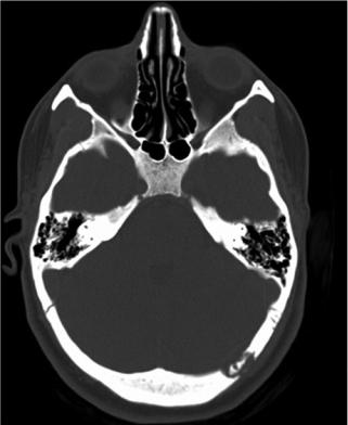
Although PLCH in adults generally presents as a single- system disease, symptoms due to extra-pulmonary localizations may be present in up 10–15% of patients. According to Tazi et al., bone lesions (20% of patients), diabetes insipidus with polyuria and polydipsia, resulting from in ltration of the posterior pituitary (5% of patients), and skin lesions are the most common extrapulmonary manifestations in PCLH (Fig. 18.1) [17]. History and physical examinations are essential to search for extrathoracic LCH involvement, as are skeletal radiographs including a dental panoramic, complete
Fig. 18.1 Skull CT scan of a patient affected by PLCH. It shows two osteolytic bone lesions in the parietal bones and a bigger one in the occipital bone
blood chemistry analysis to detect liver involvement and morning urine osmolality to screen for diabetes insipidus. Adult LCH commonly involves bones and may occur as a bone-limited disease (38%) or as a component of a multisystem disease (66%) [44]. Bone lesions manifest as pain or as a “mass” or “swelling” of the involved site.
Islinger et al. reviewed a series of LCH patients with bone lesions over a 58-year period which included 211 LCH adults. It was estimated that in adults, lesions of the skull occurred in 28% of cases, of the rib in 25%, of the pelvis in 8%, and of the spine in 3%. However, other sites can be involved in long bones and mandible [45]. The radiological appearance of bone lesions and clinical manifestations depends on the site involved and on the disease stage. Typically bone lesions are lytic or may have poorly de ned borders, and in early stages are characterized by a more aggressive pattern of osteolysis. Chronic lesions may resolve completely with or without therapy, or develop a sclerotic appearance due to periosteal new bone formation. Bone lesions of the skull are lytic, round with de ned margins and sometimes may contain a residual bone fragment. They may extend across suture lines, increase in number, or extend into adjacent soft tissue. Osseous lesions may evolve into epidural or epicranial soft tissue masses. Skull lesions can be asymptomatic or can cause headache and tenderness in the skull region involved while those of the mandible can destroy alveolar bone producing the radiological appearance of “foating teeth.” Rib involvement is demonstrated by osteolytic areas, periostitis, and fractures. Sometimes, it is possible to nd an extrapleural mass resulting from soft tissue extension, which causes pain. Pelvic involvement is characterized by poorly de ned areas of osteolysis that develop well-de ned sclerotic margins over time. Spine lesions are osteolytic and can cause the collapse of the vertebral body. In long bones, lesions are frequently intramedullary and diaphyseal and may appear aggressive. Treatment regimens differ widely and are often based on empiric observations. Surgical interventions such as curettage or total excision, radiotherapy, and chemotherapy have all been reported. Although treatment options for adults have never been validated by a clinical trial, studies in the literature compare the ef cacy of different chemotherapeutic treatments. Cantu et al. studied 58 adult LCH patients with bone lesions at a site or as a component of a multisystem disease and described improvement or resolution of bone lesions in a majority of patients treated with radiotherapy, surgery, or chemotherapy (vinblastine/prednisone, 2-chlo- rodeoxyadenosine, and cytosine arabinoside) in comparison with corticosteroids alone [46]. Lair et al. also reported that radiotherapy is a safe and effective means for providing local control of LCH involving bones [47].
Another important extrapulmonary complication of LCH is pulmonary hypertension. It tends to be more severe in
316 |
C. Vancheri and S. Puglisi |
|
|
PLCH than in other interstitial lung diseases, often characterized in later stages by dyspnea at rest and features of right- ventricular circulatory failure. Histopathologically, PH in PLCH is associated with intimal brosis and remodeling of both venous and arterial systems [48]. Dauriat et al. estimated that pulmonary hypertension is present in 92% of 36 patients evaluated for lung transplantation [49]. Because pulmonary hypertension is a poor prognostic indicator in PLCH, it is important to screen all patients, especially those with excessive dyspnea and normal lung function tests, by echocardiography [48]. In selected cases, cardiac catheterization is necessary to con rm PH. Once the diagnosis is made, therapy with vasodilators including phosphodiesterase inhibitors or endothelin receptor antagonists may be considered. Improved exercise capacity can be achieved with these agents, often with an objective reduction in pulmonary pressures, but arterial oxygenation can also worsen as a result of a greater imbalance in ventilation/perfusion due to the inhibition of hypoxic pulmonary vasoconstriction. Prostacyclin can cause severe pulmonary edema and should be used very cautiously in these patients because of the venous involvement. Le Pavec et al. reported their experiences with a group of 29 PH-PLCH patients treated with the usual pulmonary hypertension therapies: endothelin receptor antagonists, phosphodiesterase 5 inhibitors, or prostanoids demonstrating improvement in hemodynamics without oxygen worsening or pulmonary edema in most patients. Supplemental oxygen should be administered to maintain saturations greater than 90% with rest, exercise, and sleep, based on extrapolation from other diseases where the bene ts of the therapy have been demonstrated. However, more studies are needed to evaluate safety and ef cacy of all pulmonary hypertension treatments and management approaches in PH-PLCH [50].
The reported prevalence of central nervous system (CNS) complications ranges widely from 3.4% to 57% [51, 52] and can be subdivided clinically into two groups: the “mass lesion” forms presenting as space occupying lesions anywhere in the CNS; the “neurodegenerative” forms which are characterized by neural cell loss and pyramidal syndrome.
Typical LCH mass lesions may contain CD1a+ LCH cells, lymphocytes, and macrophages with histology similar to extracranial lesions, and usually involve the anterior and posterior hypothalamic pituitary regions, resulting in diabetes insipidus, growth hormone de ciency, and thyroid function abnormalities. Radiological ndings include thickening and enhancement of the pituitary stalk with loss of the posterior pituitary bright spot; enlargement and enhancement of the pineal gland; thickening and enhancement of the choroid plexus; and intraparenchymal masses, usually characterized by a nodular pattern after contrast administration. A variable degree of atrophy of the cerebellum and midbrain has also been described [51]. Magnetic resonance imaging (MRI) may show tissue expansion or
cystic changes in either the pituitary stalk or the pineal gland in up to 63% of patients [53].
“Neurodegenerative CNS LCH” is a syndrome of variable severity characterized by progressive clinical and radiological abnormalities that can occur at any point in the LCH disease course from the initial diagnosis to greater than 5 years later. The only histopathologic study available reported the absence of CD1a+ histiocytes, an infammatory collection of CD8+ lymphocytes associated with neuronal and axonal degeneration [54]. Magnetic resonance imaging (MRI) may show increased T2-weighted MRI signal in the dentate nucleus of the cerebellum, basal ganglia, and pons and PET scanning may reveal decreased or increased FDG uptake in affected regions of the brain [55]. Clinically, ataxia, and tremors may be a consequence of cerebellar involvement. Rarely, patients may develop a progressive cerebellar syndrome, with spastic tetraparesis, pseudobulbar palsy, and cognitive deterioration [56].
LCH patients should be carefully evaluated for cerebellar, pyramidal, and bulbar de cits. Several rating scales have been proposed but not yet broadly approved for LCH patients, such as the Brief Ataxia Rating Scale (BARS) which includes a subset of ve tests focusing primarily on coordination of gait, arm, leg, speech, and eye movements [57].
Very few studies are present in the medical literature and an optimal treatment for CNS localization of the disease has not been de ned. Tin et al. have described a good response to vinblastine, used in other aggressive forms of LCH, in patients with CNS mass lesions with response rates of up to 70%. No effect of this therapy has been described on neurodegenerative lesions [58].
Pulmonary Function Tests
Pulmonary function test ndings are variable in PLCH and the disease can be associated with a restrictive, obstructive, or mixed pattern. According to Tazi et al., the obstructive pattern is the most common. In this study, fow–volume curve alterations were present in 50% of patients, and the ratio of forced expiratory volume in 1 s (FEV1) to vital capacity (VC) was diminished in 20–30% of patients with recent onset of PLCH [17]. This pattern may be related to the bronchial involvement characteristic of smokers, or to bronchiolar obstruction due to peribronchiolar brosis or infammatory in ltrates [17]. On the contrary, Crausman et al. described a restrictive pattern in 11 patients of a cohort of 23 patients with an early PLCH diagnosis. However, in advanced stages, a restrictive pattern usually predominates as lung brosis progresses [18, 59]. At the time of diagnosis, up to 20% of patients may have normal pulmonary function tests, while approximately 60–90% of patients have low diffusing capacity for carbon monoxide (DLCO). Blood gas values at rest may remain normal even in advanced disease, although increased A-a gradient and hypoxemia can occur in early
Данная книга находится в списке для перевода на русский язык сайта https://meduniver.com/

18 Langerhans Cell Granulomatosis and Smoking-Related Interstitial Lung Diseases |
317 |
|
|
stages [17]. Canuet et al. correlated lung function with HRCT ndings and found that the extent of cysts was closely associated with the impairment of both lung function and gas exchange. Interestingly, a predominantly nodular pattern, suggestive of an active infammatory disease, has only moderate functional consequences [60].
Tazi et al. similarly described the correlation between lung function and HRCT lesions. They studied a group of 49 PLCH patients who experienced a deterioration of lung function in 60% of cases, including a decline of FEV1 in 40% of patients and a decline of DLCO in 50% of the patients. The DLCO reduction can herald the presence of pulmonary hypertension. However, according to other studies, the main lung function defect is airway obstruction and this nding is consistent with the bronchiolar localization of pulmonary LCH lesions. Increased profusion of cysts on HRCT scans correlated with a deterioration in lung function parameters. Serial lung function tests are the preferred method to monitor progression of disease to limit exposure to radiation [61].
Chest Radiography
Most patients with PLCH exhibit chest radiographic abnormalities. In the earlier stages of the disease, it is common to nd small nodules that typically range from 1 to 10 mm in diameter and have a bilateral and symmetric distribution on chest radiography. These nodules are characterized by irregular borders and may be single or coalescent. The distribution of nodules is typically limited to upper/middle lung zones with sparing of the lung bases, especially in the costophrenic sulci. As the disease progresses, reticulonodular abnormalities and cystic changes may predominate. As cysts become more numerous, nodules tend to diminish in number [11]. End-stage PLCH is characterized by reticular areas of opacity that may prog-
a
ress to honeycomb lung and contiguous cystic cavities up to 2 cm diameter resulting in patterns that can mimic the radiographic appearance of advanced emphysema or LAM. LAM and end-stage PLCH are the two forms of ILD that can produce hyperinfation rather than reduced lung volumes on CXR and PFTs [62]. Pneumothorax is known to be a complication of PLCH and may occur in the absence of other radiographic pulmonary abnormalities. Chest radiography has limited sensitivity and speci city for the detection and characterization of interstitial lung diseases, and in some cases of PLCH, chest X-ray may even appear to be normal. Khoor et al. reported a rare presentation of PLCH in a 45-year-old male cigarette smoker on chest radiography as a solitary pulmonary nodule. Biopsy showed the histologic and immunophenotypic characteristics of PLCH [63]. Twenty-one years after excision of the sentinel nodule, a new contralateral lung nodule appeared which remained unchanged during 36 months of observation. Another notable radiographic nding in PLCH is pulmonary artery prominence, due to pulmonary hypertension that may occasionally complicate PLCH [63].
High-Resolution Computed Tomography (HRCT)
HRCT is superior to routine chest radiography in demonstrating the morphology and distribution of lung abnormalities. Patterns differ widely based on the stage of PLCH. In the early stages of the disease, a pattern de ned by the presence of multiple nodular opacities measuring 1–5 mm in diameter or larger is often found [64]. Nodule sizes greater than 10 mm in diameter are unusual [65]. These small nodules, which are not typically apparent on chest X-rays, are characterized by irregular margins surrounded by normal lung parenchyma (Fig. 18.2). They may be profuse and are generally solid, although cavitation may occur over time.
b
Fig. 18.2 HRCT of the lungs of a patient affected by PLCH, showing a predominant nodular pattern. Centrilobular and peribronchiolar nodules and present (a), some of which are cavitated (b)

318 |
C. Vancheri and S. Puglisi |
|
|
However, the predominant characteristic of lung nodules is their distribution, with a topographical predominance in the upper and middle lung zones with relative sparing of the lung bases. Most nodules show a centrilobular or peribronchial distribution, refecting the bronchiole-centered localization of PLCH lesions in histopathologic studies. Brauner et al. has proposed a temporal progression of these pulmonary nodules into cavitary nodules and then into cysts [64], with serial scans revealing a decreasing preponderance of nodules and an increasing number of thin-walled cysts. Cystic lesions tend to be small and thick walled initially, with diameters of less than 10 mm, and then become larger and thinner walled, with diameters up to 20 mm. Conceptually, cyst formation can develop due to cavitation within a centrilobular nodular lesion or to increasing bronchiolar dilatation from granulomas destruction and brosis at the lesion margin. Cyst distribution is most prevalent in upper lung zones where they can appear as round or ovoid spherical spaces or with bizarre shapes that result from coalescence of adjacent cysts (Fig. 18.3a–c). Some of these
cystic spaces reach diameters of up to 80 mm. Advanced disease is characterized by architectural distortion by cysts with few nodules [66], while late-stage disease is marked by the presence of large areas of honeycombing, predominantly in the upper lung zones. Some studies have described full or partial resolution of lesions occurring in patients with nodular lesions, indicative of reversibility, while cystic lesions remain unchanged or worsen with time [67]. Soler et al. compared the nature of the ndings on CT scans with those of lesions on biopsy samples in PLCH patients. They found that early-stage PLCH histopathological lesions consisted of forid granulomas that were composed of typical LCs associated with macrophages and infammatory cells, particularly lymphocytes and eosinophils. In more advanced disease, necrotic granulomas were found in pulmonary samples, characterized by a prominent central cavity and fewbrotic changes, but still numerous LCs and infammatory cells in their walls. In these cases, few cavitated nodules or thin-walled cysts were seen on CT scans. In late-stage PLCH, brous cysts of variable size, demarcated by a
a |
b |
c
Fig. 18.3 HRCT of the lungs of a patient affected by PLCH showing a predominant cystic pattern. The cysts are characterized by variable wall thickness and bizarre shapes (a). Centrilobular and bronchiolocentric nodules are present, some of which are starting to cavitate (b, c)
Данная книга находится в списке для перевода на русский язык сайта https://meduniver.com/