


of Atlas
ultrasound musculoskeletal anatomy
50
Elbow
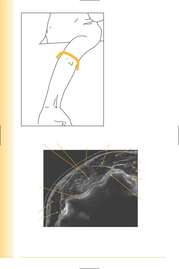
Lateral elbow
(Figures 58 and 59)
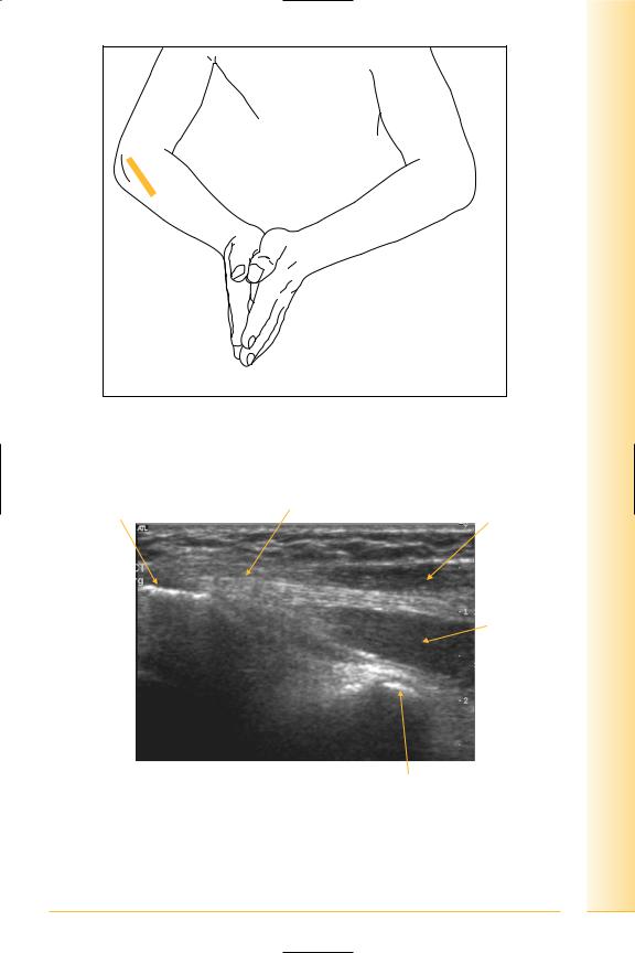
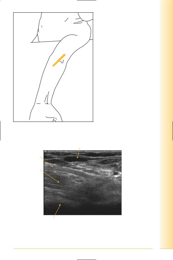
Important anatomical structures in this region of the elbow include the common extensor origin (CEO). This comprises the fused tendons of extensor carpi radialis brevis, extensor digitorum, extensor digiti minimi and extensor carpi ulnaris which attach anteriorly to the lateral epicondyle of the humerus.
The superficial group of posterior and lateral forearm muscles are brachioradialis and extensor carpi radialis longus. They originate proximal to the CEO, from the lateral supracondylar ridge of the humerus.
•Brachioradialis
Origin: lateral supracondylar ridge of humerus.
Insertion: lateral aspect distal radius.
•Extensor carpi radialis longus
Origin: lateral supracondylar ridge of humerus.
Insertion: dorsal surface base of index finger metacarpal.
Notes
limb Upper
Elbow
FIG. 58 LS, probe longitudinal to radial aspect of elbow, patient in “praying” position
Lateral humeral |
Common extensor origin |
Extensor carpi radialis |
condyle |
|
longus and brevis |
Extensor digitorum
Proximal |
Distal |
Radial head
FIG. 59 LS, common extensor origin
51

of Atlas
ultrasound musculoskeletal anatomy
52
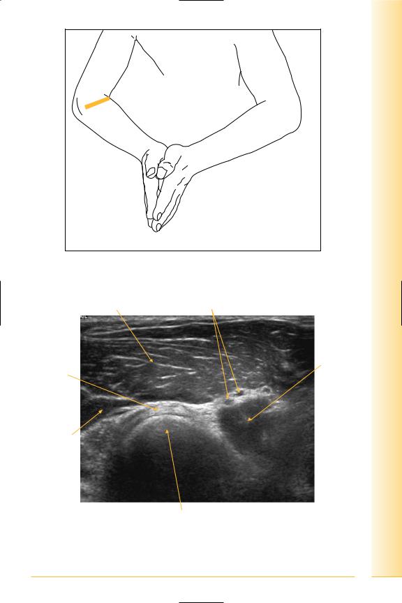
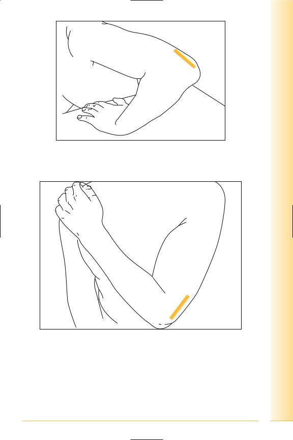
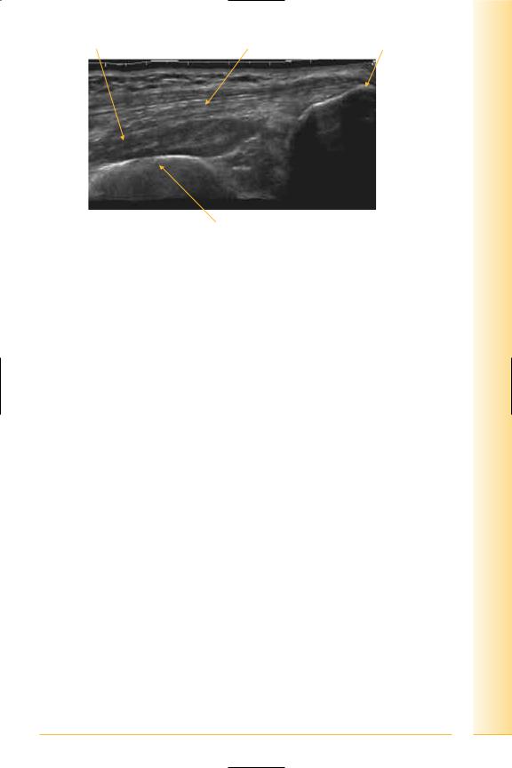
The radiocapitellar joint and annular ligament
(Figures 60–63)
Annular ligament
Encircles head of radius, attached to the anterior and posterior borders of the radial notch of the ulna.
Notes
limb Upper
Elbow
FIG. 60 TS, probe transverse to radiocapitellar joint, patient in “praying” position
Brachioradialis |
Radial neurovascular bundle |
Annular |
Brachialis |
ligament |
|
Extensor carpi radialis
Posterior |
Anterior |
Radial head
FIG. 61 TS, elbow lateral
53
of Atlas
ultrasound musculoskeletal anatomy
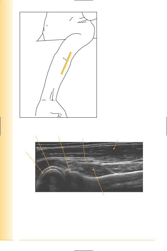
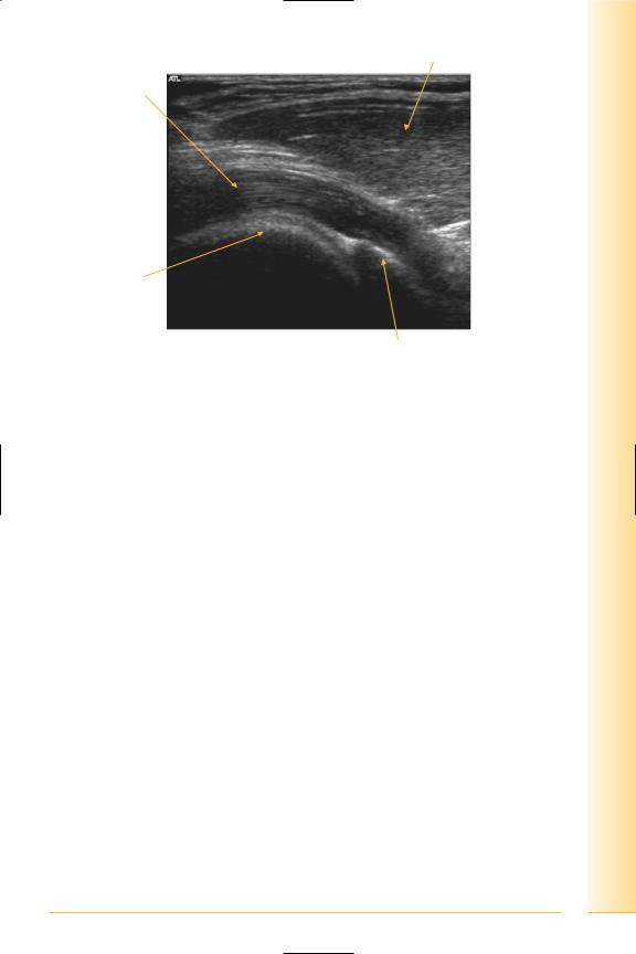
FIG. 62 LS panorma, probe longitudinal to antero-lateral elbow
Capitellum |
Radial head |
Radial neurovascular |
Brachioradialis/extensor |
|
|
bundle |
carpi radialis longus |
Articular cartilage
Proximal |
Distal |
Supinator muscle
FIG. 63 LS panorama, anterolateral elbow
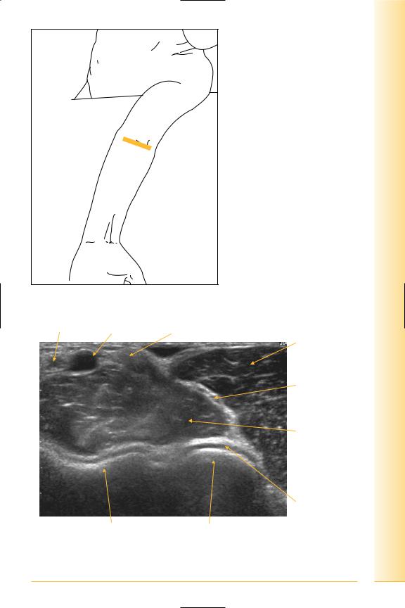
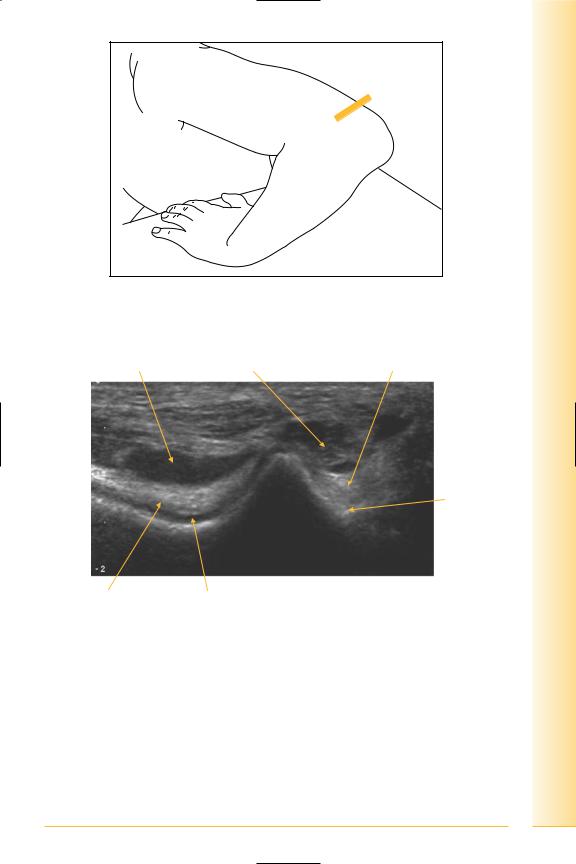
Anterior elbow
(Figures 64 and 65)
Visualizes the anterior aspect of the elbow joint, neurovascular structures and 54 biceps tendon.
limb Upper
Elbow
FIG. 64 TS, probe transverse to anterior elbow, arm extended
Brachial artery Basilic vein |
Biceps tendon |
Brachioradialis
Radial nerve
Brachialis muscle
Lateral
Articular cartilage
Medial |
Humeral trochlea |
Capitellum |
FIG. 65 TS, anterior elbow
55

of Atlas
ultrasound musculoskeletal anatomy
56
Biceps tendon
(Figures 66–70)
It inserts onto the tuberosity of the radius, and a bursa separates bone and tendon just proximal to the insertion. Further insertions are via the bicipital aponeurosis into the deep fascia on the ulnar aspect of the forearm and posterior subcutaneous border of the ulna.
It can be difficult to demonstrate the tendon due to anisotropy as it travels deeper to its insertion.
Notes
limb Upper
Elbow
FIG. 66 LS, probe longitudinal to distal biceps tendon, slightly oblique to long axis of upper limb
Median cubital vein
Biceps tendon
Flexor muscle  group
group
Brachialis
Proximal |
Distal |
Distal humerus
FIG. 67 LS, anterior elbow
57
of Atlas
ultrasound musculoskeletal anatomy
58
Brachialis |
Median cubital vein |
Proximal |
Capitellum |
Radial head |
Distal |
FIG. 68 LS, biceps tendon
FIG. 69 LS, probe longitudinal to antero-medial aspect of elbow
Flexor muscle group
Brachialis
Proximal |
Distal |
Trochlea of humerus
Coronoid process of ulna
FIG. 70 LS, antero-medial elbow
limb Upper
Elbow
59

of Atlas
ultrasound musculoskeletal anatomy
60
Medial elbow
(Figures 71 and 72)
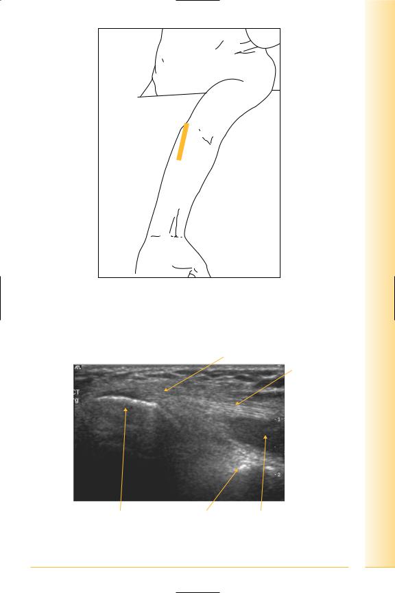
Pathologically and anatomically important structures here include the common flexor origin (CFO), ulnar collateral ligament and medial aspect of the elbow joint.
The CFO is situated anteriorly on the medial epicondyle of the humerus, and gives origin to the superficial muscle group of pronator teres, flexor carpi radialis, flexor digitorum superficialis, palmaris longus and flexor carpi ulnaris. These muscles form the medial border of the cubital fossa.
The deep forearm muscles include flexor pollicis longus, flexor digitorum profundus and pronator quadratus.
Notes
limb Upper
Elbow
FIG. 71 LS, probe longitudinal to antero-medial elbow, access to which is improved if the patient leans to that side
Common flexor origin
Common flexor tendon
Proximal |
Distal |
Medial epicondyle |
Coronoid process |
Flexor muscle |
of humerus |
of ulna |
group |
FIG. 72 LS, common flexor origin
61

of Atlas
ultrasound musculoskeletal anatomy
62
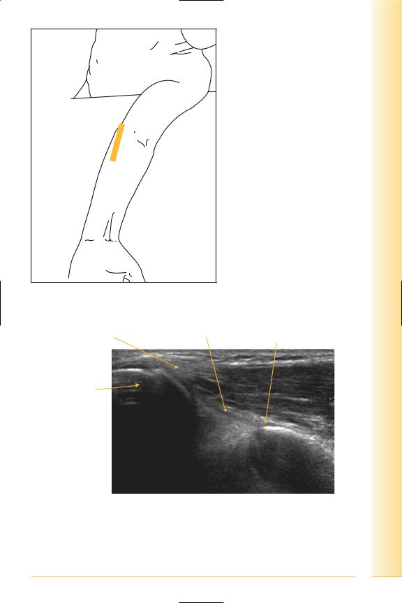
Ulnar collateral ligament (UCL)
(Figures 73–76)
This triangular ligament has three parts:
•The strongest is the anterior band, which can be seen deep to the CFO, running from the medial epicondyle of the humerus to the coronoid process of the ulna (the “sublime” tubercle).
•The posterior band runs posteriorly from the sublime tubercle to the olecranon.
•The middle band spans anterior and posterior.
Notes
limb Upper
Elbow
FIG. 73 LS, probe longitudinal to medial elbow (similar position to CFO)
Common flexor tendon |
Anterior band of UCL Sublime tubercle of |
|
coranoid process of ulna |
Medial condyle of humerus
Proximal |
Distal |
FIG. 74 LS, medial elbow showing UCL
63
of Atlas
ultrasound musculoskeletal anatomy
|
|
FIG. 75 TS panorama, anterior |
|
|
elbow |
Biceps muscle Brachialis |
Radial nerve |
|
and tendon |
Brachial artery/median nerve |
Brachioradialis |
|
||
|
|
Extensor carpi |
|
|
radialis |
|
|
longus and |
|
|
brevis |
|
|
Common |
|
|
extensor origin |
Trochlea of |
|
|
humerus |
|
|
|
|
Capitellum |
Pronator |
|
|
teres and |
|
|
flexor |
|
Lateral |
carpi radialis |
|
|
Medial
FIG. 76 TS panorama, anterior elbow
64


of Atlas
ultrasound musculoskeletal anatomy
66
Posterior elbow
(Figures 77–81)
The triceps tendon attaches to the olecranon of the ulna.
The ulnar nerve can be seen in a groove posterior to medial humeral epicondyle.
Examination of the posterior elbow is facilitated by placing the joint in one of the two positions
•Patient in “crab” position.
•Patient holding contralateral shoulder.
Notes
limb Upper
Elbow
FIG. 77 LS, probe longitudinal to posterior elbow, patient in “crab” position
FIG. 78 LS, probe longitudinal to posterior elbow, patient holding contralateral shoulder
67
of Atlas
ultrasound musculoskeletal anatomy
68
Medial head of triceps muscle |
Triceps tendon Olecranon process of ulna |
Proximal |
Distal |
FIG. 79 LS, posterior elbow
FIG. 80 LS panorama, posterior elbow
Medial head of triceps |
Triceps tendon |
Olecranon process of ulna |
Proximal |
Distal |
Distal humerus
FIG. 81 LS panorama, triceps
limb Upper
Elbow
69

of Atlas
ultrasound musculoskeletal anatomy
70
Ulnar nerve
(Figures 82 and 83)
Notes
limb Upper
Elbow
FIG. 82 TS, probe transverse to posterior elbow
Triceps tendon Flexor carpi ulnaris muscle |
Ulnar nerve |
Lateral
Ulnar groove
Medial
Posterior fat pad |
Olecranon fossa |
|
of humerus |
FIG. 83 TS, posterior elbow
71