

of Atlas
ultrasound musculoskeletal anatomy
8
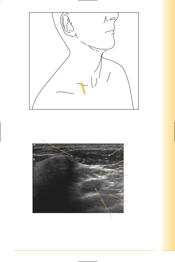
Infraclavicular fossa
(Figures 10 and 11)
Notes
Chest
fossa Infraclavicular
FIG. 10 LS, probe inferior to the clavicle
Inferior border of clavicle
|
Pectoralis |
|
major |
|
Pectoralis |
|
minor |
|
Subclavius |
Superior |
Inferior |
|
Pleura |
|
Subclavian vein |
FIG. 11 LS, infraclavicular fossa |
|
9

of Atlas
ultrasound musculoskeletal anatomy
10
Sternoclavicular joint
(Figures 12 and 13)
This is an atypical synovial joint, like the acromioclavicular joint, as the articular surfaces are covered with fibrocartilage. The medial end of the clavicle articulates with the manubrium and first costal cartilage. The capsule is thickened anteriorly and posteriorly to form the sternoclavicular ligaments. Further ligaments attach to the first rib and contralateral clavicle.
Notes

Chest
joint Sternoclavicular
FIG. 12 Probe longitudinal to joint, angled at 45 degrees to midline
|
Capsule and anterior |
|
Clavicle |
sternoclavicular ligament |
Manubrium |
SC joint containing articular disc
Lateral |
Medial |
FIG. 13 LS, sternoclavicular joint
11