


Abdomen and pelvis
Anterior wall . . . . . . . . . . . . . . . . . . . . . . . . . . . . . . . . . . . . . . 116 Posterior wall . . . . . . . . . . . . . . . . . . . . . . . . . . . . . . . . . . . . . . 126 Groin . . . . . . . . . . . . . . . . . . . . . . . . . . . . . . . . . . . . . . . . . . . . 134 Hip . . . . . . . . . . . . . . . . . . . . . . . . . . . . . . . . . . . . . . . . . . . . . . 146
115

of Atlas
ultrasound musculoskeletal anatomy
116
Anterior wall
(Figures 142–155)
•Rectus sheath
Aponeurosis of three muscles (external oblique, internal oblique, transversus abdominis) to form linea alba in midline.
•Proximal attachment
Costal margin, xiphisternum.
•Distal attachment
Pubic symphysis and crest.
Notes

and Abdomen
pelvis wall Anterior
FIG. 142 TS, midline
Linea alba |
Fat |
Rectus abdominis
Bowel
Right |
Left |
Peritoneum
FIG. 143 TS, rectus sheath – mid-abdomen
117
of Atlas
ultrasound musculoskeletal anatomy
FIG. 144 TS, probe right of midline
Subcutaneous fat
Rectus
Medial
Lateral
Bowel wall
Peritoneum
FIG. 145 TS, rectus abdominis
118
and Abdomen
pelvis wall Anterior
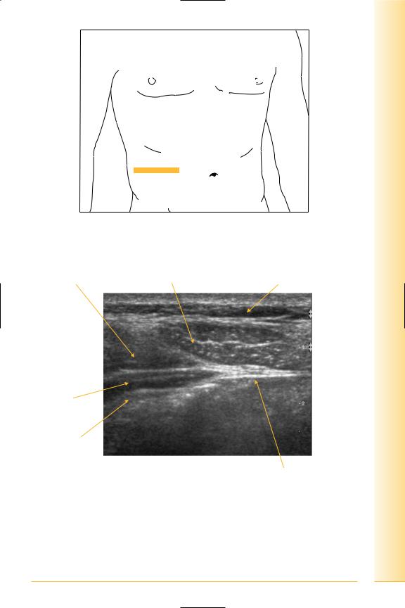
FIG. 146 TS, probe over flank/anterior wall
External oblique |
Rectus abdominis |
Fat |
Lateral |
Medial |
Internal oblique
Transversus abdominis
Peritoneum
FIG. 147 TS, anterior abdominal wall – mid-abdomen
119

of Atlas
ultrasound musculoskeletal anatomy
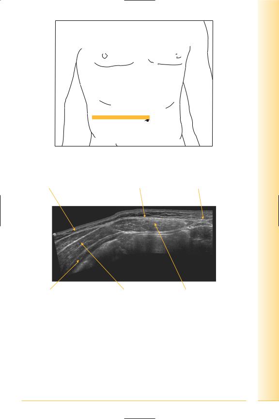
FIG. 148 TS, probe over flank
External oblique
Lateral
Transversus  abdominis
FIG. 149 TS, anterior abdominal wall – flank
120
Fat
Medial
Internal oblique
Peritoneum
and Abdomen
pelvis wall Anterior
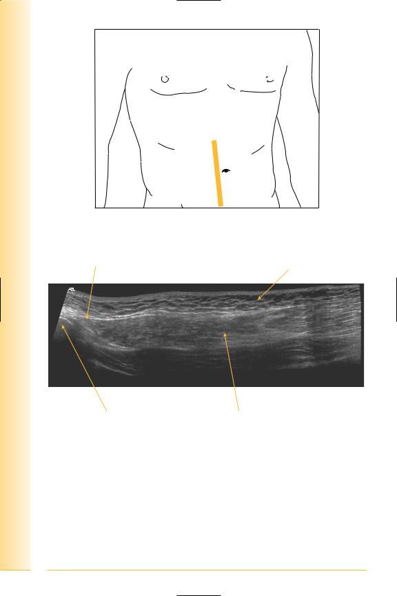
FIG. 150 TS panorama, rectus sheath
External oblique |
Rectus sheath |
Linear alba |
Lateral |
Medial |
Transversus |
Internal oblique |
Rectus abdominis |
abdominis |
|
|
FIG. 151 TS panorama, anterior abdominal wall
121
of Atlas
ultrasound musculoskeletal anatomy
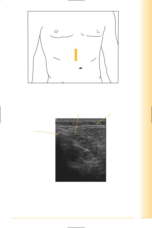
FIG. 152 LS panorama, right of midline
Rectus insertion |
Fat |
|
Proximal |
Distal |
|
|
Xiphisternum |
Rectus abdominis |
FIG. 153 LS panorama, rectus origin from xiphisternum
122
and Abdomen
pelvis wall Anterior
FIG. 154 LS, probe over xiphisternum. Proximal insertion is normally ill defined and appearance of xiphisternum depends on the degree of calcification
Rectus origin |
Fat |
Xiphisternum
Superior
Inferior
FIG. 155 LS, rectus origin
123

of Atlas
ultrasound musculoskeletal anatomy
124
Distal rectus insertion
(Figures 156 and 157)
Notes
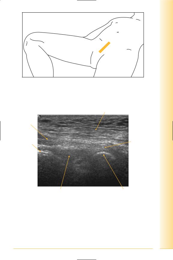
FIG. 156 LS, probe over symphysis
and Abdomen
pelvis wall Anterior
Subcutaneous fat
Rectus muscle
Peritoneum |
Rectus tendon |
|
|
Proximal |
Distal |
Fat |
Body of pubis |
FIG. 157 LS, distal rectus insertion
125