

of Atlas
ultrasound musculoskeletal anatomy
12
Chest wall
Anterior
The thoracic wall muscles lie in three layers analogous to those in the abdomen, but separated by ribs. The outer two layers (external and internal intercostal) are usually visible in a rib space, deep to which can be seen the pleural space and lung. The neurovascular bundle lies deep to the second layer at the superior aspect of the intercostal space.
Ribs and costal cartilages
(Figures 14–17)
The anterior aspect of a rib articulates with a costal cartilage via a cartilaginous joint at which no movement is possible. The rib is deeply concave, and cartilage convex. The second to seventh costal cartilages articulate with the sternum via synovial joints. Calcification within costal cartilages is highly variable, and causes foci of hyper-echogenicity.
Notes
Chest
wall Chest
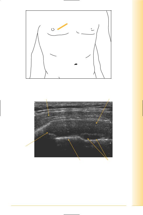
FIG. 14 Probe longitudinal to costal cartilage
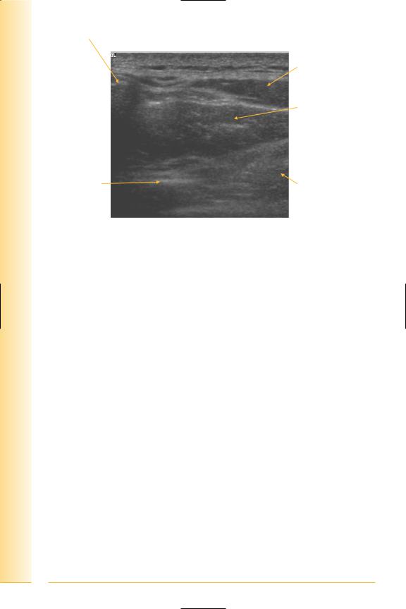
Pectoralis major |
Costal cartilage |
Lateral |
Medial |
Costo-chondral junction
Pleura |
Internal thoracic |
|
artery and vein |
FIG. 15 LS, chest wall parasternal
13
of Atlas
ultrasound musculoskeletal anatomy
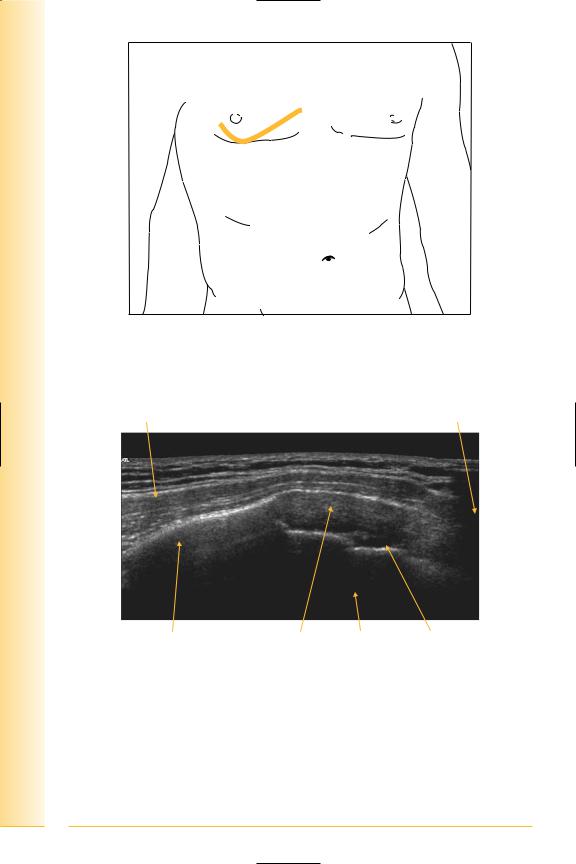
FIG. 16 LS panorama of rib and costal cartilage
Pectoralis major |
Sternum |
Lateral |
Medial |
Rib |
Costal cartilage Lung Internal thoracic vessels |
FIG. 17 Panorama, anterior chest wall
14

of Atlas
ultrasound musculoskeletal anatomy
16
Lateral chest wall
(Figures 18 and 19)
External and internal intercostals
•Origin: lower border of superior rib.
•Insertion: upper border of inferior rib. Internal intercostals deep to external.
Serratus anterior
•Origin: upper eight ribs, overlying the lateral chest wall.
•Insertion: inferior angle and costal margin of the scapula. It forms the medial wall of the axilla.
Notes

Chest
wall Chest
FIG. 18 TS, rib space on lateral aspect of chest
Serratus anterior |
Rib |
Fat |
Superior |
Inferior |
External |
Internal |
intercostal |
intercostal |
Lung |
Pleura |
FIG. 19 TS of chest wall – intercostals |
|
17
of Atlas
ultrasound musculoskeletal anatomy
18
Posterior chest wall
(Figures 20–22)
Trapezius muscle covers the postero-medial aspect of the upper chest:
•Origin: from skull to the T12 vertebra in the midline.
•Insertion: clavicle, acromion and spine of the scapula.
Deep to trapezius are the muscles that extend from the vertebral column to the medial aspect of the scapula – levator scapulae superiorly and the rhomboids inferiorly. Inferiorly, trapezius covers the superior aspect of latissimus dorsi. The erector spinae muscles are deep to the rhomboids.
•Levator scapulae
Origin: posterior tubercles of transverse processes of upper four cervical vertebrae.
insertion: superior angle, medial border of scapula.
•Rhomboids
Origin: lower part of ligamentum nuchae and spines of cervical and upper four thoracic vertebrae.
Insertion: medial border scapula, major inferiorly, and minor between levator scapulae and major.
•Latissimus dorsi
Origin: spines of lower six thoracic vertebrae, lumbar fascia, lower four ribs and posterior iliac crest.
Insertion: floor of bicipital groove of humerus.
Notes

Chest
wall Chest
FIG. 20 TS of posterior chest wall, probe at medial border of scapula
Medial border of scapula
Trapezius
|
Rhomboid major |
Lateral |
Medial |
|
Rib |
FIG. 21 TS, posterior chest wall
19
of Atlas
ultrasound musculoskeletal anatomy
20
Medial border of scapula |
|
|
Trapezius |
|
Rhomboid |
|
major muscle |
Lateral |
Medial |
Rib |
Erector spinae |
FIG. 22 TS, inferoposterior chest wall
