


of Atlas
ultrasound musculoskeletal anatomy
96
Hand
Palm
Palmar spaces
(Figures 114 and 115)
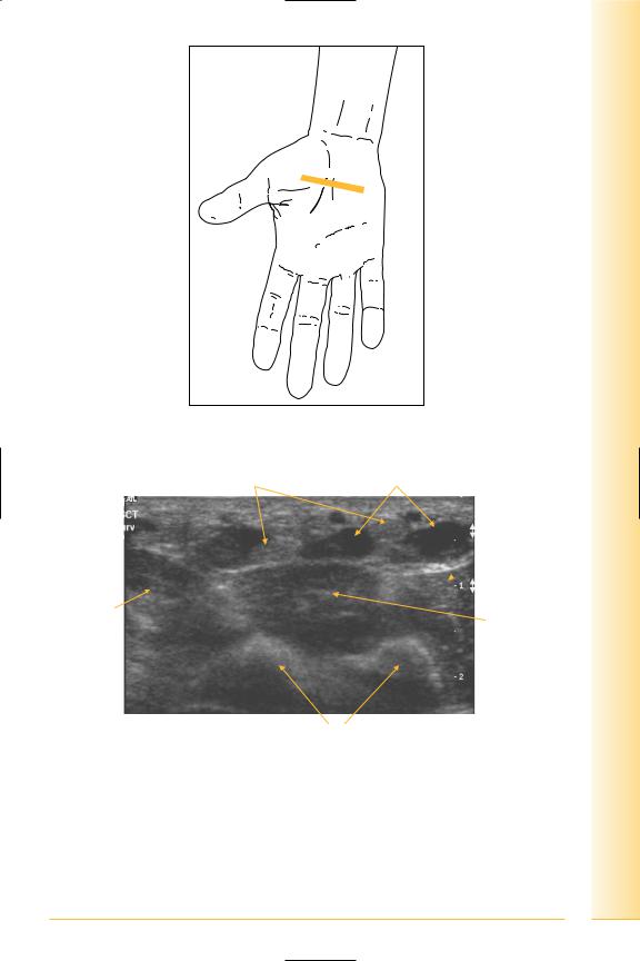
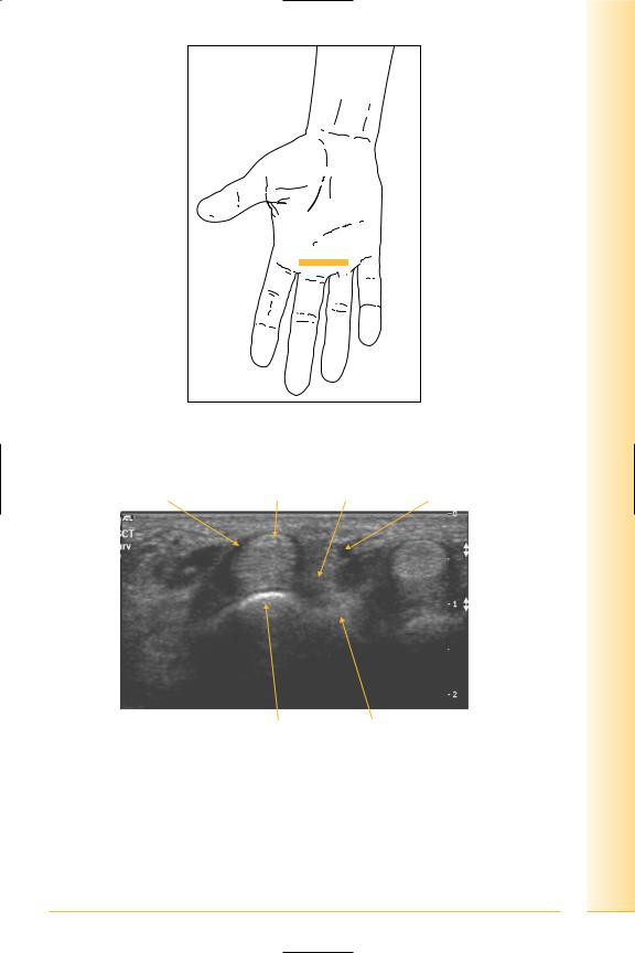
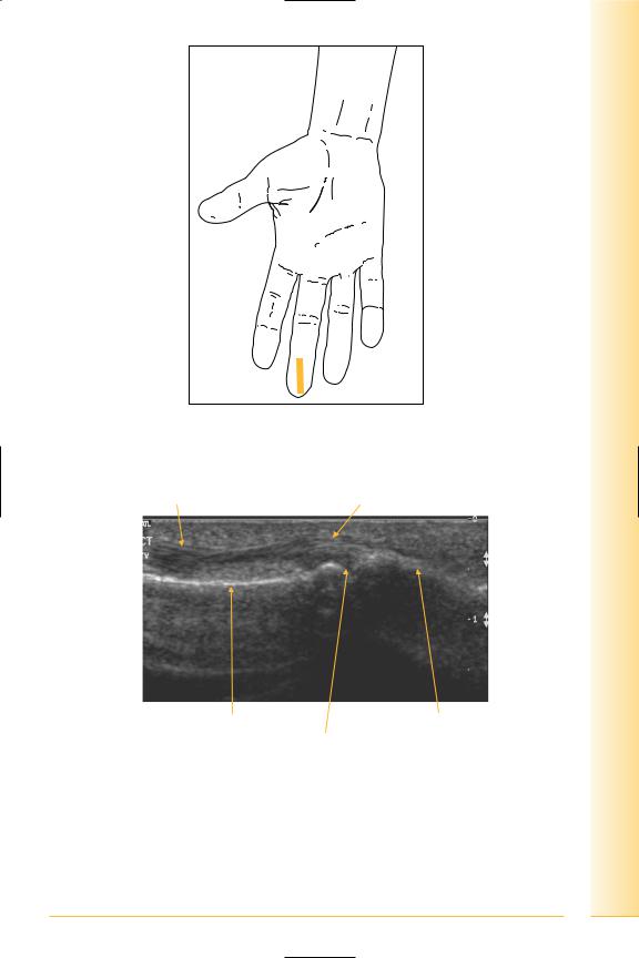
The palm is divided into three spaces by two septa passing from the palmar aponeurosis to the thumb and little finger metacarpals. The lateral space contains thenar muscles; the medial contains hypothenar muscles, and the central contains long flexor tendons, lumbricals, the superficial and deep palmar arches and median nerve.
Central palmar space
Notes
limb Upper
Hand
FIG. 114 TS, probe transverse to flexor tendons in proximal palm
Flexor tendons |
Lumbricals |
Opponens
 digiti minimi
Adductor |
Interossei |
pollicis |
Lateral |
Metacarpals |
Medial |
FIG. 115 TS, central palmar space
97
of Atlas
ultrasound musculoskeletal anatomy
98
Medial (hypothenar) and lateral (thenar) palmar spaces
(Figures 116–121)
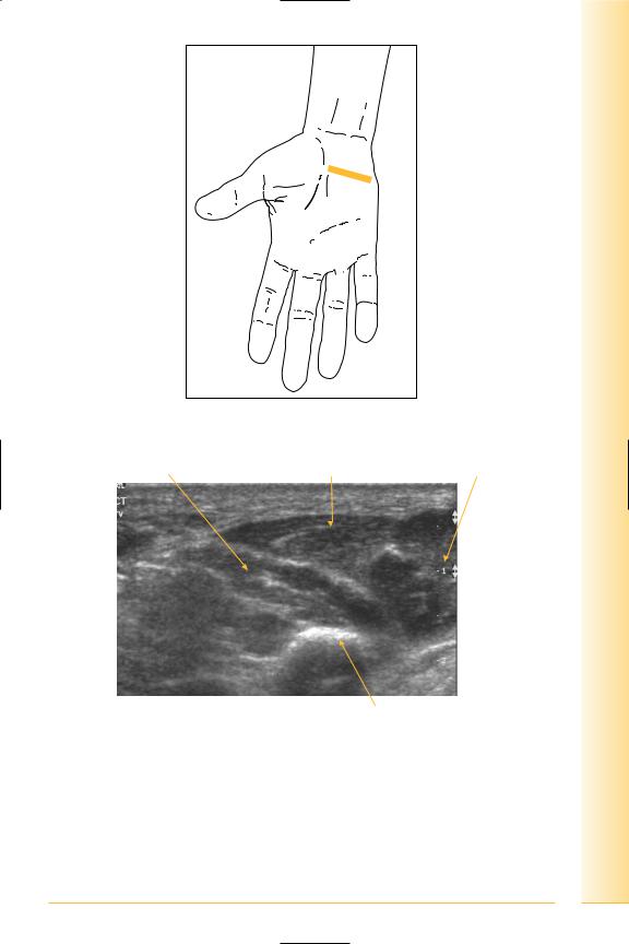
•The muscles in the hypothenar eminence are abductor digiti minimi, opponens digiti minimi and flexor digiti minimi.
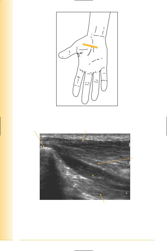
•The muscles in the thenar eminence are abductor pollicis brevis, opponens pollicis and flexor pollicis brevis.
Notes
limb Upper
Hand
FIG. 116 TS, probe transverse on hypothenar eminence
Opponens digiti minimi |
Flexor digiti minimi |
Abductor digiti minimi |
Lateral |
Medial |
|
Little finger metacarpal
FIG. 117 TS, hypothenar eminence
99
of Atlas
ultrasound musculoskeletal anatomy
FIG. 118 TS, probe on thenar eminence
Thumb metacarpal |
Abductor pollicis brevis |
Flexor pollicis brevis
 Opponens
pollicis
Lateral |
Medial |
Adductor pollicis
FIG. 119 TS, thenar eminence
100
limb Upper
Hand
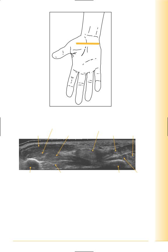
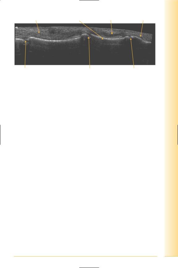
FIG. 120 TS panorama, thenar and hypothenar eminences
Opponens pollicis |
Flexor tendons in |
|
|
|
distal carpal canal |
Abductor pollicis brevis |
Flexor pollicis brevis |
Flexor and abductor digiti minimi |
Thumb metacarpal |
Adductor pollicis |
Little finger |
Opponens |
|
|
metacarpal |
digiti minimi |
Lateral |
|
Medial |
|
FIG. 121 TS panorama, palmar spaces
101

of Atlas
ultrasound musculoskeletal anatomy
102
Flexor tendons
(Figures 122–138)
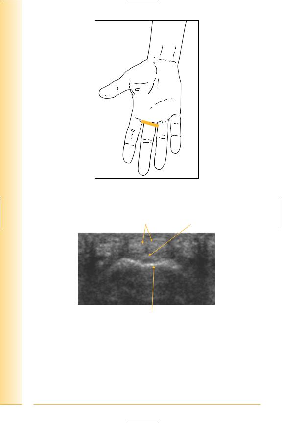
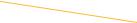
The superficial flexor tendons pass deep to the flexor retinaculum at the wrist. In the palm they are contained within a common flexor sheath, superficial to the profundus tendons. This relationship continues in the common synovial sheath of the finger. The superficialis tendon splits at the level of the proximal phalanx, and is pierced by the profundus tendon, which is therefore the most superficial tendon at the distal part of the proximal phalanx. The superficial tendon inserts onto the sides of the palmar surface of the middle phalanx, and the deep tendon continues to the base of the distal phalanx.
Notes
limb Upper
Hand
FIG. 122 TS, probe transverse to flexor tendons, distal palm
Flexor tendon sheath |
Flexor tendon Lumbrical Neurovascular bundle |
Lateral |
Metacarpal |
Interossei |
Medial |
FIG. 123 TS, flexor tendons
103
of Atlas
ultrasound musculoskeletal anatomy
FIG. 124 TS, probe over proximal phalanx
Flexor digitorum |
Flexor digitorum |
superficialis tendon |
profundus tendon |
Lateral |
Medial |
Proximal phalanx
FIG. 125 TS, flexor tendon, level of proximal phalanx
104

limb Upper
Hand
FIG. 126 TS, level of distal proximal phalanx
Superficial flexor tendons
 Deep flexor tendon
 Proximal phalanx
Lateral |
Medial |
FIG. 127 TS of finger flexor tendons, level of proximal phalanx
105

of Atlas
ultrasound musculoskeletal anatomy
FIG. 128 TS, level of middle phalanx
Deep flexor tendon
|
Middle phalanx |
Lateral |
Medial |
FIG. 129 TS, finger flexor tendon, level of middle phalanx
106
limb Upper
Hand
FIG. 130 LS, probe longitudinal to flexor tendons, level of metacarpophalangeal joint. Dynamic assessment with finger flexion and extension
Superficial and deep flexor tendon |
Articular cartilage |
Proximal |
Metacarpal |
Proximal phalanx |
Distal |
FIG. 131 LS, finger flexor tendon
107
of Atlas
ultrasound musculoskeletal anatomy
FIG. 132 LS, probe longitudinal to proximal interphalangeal joint
Volar plate
Proximal |
Distal |
Flexor digitorum (profundus |
PIP joint |
and superficialis) tendons |
|
FIG. 133 LS, flexor tendon
108
limb Upper
Hand
FIG. 134 LS, probe longitudinal to distal interphalangeal joint
Flexor digitorum tendons |
Flexor digitorum profundus tendon |
Proximal |
Distal |
Middle phalanx |
Distal phalanx |
Distal interphalangeal joint |
|
FIG. 135 LS, finger
109

of Atlas
ultrasound musculoskeletal anatomy
FIG. 136 LS panorama, finger flexors
Superficial and deep flexor tendons |
Deep flexor tendon |
Tendon insertion |
Proximal |
Middle phalanx |
Distal phalanx |
Distal |
FIG. 137 LS panorama, flexor digitorum profundus insertion
110
Superficial and deep |
Site of superficialis |
Profundus |
Insertion of |
flexor tendons |
insertion |
tendon |
profundus tendon |
limb Upper
Hand
MCPJ |
PIPJ |
DIPJ |
Proximal |
|
Distal |
FIG. 138 Panorama, flexor digitorum tendons
111

of Atlas
ultrasound musculoskeletal anatomy
112
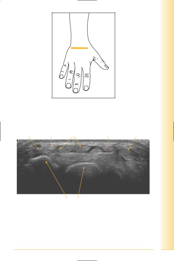
Dorsum of the hand
(Figures 139–141)
Notes
limb Upper
Hand
FIG. 139 TS, probe transverse on dorsum of hand
Extensor carpi |
Extensor |
Extensor digitorum |
Extensor |
Extensor |
ulnaris |
digiti minimi |
tendons |
indicis |
pollicis longus |
Medial |
Carpal bones |
Lateral |
FIG. 140 TS, dorsum of hand
113
of Atlas
ultrasound musculoskeletal anatomy
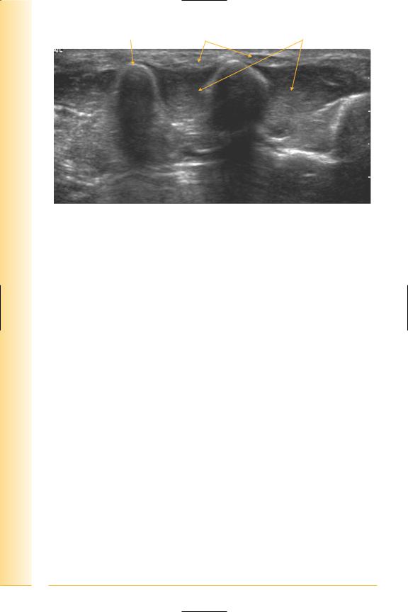
Metacarpal |
Extensor tendons |
Dorsal interossei |
Medial |
Lateral |
FIG. 141 TS mid dorsum of hand
114