
of Atlas
ultrasound musculoskeletal anatomy
134
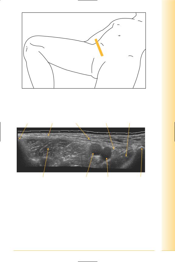
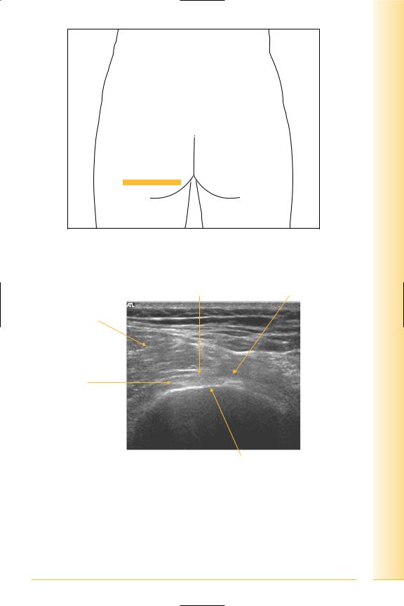
Groin
(Figures 170 and 171)
•Inguinal ligament
The lower free border of the external oblique aponeurosis between pubic tubercle and anterior superior iliac spine.
•Inguinal canal
Anterior wall: external oblique aponeurosis, reinforced by internal oblique.
Posterior wall: transversalis fascia, reinforced by conjoint tendon medially.
Contents: spermatic cord or round ligament, genito-femoral, ilio-inguinal and sympathetic nerves, testicular, cremasteric, and ductus deferens arteries.
Notes
and Abdomen
pelvis Groin
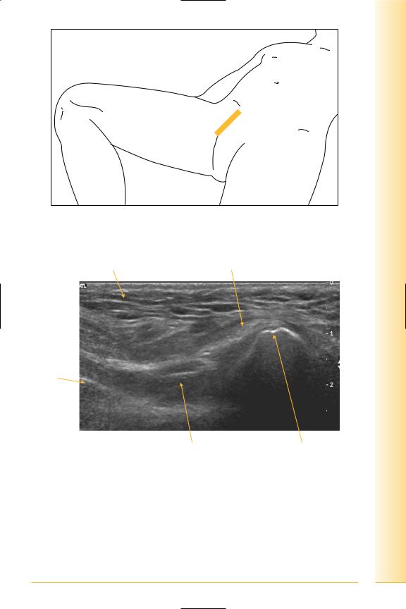
FIG. 170 TS panorama, along inguinal ligament
Anterior superior |
|
|
|
iliac spine |
Inguinal ligament |
Femoral canal |
Pectineus |
Ilio-psoas |
Femoral artery |
Femoral vein |
Pubic tubercle |
Lateral |
|
|
Medial |
FIG. 171 TS panorama, inguinal ligament
135

of Atlas
ultrasound musculoskeletal anatomy
136
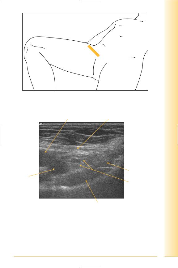
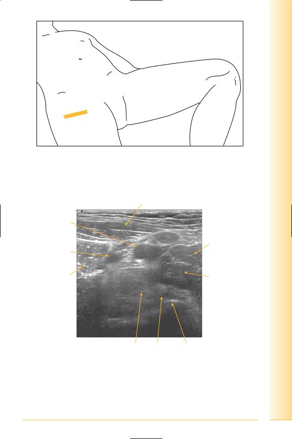
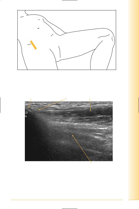
Femoral triangle: boundaries
(Figures 172 and 173)
•Superior: inguinal ligament.
•Lateral: sartorius.
•Medial: adductor longus.
•Floor: adductor longus and pectineus.
Contents: femoral sheath, femoral nerve.
Femoral sheath is a downward extension of the extraperitoneal fascia into the thigh.
Contents
•Lateral: femoral artery.
•Central: femoral vein.
•Medial: fat, lymphatics (femoral canal). This communicates superiorly via the femoral ring with abdominal extraperitoneal fascia.
Notes
and Abdomen
pelvis Groin
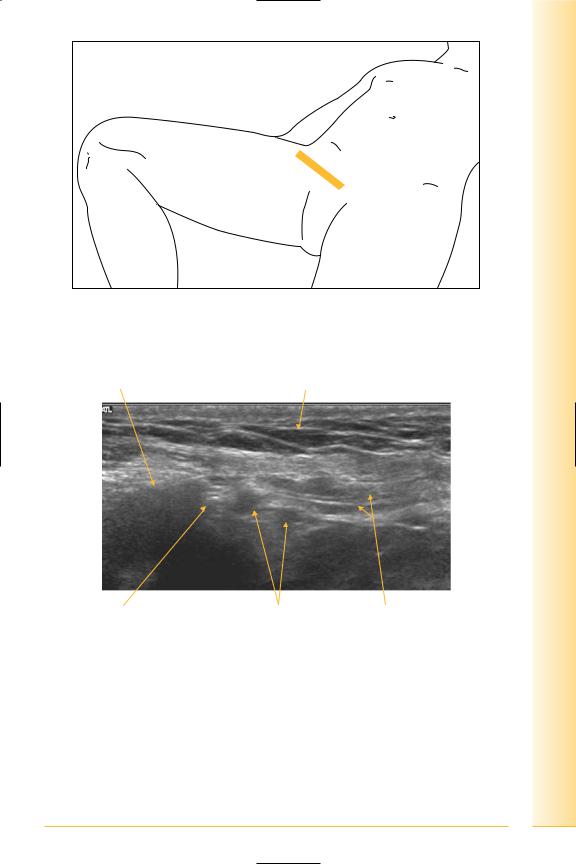
FIG. 172 TS of femoral triangle. Leg abducted or adducted
Femoral artery |
Inguinal ligament |
Lateral |
Medial |
Adductor longus
Femoral vein
Femoral canal
Pectineus
FIG. 173 TS, femoral sheath
137
of Atlas
ultrasound musculoskeletal anatomy
138
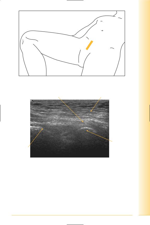
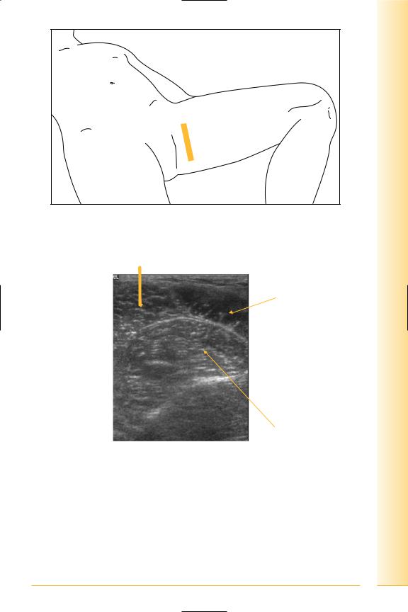
Rectus insertion
(Figures 174 and 175)
Notes
and Abdomen
pelvis Groin
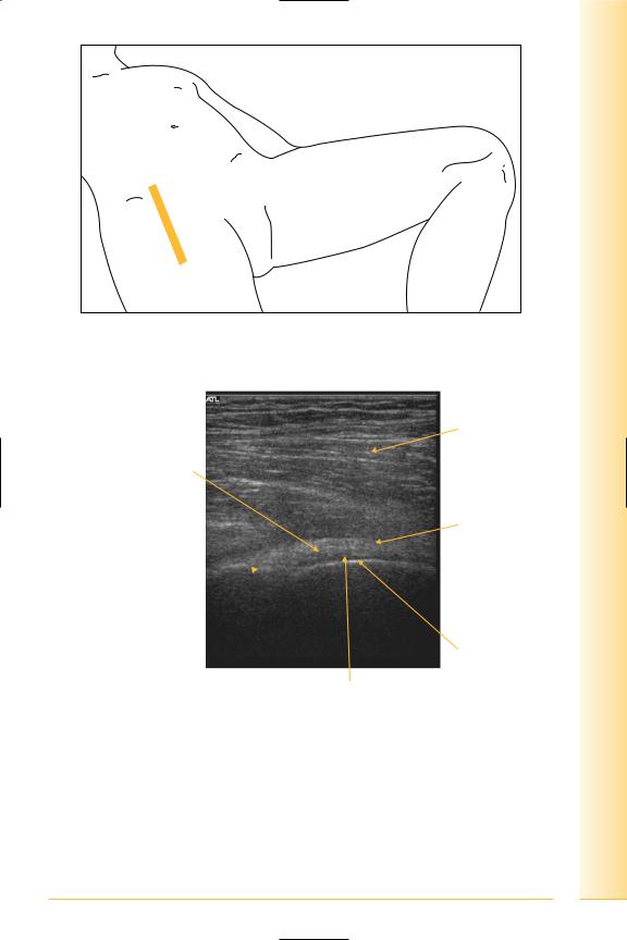
FIG. 174 LS, probe over symphysis
Rectus insertion |
Fat |
Proximal |
Distal |
Pubic ramus
Bowel
FIG. 175 LS, rectus insertion at symphysis
139

of Atlas
ultrasound musculoskeletal anatomy
140
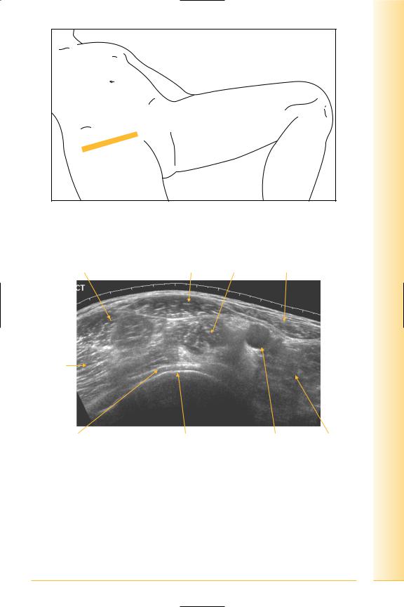
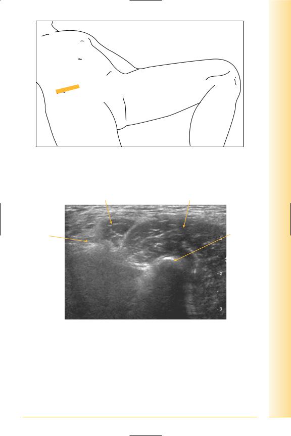
Symphysis pubis
(Figures 176 and 177)
Symphysis pubis is united by fibrocartilaginous disc and interpubic ligaments.
Notes
and Abdomen
pelvis Groin
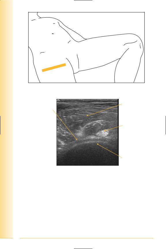
FIG. 176 TS, probe over symphysis
Distal rectus insertion |
Interpubic ligament |
Fat |
Right |
Left |
Symphysis |
Pubic body |
FIG. 177 TS, distal rectus insertion
141

of Atlas
ultrasound musculoskeletal anatomy
142
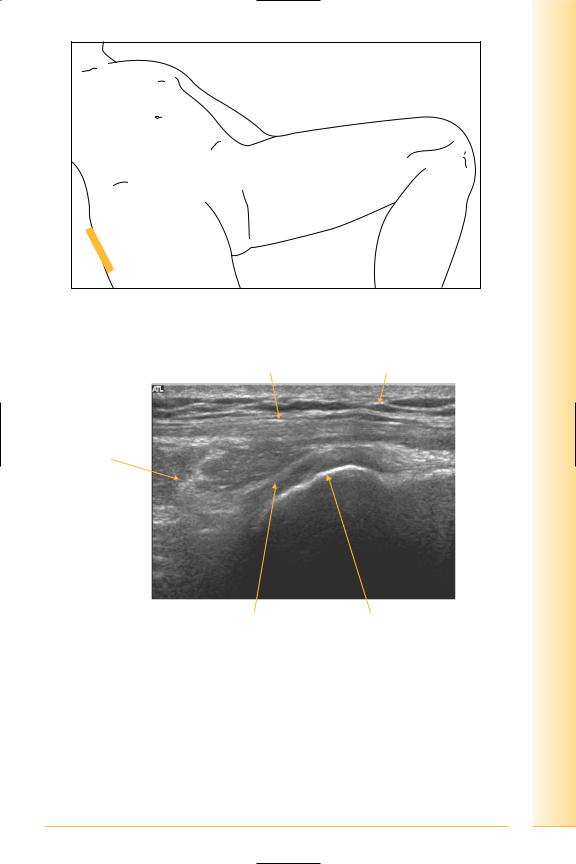
Conjoint tendon/superficial ring
(Figures 178 and 179)
The superficial ring is a deficiency in the external oblique aponeurosis, at the midpoint of the inguinal ligament, lateral to the conjoint tendon.
Notes
and Abdomen
pelvis Groin
FIG. 178 LS, probe lateral to rectus tendon
Fat |
Conjoint tendon |
Bowel
Proximal
Superficial ring |
Pubic tubercle Distal |
FIG. 179 LS, conjoint tendon
143

of Atlas
ultrasound musculoskeletal anatomy
144
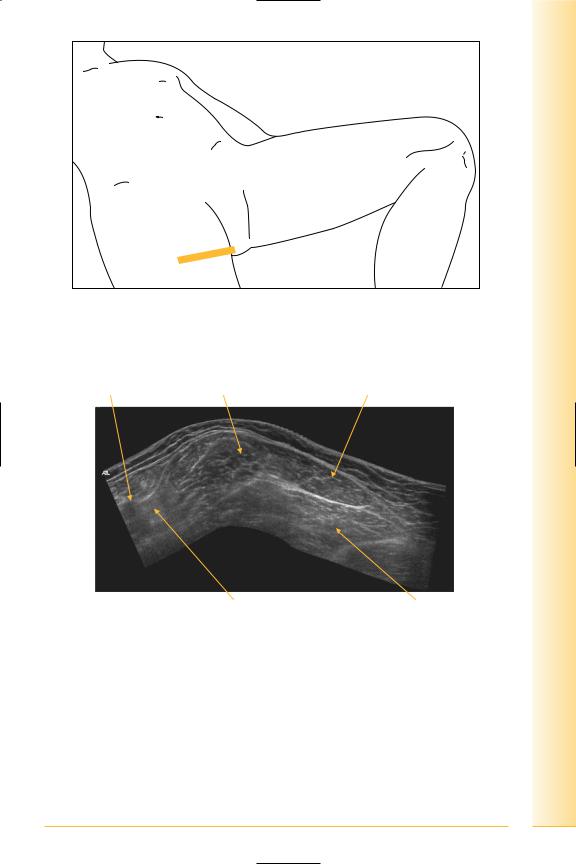
Deep ring: lateral to inferior epigastric vessels
(Figures 180 and 181)
Notes
and Abdomen
pelvis Groin
FIG. 180 TS, probe superior to inguinal ligament angled parallel to ligament
Bowel gas |
Fat |
Lateral |
Medial |
Deep ring |
Inferior epigastric |
Spermatic cord |
|
artery and vein |
|
FIG. 181 TS, oblique deep ring
145

of Atlas
ultrasound musculoskeletal anatomy
146
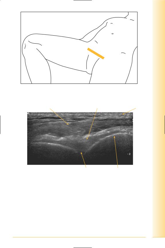
Hip
Synovial ball and socket joint
(Figures 182 and 183)
Anterior
• Ilio-psoas and pectineus separate joint from femoral vessels and nerve.
Notes
and Abdomen
pelvis Hip
FIG. 182 LS, supine, leg straight
Ilio-psoas
Synovium/capsule
and iliofemoral ligament
Fat
Proximal |
Distal |
Anterior lip  acetabulum
Femoral head
Hyaline cartilage
FIG. 183 LS, anterior hip
147
of Atlas
ultrasound musculoskeletal anatomy
148
Femoral neck
(Figures 184–187)
Notes

and Abdomen
pelvis Hip
FIG. 184 LS, supine, leg straight, probe slightly distal to femoral head, angled to femoral neck
Ilio-psoas
Synovium/capsule
and iliofemoral ligament
Proximal |
Distal |
Femoral neck
FIG. 185 LS, anterior femoral neck
149
of Atlas
ultrasound musculoskeletal anatomy
150
FIG. 186 TS, supine, leg straight, probe slightly distal to femoral head
Ilio-psoas
Synovium/ iliofemoral ligament
Ilio-psoas tendon
Lateral |
Medial |
Femoral neck
FIG. 187 TS, anterior femoral neck
Ilio-psoas
(Figures 188 and 189)
•Distal insertion: lesser trochanter.
•Sartorius: proximal attachment is at the anterior superior iliac spine, distal insertion is antero-medial tibia.
and Abdomen
pelvis Hip
FIG. 188 TS, supine, leg straight
Sartorius
Femoral vein
Pectineus
Femoral artery
Rectus femoris |
Adductor brevis |
Lateral |
Medial |
Muscle, tendon |
Lesser trochanter |
ilio-psoas |
|
FIG. 189 TS, distal psoas insertion
151

of Atlas
ultrasound musculoskeletal anatomy
152
Panorama of anterior hip
(Figures 190–193)
Notes
and Abdomen
pelvis Hip
FIG. 190 TS, supine
Tensor fasciae latae |
Rectus femoris |
Ilio-psoas Sartorius |
muscle |
|
|
Gluteal muscles
Lateral |
Medial |
Femoral head cartilage |
Femoral head |
Femoral artery |
Adductors |
FIG. 191 TS panorama, anterior hip
153

of Atlas
ultrasound musculoskeletal anatomy
154
FIG. 192 TS, probe over lesser trochanter
Rectus femoris
Tensor fasciae latae
Ilio-psoas tendon
Lateral |
Medial |
Lesser trochanter
FIG. 193 TS, hip – lesser trochanter
Greater trochanter
(Figures 194 and 195)
•Tensor fasciae latae
Origin: iliac crest.
Insertion: ilio-tibial tract.
•Gluteus maximus
Origin: ilium, sacrum, coccyx.
Insertion: ilio-tibial tract, gluteal tuberosity of femur.
•Trochanteric bursa
Deep to fascia lata and gluteus.
and Abdomen
pelvis Hip
FIG. 194 LS, supine, probe lateral overlying greater trochanter
Fascia lata |
Fat |
Gluteus maximus
Proximal |
Distal |
Trochanteric bursa |
Greater trochanter |
FIG. 195 LS, greater trochanter
155

of Atlas
ultrasound musculoskeletal anatomy
156
Adductors
(Figures 196 and 197)
•Adductor longus
Origin: anterior body of pubis.
•Adductor brevis
Origin: body and inferior ramus of pubis.
•Adductor magnus
Origin: ischiopubic ramus.
Notes
and Abdomen
pelvis Hip
FIG. 196 TS, probe antero-medial thigh. The leg may be semi-flexed and abducted as an alternative position
Femoral vessels |
Adductor longus |
Adductor brevis |
Anterior |
Posterior |
Femoral triangle |
Adductor magnus |
FIG. 197 TS panorama, hip adductors
157
of Atlas
ultrasound musculoskeletal anatomy
158
Adductor origin – LS
(Figures 198 and 199)
Notes
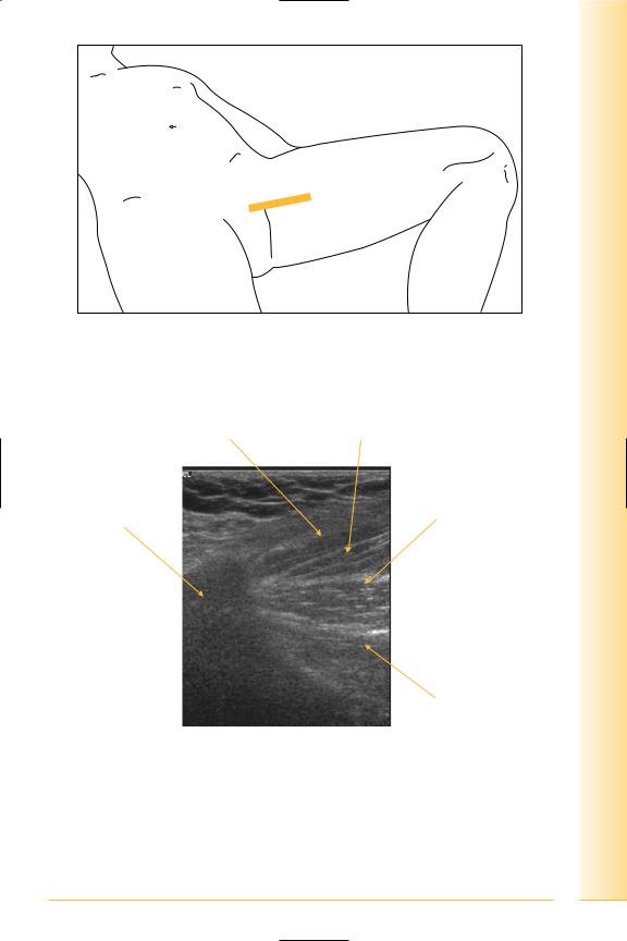
FIG. 198 LS, leg abducted
Pectineus
Pubis
Proximal
FIG. 199 LS, hip adductors origin
and Abdomen
pelvis Hip
Adductor longus
Adductor brevis
Distal
Adductor magnus
159

of Atlas
ultrasound musculoskeletal anatomy
160
Adductor origin/antero-medial thigh
(Figures 200 and 201)
•Pectineus
Origin: pectineal line, pubis to lesser trochanter/linea aspera.
•Adductor longus
Anterior pubis to linea aspera.
•Adductor brevis
Body and inferior ramus of pubis to linea aspera.
•Adductor magnus
Ramus and ischial tuberosity to linea aspera and adductor tubercle on medial femoral condyle.
•Gracilis
Pubic ramus to antero-medial tibia.
Notes
and Abdomen
pelvis Hip
FIG. 200 TS, leg abducted
Adductor longus
Adductor brevis
Anterior |
Posterior |
Adductor magnus
FIG. 201 TS, hip adductors
161

of Atlas
ultrasound musculoskeletal anatomy
162
Rectus femoris
(Figures 202 and 203)
•Origin: anterior inferior iliac spine and ilium superior to acetabulum.Insertion: upper border of patella.
Notes
and Abdomen
pelvis Hip
FIG. 202 LS, supine, proximal to hip joint
Anterior inferior iliac spine |
Rectus femoris |
|
tendon and |
muscle |
Proximal |
Distal |
Vastus intermedius
FIG. 203 LS, proximal rectus femoris insertion
163

of Atlas
ultrasound musculoskeletal anatomy
164
Rectus femoris – TS
(Figures 204 and 205)
Notes
and Abdomen
pelvis Hip
FIG. 204 TS, supine, proximal to hip joint
Rectus femoris |
Ilio-psoas |
Anterior inferior |
Ilio-pubic |
iliac spine |
eminence |
Lateral |
Medial |
FIG. 205 TS, proximal rectus femoris
165

of Atlas
ultrasound musculoskeletal anatomy
166
Hamstrings
(Figures 206 and 207)
•Origin: (lateral to medial).
•Semimembranosus: ischial tuberosity.
•Biceps femoris and semitendinosus: common tendon from the ischial tuberosity (short head of biceps from linea aspera).
Notes
and Abdomen
pelvis Hip
FIG. 206 TS, patient prone, probe over ischial tuberosity
Semitendinosus tendon |
Biceps tendon |
Gluteus maximus
Semimembranosus tendon
Lateral |
Medial |
Ischial tuberosity
FIG. 207 TS, hamstring insertion
167
of Atlas
ultrasound musculoskeletal anatomy
168
Biceps femoris
(Figures 208 and 209)
Origin – LS.
Notes

and Abdomen
pelvis Hip
FIG. 208 LS, prone, probe over mid-ischial tuberosity
Proximal |
Tendons |
Distal |
Biceps
Ischial tuberosity
FIG. 209 LS, hamstring insertion
169