


Upper limb
Shoulder . . . . . . . . . . . . . . . . . . . . . . . . . . . . . . . . . . . . . . . 28 Upper arm . . . . . . . . . . . . . . . . . . . . . . . . . . . . . . . . . . . . . 46 Elbow . . . . . . . . . . . . . . . . . . . . . . . . . . . . . . . . . . . . . . . . . 50 Forearm . . . . . . . . . . . . . . . . . . . . . . . . . . . . . . . . . . . . . . . 72 Wrist . . . . . . . . . . . . . . . . . . . . . . . . . . . . . . . . . . . . . . . . . . 82 Hand . . . . . . . . . . . . . . . . . . . . . . . . . . . . . . . . . . . . . . . . . 96
27
of Atlas
ultrasound musculoskeletal anatomy
28
Shoulder
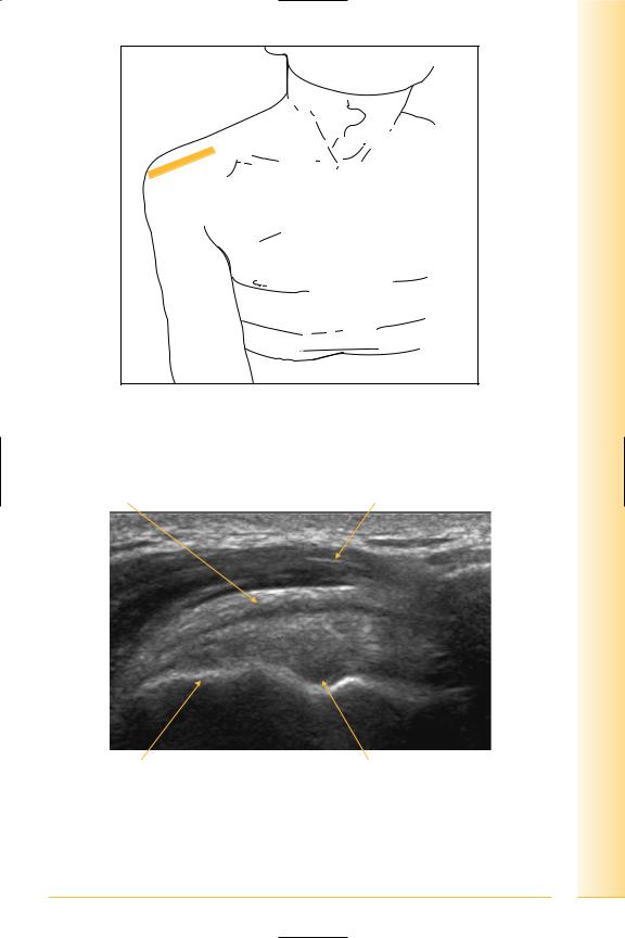
Acromioclavicular joint
(Figures 28–30)
Atypical synovial joint (articular surfaces lined with fibrocartilage), containing an incomplete articular disc. Surrounding capsule thickened superiorly to form acromioclavicular ligament.
Notes
limb Upper
Shoulder
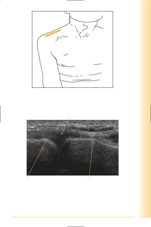
FIG. 28 Probe coronal adjacent to superior aspect of joint. Arm adducted
Acromioclavicular ligament
Lateral |
Medial |
Acromion process |
Clavicle |
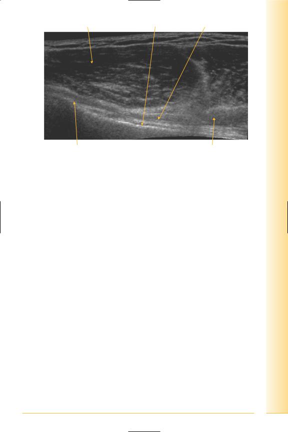
FIG. 29 LS, acromioclavicular joint
29
of Atlas
ultrasound musculoskeletal anatomy
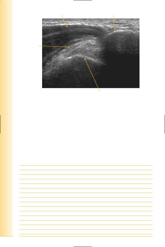
30
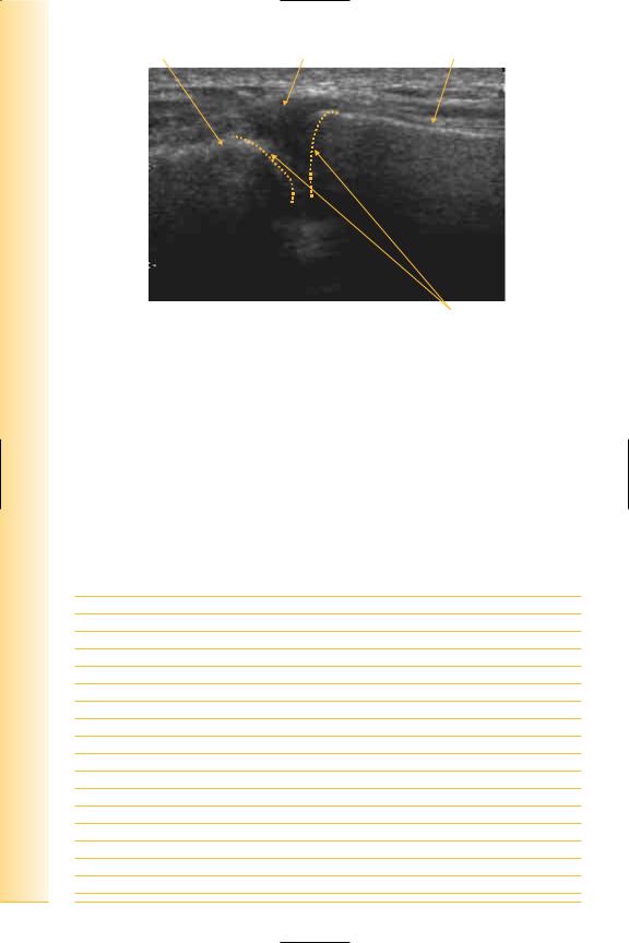
Acromion |
Acromioclavicular ligament |
Clavicle |
Lateral |
Medial |
Articular surfaces
FIG. 30 LS, acromioclavicular joint
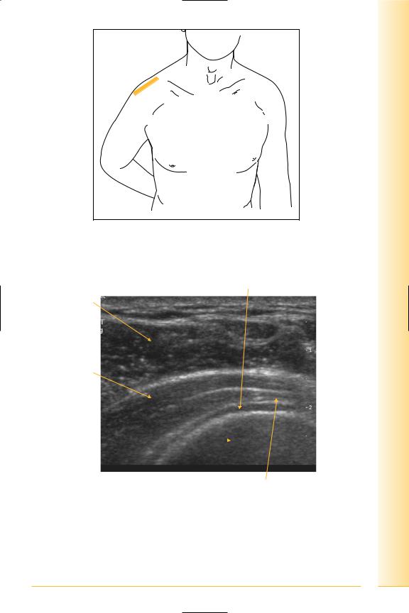
Long head of biceps
(Figures 31–35)
It arises from the supraglenoid tubercle and adjacent glenoid labrum (biceps–labral complex) and traverses the glenohumeral joint surrounded by synovium to enter the bicipital groove. It is rarely visible within the joint, but is reliably seen adjacent to the proximal humerus where it is contained within its groove by the transverse ligament.
Notes
limb Upper
Shoulder
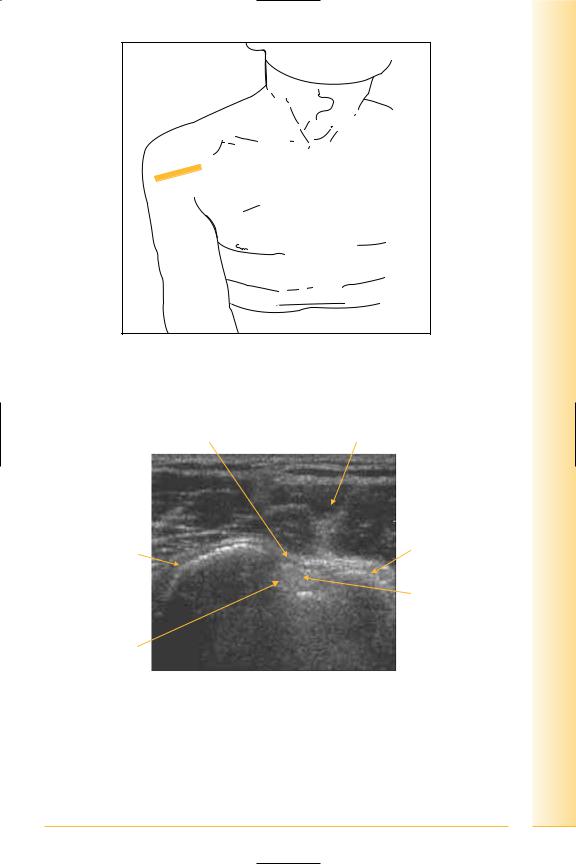
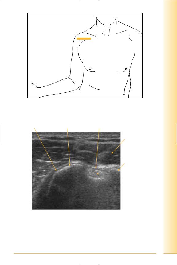
FIG. 31 TS, probe transverse across superior aspect of bicipital groove. Arm adducted, hand supinated. Examination of the rotator cuff is typically conducted from behind the patient
Transverse ligament |
Deltoid muscle |
Lesser tuberosity |
Greater tuberosity |
|
Biceps tendon |
Floor of groove
Medial |
Lateral |
FIG. 32 TS, long head of biceps tendon
31
of Atlas
ultrasound musculoskeletal anatomy
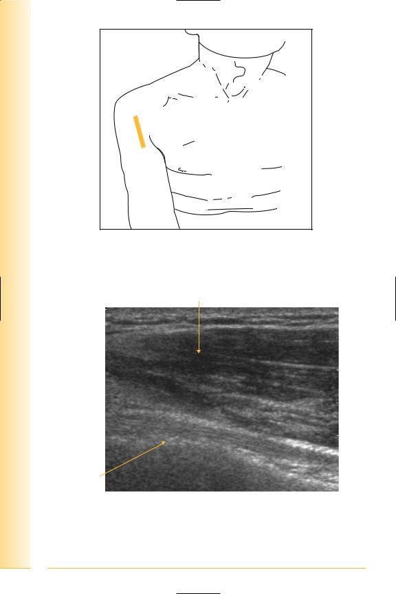
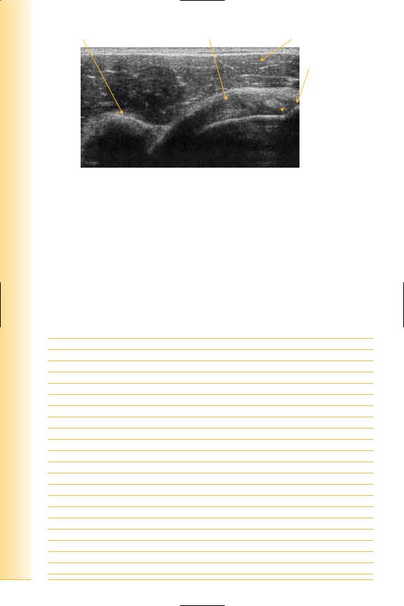
FIG. 33 LS, probe longitudinal to long head of biceps tendon. Arm adducted, hand supinated. Dynamic examination for subluxation of the tendon using internal and external rotation of the glenohumeral joint
Deltoid muscle
Proximal |
Distal |
Biceps tendon 
Floor of groove
FIG. 34 LS, long head of biceps tendon
32
Proximal |
Deltoid muscle |
Humerus |
Biceps tendon |
limb Upper
Shoulder
Floor of groove |
Biceps muscle Distal |
FIG. 35 LS panorama, long head of biceps
33

of Atlas
ultrasound musculoskeletal anatomy
34
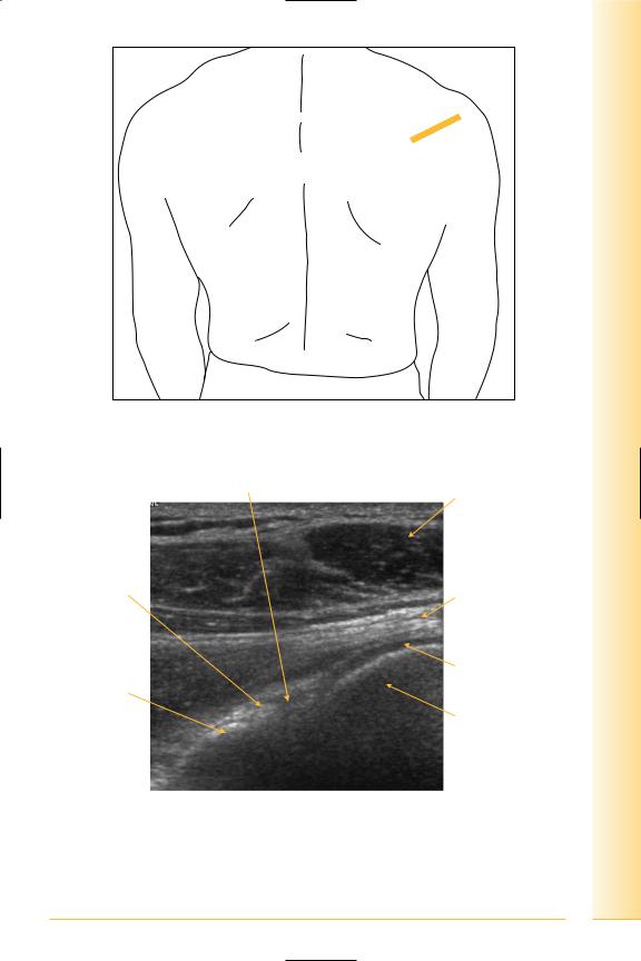
Subscapularis
(Figures 36–38)
It is a multipennate muscle, originating from the costal surface of the scapula, whose tendon inserts into the lesser tuberosity of the humerus. It is separated from the shoulder joint by its bursa, which generally communicates with the joint cavity. Forms part of posterior wall of axilla.
Notes
limb Upper
Shoulder
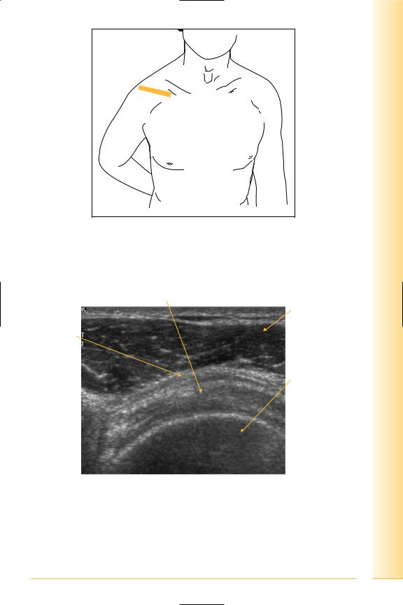
FIG. 36 LS, probe longitudinal to the subscapularis muscle (transverse to anterior shoulder). Arm externally rotated with elbow kept against chest wall. Dynamic examination using internal and external rotation of the glenohumeral joint
Subscapularis tendon Lesser tuberosity Transverse ligament
Deltoid muscle
Greater tuberosity
 Long head of biceps tendon
Medial |
Lateral |
FIG. 37 TS, subscapularis tendon
35
of Atlas
ultrasound musculoskeletal anatomy
36
Coracoid process |
Subscapularis tendon |
Deltoid muscle |
Greater tuberosity
 Long head of biceps tendon
Medial |
Lateral |
FIG. 38 TS, subscapularis tendon
Supraspinatus
(Figures 39–45)
Arises from the supraspinous fossa of the scapula and scapular spine. The tendon passes over the superior aspect of the shoulder joint to insert into the uppermost facet of the greater tuberosity of the humerus. The normal tendon shows a smooth, convex superior surface.
Notes
limb Upper
Shoulder
FIG. 39 TS, probe transverse to supraspinatus tendon, with shoulder extended and internally rotated. Shoulder extension with internal rotation is required for clear visualization (back of hand in small of back, or “hand-in wallet” position, elbow pointing posteriorly)
Supraspinatus tendon
Deltoid muscle
Peribursal fat
Humeral head
Coracoid  process
Medial |
Lateral |
FIG. 40 TS, supraspinatus
37
of Atlas
ultrasound musculoskeletal anatomy
FIG. 41 LS, probe longitudinal to supraspinatus tendon, with shoulder extended and internally rotated
Peribursal fat |
Articular cartilage |
Deltoid muscle
|
|
Supraspinatus |
|
|
tendon |
Anatomical |
|
Greater tuberosity |
|
|
|
neck of |
|
|
humerus |
|
|
Medial |
Humerus |
Lateral |
FIG. 42 LS, supraspinatus
38
limb Upper
Shoulder
FIG. 43 Dynamic assessment of supraspinatus can be useful in the further evaluation of impingement and cuff tears. LS, probe over supraspinatus whilst abducting and adducting arm. This can be performed either from the front or back
Peribursal fat |
Deltoid |
Lateral |
Medial |
Greater tuberosity of humerus |
Anatomical neck of humerus |
FIG. 44 LS, supraspinatus tendon in adduction
39
of Atlas
ultrasound musculoskeletal anatomy
40
Deltoid muscle |
Acromion process |
Supraspinatus tendon
Lateral |
Medial |
Greater tuberosity of humerus
FIG. 45 LS, supraspinatus tendon in abduction
Infraspinatus
(Figures 46 and 47)
Arises from the infraspinous fossa on the posterior aspect of the scapula, inserting onto the middle facet of the greater tuberosity of the humerus. The muscular fibres extend laterally for a greater distance, which occasionally allows distinction of this tendon from the adjacent supraspinatus and teres minor, which form a continuous cuff tendon.
Notes
limb Upper
Shoulder
FIG. 46 LS, probe longitudinal to infraspinatus tendon with shoulder extended and internally rotated
Humeral articular cartilage
Deltoid muscle
Infraspinatus muscle fibres
Humeral head 
Medial |
Lateral |
Infraspinatus tendon
FIG. 47 LS, infraspinatus
41

of Atlas
ultrasound musculoskeletal anatomy
42
Posterior joint
(Figures 48–51)
Visualizes infraspinatus and teres minor.
Teres minor
•Origin: upper two-thirds lateral border of scapula.
•Insertion, lower facet of greater tuberosity of humerus.
Notes
limb Upper
Shoulder
FIG. 48 LS, oblique probe longitudinal to infraspinatus. Arm adducted
Glenohumeral joint
Deltoid muscle
Glenoid labrum
Infraspinatus tendon
Articular cartilage
Posterior glenoid
Humeral head
Medial |
Lateral |
FIG. 49 Posterior shoulder
43

of Atlas
ultrasound musculoskeletal anatomy
FIG. 50 TS panorama of rotator cuff
|
Merging tendons of supraand infraspinatus |
|
Humeral |
|
articular |
Deltoid |
cartilage |
 Infraspinatus
muscle
Anterior |
Humeral head Teres minor Posterior |
FIG. 51 TS panorama, rotator cuff
44
