


of Atlas
ultrasound musculoskeletal anatomy
214
Calf
Anterior, lateral and posterior compartments divided by tibia, interosseous membrane, and anterior and posterior intermuscular septa. The anterior septum passes to the anterior border of the fibula and separates the anterior (dorsi-flexor) from the lateral (evertor) compartment.
Anterior compartment
(Figures 265 and 266)
•Tibialis anterior
Origin: proximal two-thirds of tibia.
Insertion: medial cuneiform and first metatarsal.
•Extensor hallucis longus
Origin: anterior proximal fibula.
Insertion: distal phalanx great toe.
•Extensor digitorum longus
Origin: anterior proximal fibula.
Insertion: dorsum of middle and terminal phalanges.
•Peroneus tertius
Origin: lower anterior fibula.
Insertion: fifth metatarsal.
Notes
limb Lower
Calf
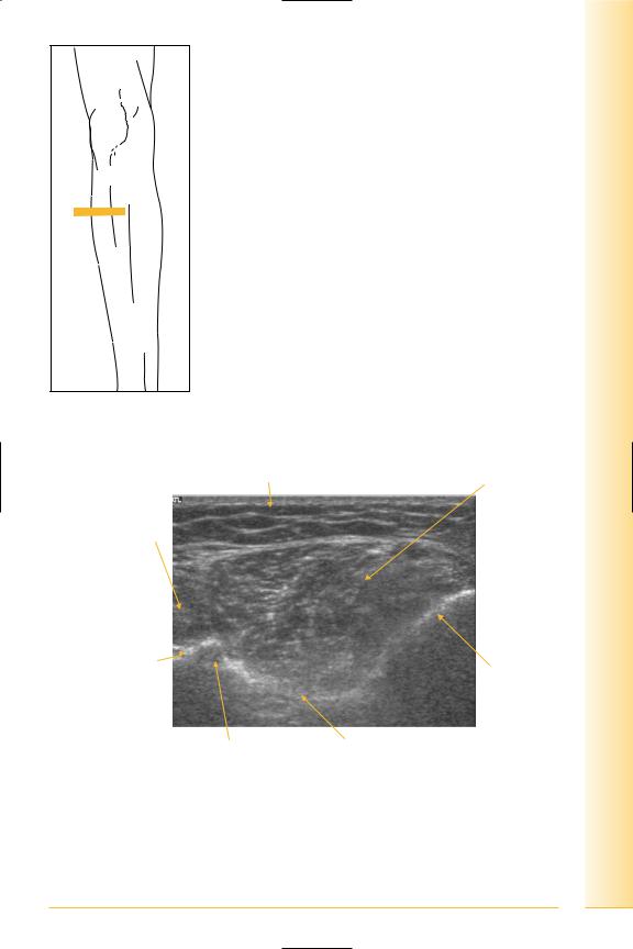
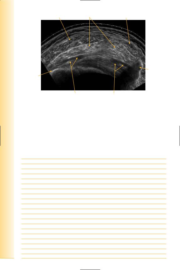
FIG. 265 TS, probe lateral to tibia
Fat |
Tibialis anterior |
Extensor hallucis longus
Lateral |
Medial |
Fibula |
Tibia |
|
Anterior tibial artery |
Interosseus membrane |
FIG. 266 TS, mid-calf ant/lat
215

of Atlas
ultrasound musculoskeletal anatomy
216
Lateral compartment
(Figures 267–270)
•Peroneus longus
Origin: proximal lateral fibula.
Insertion: first metatarsal and medial cuneiform.
•Peroneus brevis
Origin: lower lateral fibula.
Insertion: fifth metatarsal.
Notes
limb Lower
Calf
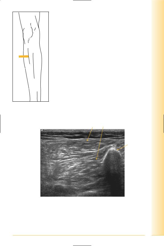
FIG. 267 TS, probe lateral to fibula, mid-calf
Peroneus longus and brevis
Fibula
Soleus 
Flexor hallucis  longus
Posterior |
Anterior |
FIG. 268 TS, peroneal compartment
217
of Atlas
ultrasound musculoskeletal anatomy
218
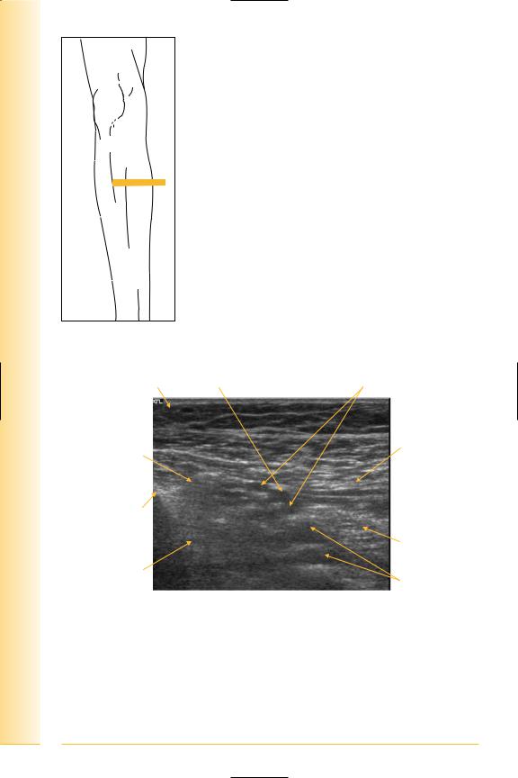
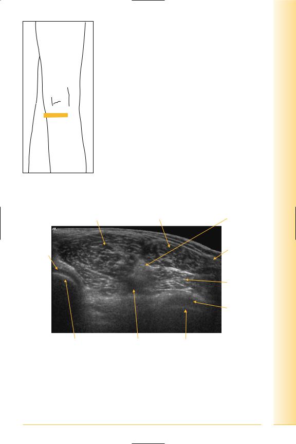
|
Extensor hallucis longus |
|
Peroneus longus and brevis |
and digitorum longus |
Tibialis anterior |
|
|
Interosseus |
|
|
membrane |
Soleus |
|
|
Lateral |
|
Medial |
Fibula |
Tibialis posterior |
Tibia |
FIG. 269 TS panorama, antero-lateral calf |
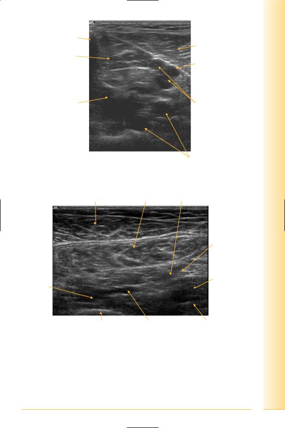
|
|
Peroneus longus and brevis |
Extensor hallucis longus and digitorum |
|
Tibialis anterior
Lateral |
Medial |
Fibula
FIG. 270 TS panorama, peroneal compartment
Posterior compartment
(Figures 271–275)
Superficial muscles
•Gastrocnemius
Origin: medial and lateral femoral condyles.
Insertion: soleus and tendo-achilles.

•Soleus
Origin: soleal line tibia and posterior fibula.
Insertion: tendo-achilles.
•Plantaris
Origin: lateral supracondylar line.
Insertion: tendo-achilles.
Deep muscles
•Popliteus
Origin: posterior tibia proximal to soleal line.
Insertion: lateral femoral epicondyle.
•Flexor digitorum longus
Origin: medial posterior tibia.
Insertion: terminal phalanges lateral four toes.
•Tibialis posterior
Origin: posterior interosseus membrane, tibia and fibula.
Insertion: navicular.
•Flexor hallicus longus
Origin: posterior distal fibula.
Insertion: distal phalanx great toe.
Notes
limb Lower
Calf
219
of Atlas
ultrasound musculoskeletal anatomy
FIG. 271 TS, probe medial to tibia
Fat |
Posterior tibial artery |
Posterior tibial veins |
Medial head of Flexor digitorum gastrocnemius longus
Anterior
Posterior
Tibia
Soleus
Tibialis posterior
Peroneal veins
FIG. 272 TS, mid-calf – medial
220
Tibia
Soleus
Flexor digitorum longus
Posterior tibial artery
 Posterior tibial nerve
Tibialis posterior |
Posterior tibial veins |
|
Anterior |
Posterior |
Peroneal veins
FIG. 273 TS, medial calf
Gastrocnemius |
Soleus |
Peroneal artery |
|
|
Posterior |
|
|
tibial nerve |
Flexor |
|
Flexor hallicus |
|
longus |
|
digitorum |
|
|
|
|
|
longus |
|
|
Anterior |
|
Posterior |
|
|
|
Tibia |
Posterior tibial artery |
Fibula |
FIG. 274 TS, proximal posterior calf
limb Lower
Calf
221
of Atlas
ultrasound musculoskeletal anatomy
222
Medial head gastrocnemius Soleus |
Lateral head gastrocnemius |
|
Fibula |
Tibia |
|
Medial |
Lateral |
Posterior tibial vessels |
Peroneal vessels |
FIG. 275 TS panorama, posterior calf
Posterior calf (popliteal fossa)
(Figures 276 and 277)
Notes
limb Lower
Calf
FIG. 276 TS, patient prone
Gastrocnemius medial head |
Lateral head |
Tibial nerve |
|
|
|
|
Common |
Hyaline |
|
|
peroneal |
cartilage |
|
|
nerve |
|
|
|
Plantaris |
|
|
|
Hyaline |
|
|
|
cartilage |
|
|
|
Lateral |
Medial |
Popliteal artery Lateral femoral condyle |
|
|
Medial femoral condyle |
|
||
FIG. 277 TS, proximal gastrocnemius
223