


of Atlas
ultrasound musculoskeletal anatomy
224
Ankle
Ankle: posterior
(Figure 278–285)
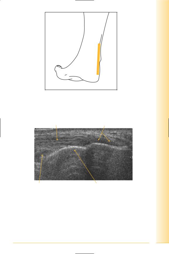
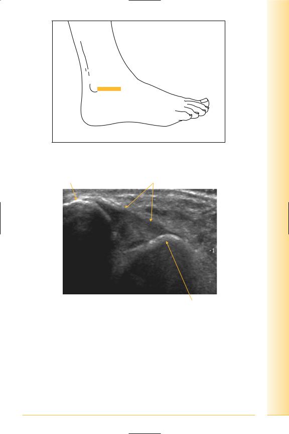
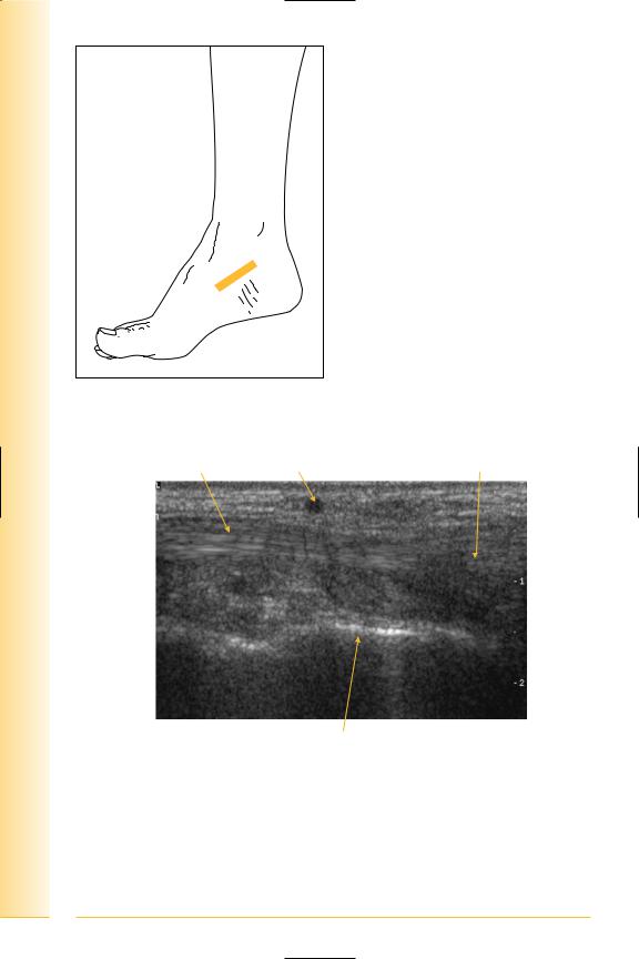
Tendo-achilles: formed by gastrocnemius and soleus to attach distally to the posterior superior calcaneum. There is no synovial sheath but it has an hyperechoic paratenon. Deep to distal tendon is Kager’s fat pad and pre-achilles bursa. Retro-calcaneal bursa lies posterior to the tendon attachment.
Notes
limb Lower
Ankle
FIG. 278 LS, patient prone. Stand-off medium is sometimes useful. Dynamic examination should be performed by passively and actively dorsiand plantarflexing the foot. Dorsi-flexion straightens the tendon to avoid anisotropy
Tendon fibrils |
Insertion |
Proximal |
Distal |
Pre-achilles bursa |
Calcaneum |
FIG. 279 LS, distal tendo-achilles insertion
225
of Atlas
ultrasound musculoskeletal anatomy
226
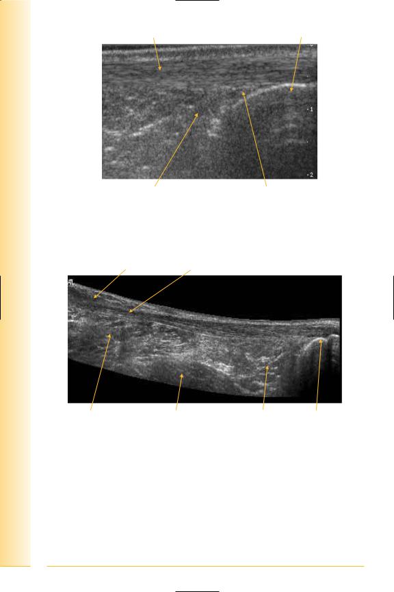
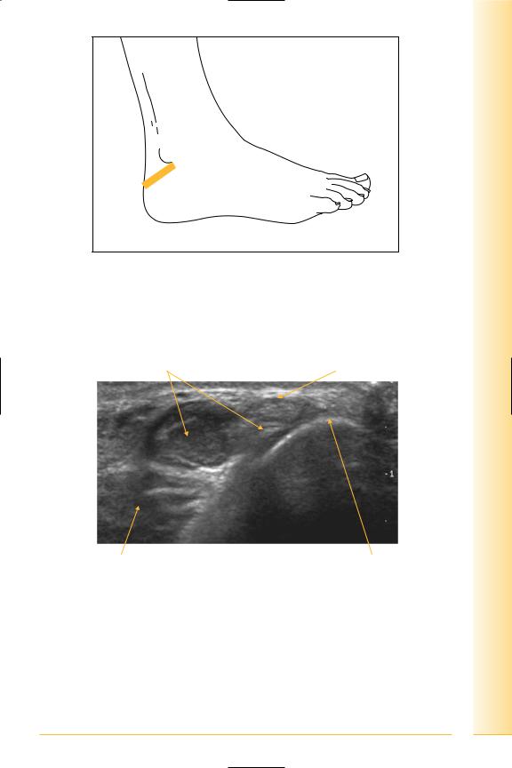
Tendon |
Calcaneum |
Proximal |
Distal |
Kager’s fat pad |
Pre-achilles bursa |
FIG. 280 LS, body of tendo-achilles
Proximal Gastrocnemius Musculotendonous insertion |
Distal |
Soleus |
Flexor hallucis longus |
Kager’s fat pad Calcaneum |
FIG. 281 LS, panorama of tendo-achilles
limb Lower
Ankle
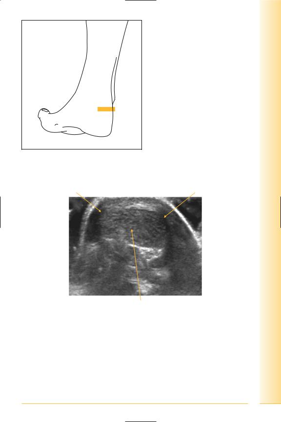
FIG. 282 TS, prone with probe over tendo-achilles. Angle medially and laterally for paratenon
Lateral paratenon |
Medial paratenon |
Lateral |
Medial |
Tendon fibrils
FIG. 283 TS, tendo-achilles. Due to edge effect, the medial and lateral paratenon is difficult to visualize unless the probe is angled to assess them individually
227
of Atlas
ultrasound musculoskeletal anatomy
228
Paratenon |
Tendon fibrils |
Lateral |
Medial |
Flexor hallicus longus
FIG. 284 TS, lateral paratenon. A stand-off pad is often helpful for assessment of the tendo-achilles
Paratenon |
Tendon fibres |
Medial |
Lateral |
Fat
FIG. 285 TS, distal tendo-achilles


of Atlas
ultrasound musculoskeletal anatomy
230
Ankle: lateral
Ligaments – antero-lateral complex composed of three separate parts:
•calcaneo-fibular,
•posterior talo-fibular and
•anterior talo-fibular ligaments.
Calcaneo-fibular ligament
(Figures 286 and 287)
Passes posteriorly from the tip of the lateral malleolus to the lateral border of the calcaneum.
Posterior talo-fibular ligament
Posterior talo-fibular is not successfully imaged on ultrasound.
Notes
limb Lower
Ankle
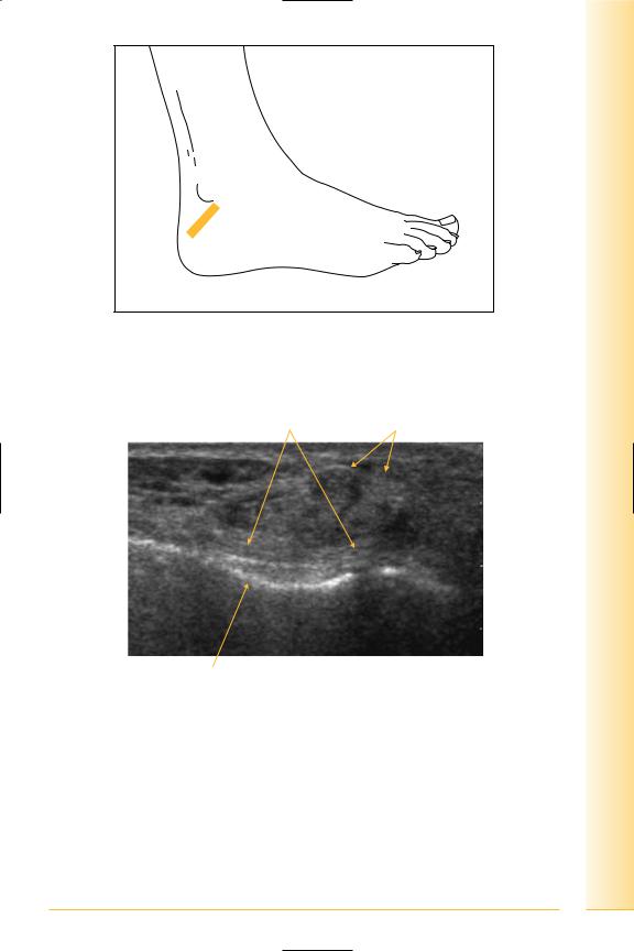
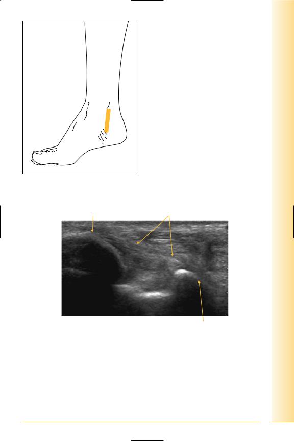
FIG. 286 LS, foot may be internally rotated, probe posterior and inferior to lateral malleolus. Foot eversion and inversion for dynamic examination
CF ligament |
Peroneal tendons |
Posterior |
Anterior |
Lateral border of os calcis
FIG. 287 LS, calcaneo-fibular ligament
231

of Atlas
ultrasound musculoskeletal anatomy
232
Anterior talo-fibular ligament
(Figures 288 and 289)
Passes horizontally from lateral malleolus to neck of talus. Foot eversion and inversion for dynamic examination.
Notes
limb Lower
Ankle
FIG. 288 LS, probe anterior and inferior to lateral malleolus
Lateral malleolus |
Anterior talo-fibular ligament |
Posterior |
Anterior |
Talus
FIG. 289 LS, anterior talo-fibular ligament
233

of Atlas
ultrasound musculoskeletal anatomy
234
Tendons
(Figures 290 and 291)
Peroneus longus and brevis. Brevis is first medial, then anterior to longus and both should be posterior to the lateral malleolus.
•Peroneus longus
Origin: proximal lateral fibula.
Insertion: first metatarsal and medial cuneiform.
•Peroneus brevis
Origin: distal lateral fibula.
Insertion: fifth metatarsal.
Notes
limb Lower
Ankle
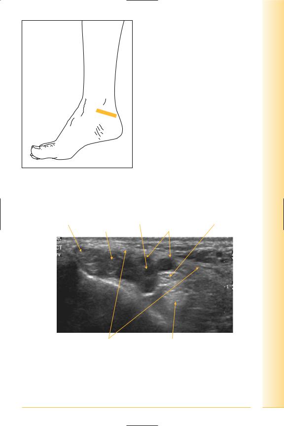
FIG. 290 TS, probe posterior and inferior to lateral malleolus. Plantar-flexing the foot can “straighten” the tendons. Dynamic examination using foot inversion and eversion
Peroneus brevis tendon and muscle |
Peroneus longus tendon |
Posterior |
Anterior |
Flexor hallicus longus |
Lateral malleolus |
FIG. 291 TS, peroneal tendons
235

of Atlas
ultrasound musculoskeletal anatomy
236
Distal peroneus brevis insertion
(Figures 292 and 293)
Notes

limb Lower
Ankle
FIG. 292 LS, probe over base of fifth metatarsal
Peroneus brevis tendon |
Base of fifth metatarsal |
Proximal |
Distal |
FIG. 293 LS, peroneus brevis insertion
237
of Atlas
ultrasound musculoskeletal anatomy
238
Ankle: medial
Ligaments: deltoid
(Figures 294 and 295)
Deltoid ligament: Triangular shaped with deep and superficial layers. The superficial part attaches to the sustentaculum tali. The deep layer extends to the navicular and neck of talus.
Notes
limb Lower
Ankle
FIG. 294 LS, probe inferior to medial malleolus
Medial malleolus |
Deltoid ligament |
Proximal |
Distal |
Talus
FIG. 295 LS, deltoid ligament
239

of Atlas
ultrasound musculoskeletal anatomy
240
Tendons
(Figures 296–299)
Tibialis posterior, flexor digitorum longus, flexor hallucis longus from anterior to posterior.
•Tibialis posterior
Origin: posterior interosseous membrane, tibia and fibula.
Insertion: navicular.
•Flexor digitorum longus
Origin: medial posterior tibia.
Insertion: terminal phalanges lateral four toes.
•Flexor hallucis longus
Origin: posterior distal fibula.
Insertion: distal phalanx great toe.
Posterior tibial nerve: divides into lateral and medial plantar nerves.
•Lateral plantar – under flexor retinaculum passes along the sole of the foot to the fifth metatarsal. Sensory innervation lateral foot and toes, motor to intrinsic foot muscles.
•Medial plantar – under flexor retinaculum to sole. Sensory and motor to medial sole and toes.
Notes
limb Lower
Ankle
FIG. 296 TS, probe over medial malleolus. Dynamic examination using foot inversion/eversion
Tibialis posterior |
Posterior tibial artery |
Posterior tibial nerve |
|
|
Flexor digitorum |
Veins |
|
Anterior |
Posterior |
Flexor retinaculum |
Flexor hallucis longus |
FIG. 297 TS, medial ankle
241
of Atlas
ultrasound musculoskeletal anatomy
242
FIG. 298 LS, probe over navicular. Distal attachment always appears more ill defined, expanded and hypo-echoic compared to the rest of the tendon
Tendon |
Vein |
Distal expansion |
Proximal |
Distal |
Talus
FIG. 299 LS, distal tibialis posterior insertion
