


of Atlas
ultrasound musculoskeletal anatomy
184
Knee
Modified hinge synovial joint.
Anterior knee
(Figures 226–233)
Quadriceps tendon – rectus femoris, vastus lateralis, medialis, intermedius. Patella tendon – single musculotendinous expansion from lower patella to tibial tuberosity.
Bursae
•Superficial pre-patellar
superficial to lower patella and proximal patellar tendon.
•Deep infrapatellar
deep to patella tendon, separating it from tibia.
Notes

limb Lower
Knee
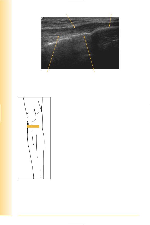
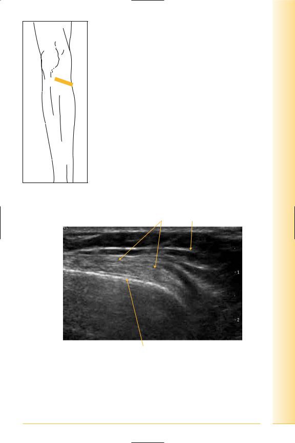
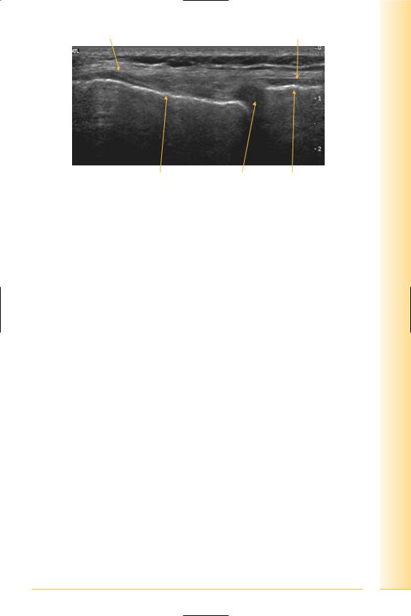
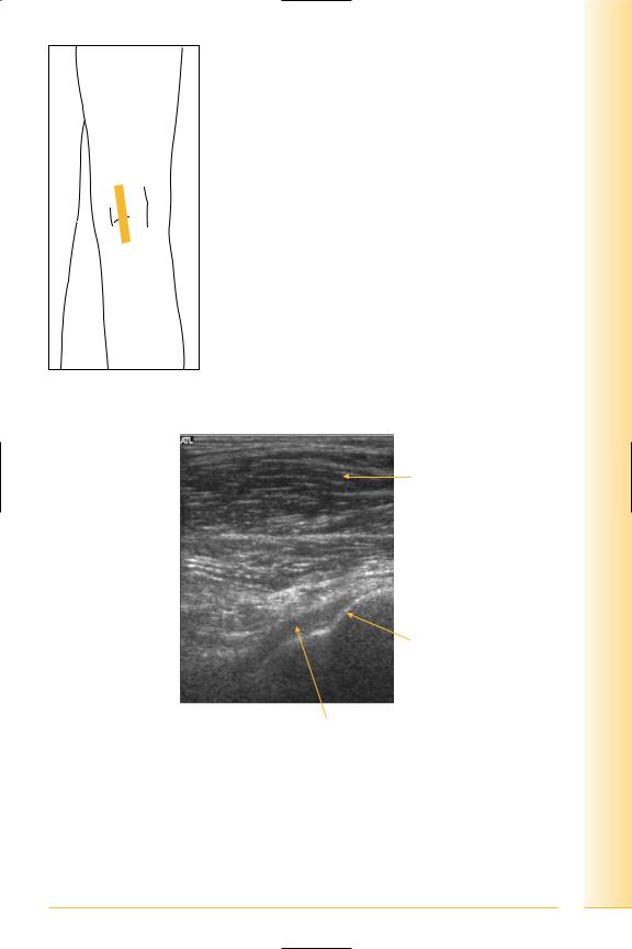
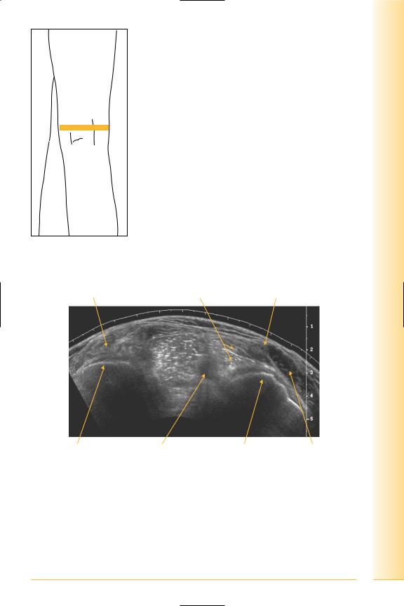
FIG. 226 LS, probe distal to patella. Contract quads or flex knee to straighten tendon, avoiding anisotropy
Tendon
Proximal |
Distal |
Lower pole patella |
Hoffa’s fat pad |
Tibia |
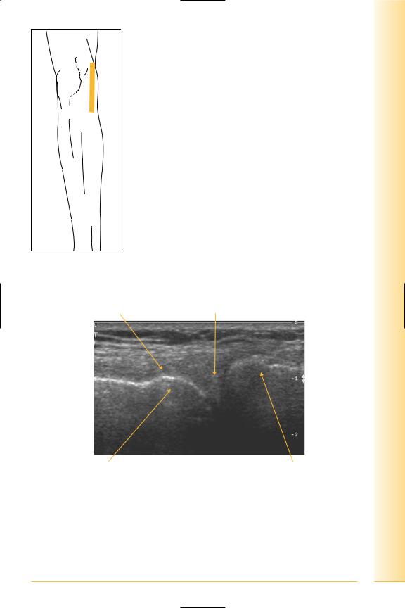
FIG. 227 LS, patellar tendon proximal insertion
185
of Atlas
ultrasound musculoskeletal anatomy
186
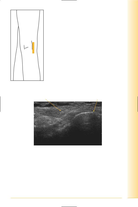
Tendon |
Tibial tuberosity |
Proximal |
Distal |
Deep infrapatellar bursa |
Tibia |
FIG. 228 LS, patellar tendon, tibial insertion
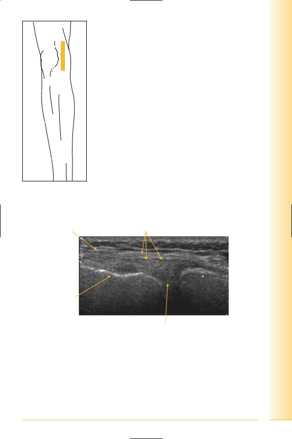
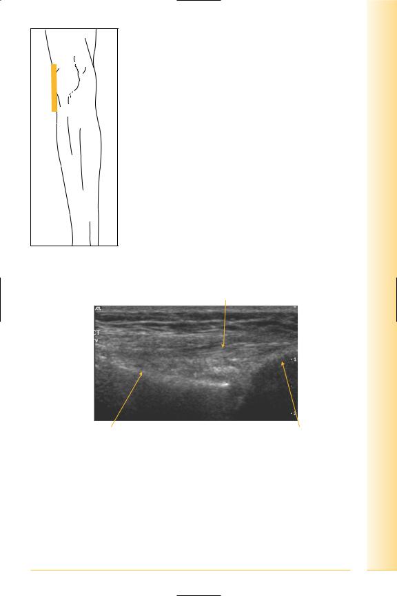
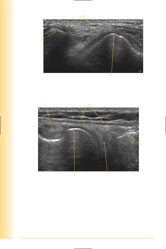
FIG. 229 TS, probe proximal to tibial tuberosity
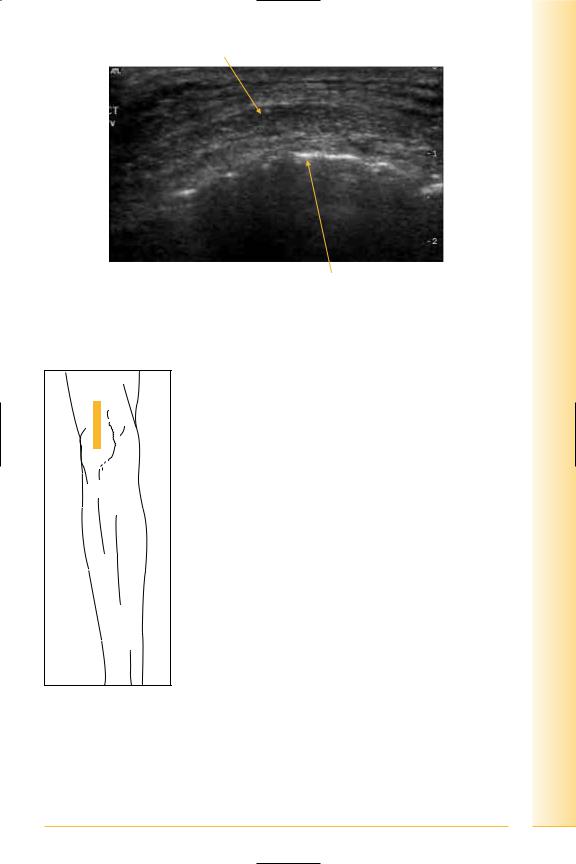
Tendon
Lateral |
Medial |
Tibia
FIG. 230 TS, patellar tendon, tibial insertion
limb Lower
Knee
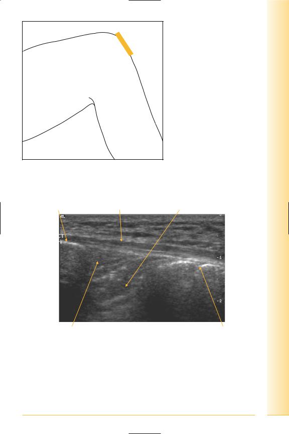
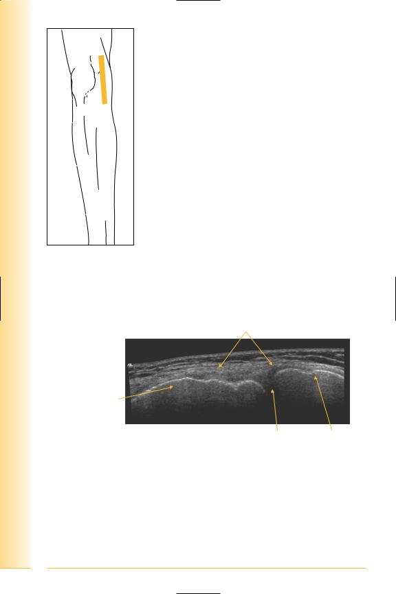
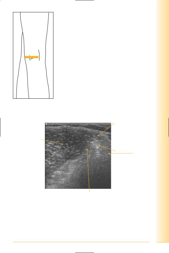
FIG. 231 LS quadriceps tendon, probe proximal to upper pole of patella
187
of Atlas
ultrasound musculoskeletal anatomy
188
Fat
Proximal |
Quadriceps tendon |
Distal |
Suprapatellar pouch |
|
Upper pole patella |
FIG. 232 LS, distal quadriceps tendon
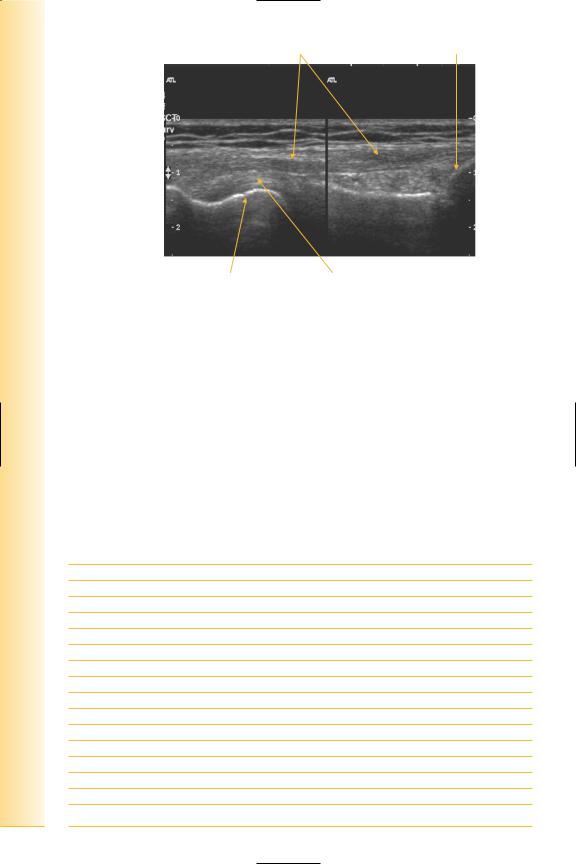
Quads tendon |
Patella |
Patellar tendon |
Proximal |
Tibia |
Distal |
FIG. 233 LS panorama, extensor compartment
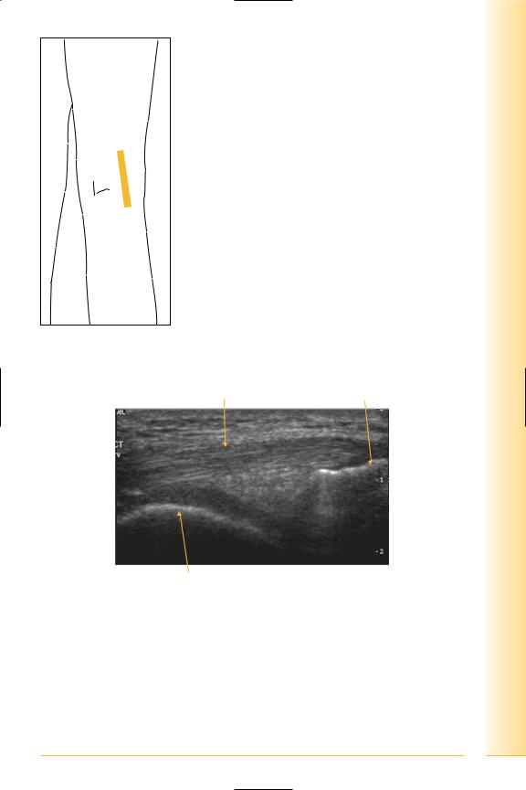
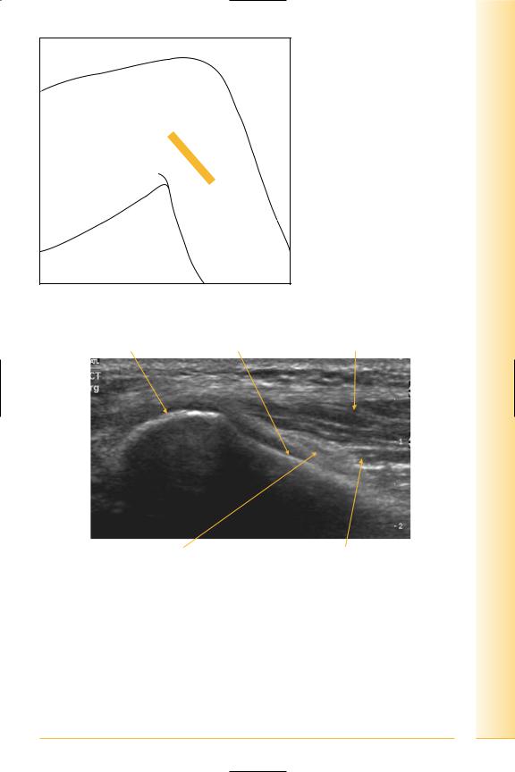
Anterior cruciate ligament
(Figures 234 and 235)
The anterior cruciate ligament (ACL) attaches on the antero-medial tibial intercondylar area and inserts on the medial surface of the lateral femoral condyle.
limb Lower
Knee
FIG. 234 LS ACL, probe midline over patellar tendon
Patella |
Patellar tendon |
ACL-tibial insertion |
Proximal |
Distal |
Hoffa’s fat pad |
Tibial tuberosity |
FIG. 235 LS of anterior knee, ACL
189

of Atlas
ultrasound musculoskeletal anatomy
190
Trochlear groove
(Figures 236 and 237)
Notes

limb Lower
Knee
FIG. 236 TS, knee flexed, probe distal to patella
Hyaline cartilage |
Fat |
Lateral |
Medial |
Trochlear groove |
Femoral cortex |
FIG. 237 TS, trochlear groove
191

of Atlas
ultrasound musculoskeletal anatomy
192
Semimembranosus – inserts postero-medial tibial condyle
(Figures 238 and 239)
Notes
limb Lower
Ahead
FIG. 238 LS, leg extended
Semimembranosus tendon |
Tibia |
Proximal |
Femoral condyle |
Distal |
FIG. 239 LS, semimembranosus tendon
193

of Atlas
ultrasound musculoskeletal anatomy
194
Pes anserinus
(Figures 240 and 241)
Insertion of sartorius, gracilis and semitendinosus. Semitendinosus inserts onto antero-medial tibia shaft, posterior to gracilis and sartorius. A bursa (anserine bursa) separates gracilis and semitendinosus from the tibia, with another bursa between them and sartorius.
Notes
limb Lower
Knee
FIG. 240 TS, leg extended
Pes anserinus |
Fat |
Lateral
Medial
Tibia
FIG. 241 TS, pes anserinus
195

of Atlas
ultrasound musculoskeletal anatomy
196
Medial knee
Medial meniscus
(Figures 242–244)
Notes
limb Lower
Knee
FIG. 242 LS, leg straight. Valgus strain may be applied to assess stability
Deep medial collateral ligament |
Meniscus |
Proximal |
Distal |
Medial femoral condyle |
Medial tibial plateau |
FIG. 243 LS, medial meniscus
197
of Atlas
ultrasound musculoskeletal anatomy
198
Medial collateral ligament
Femoral condyle 
|
Tibia |
Proximal |
Distal |
|
Medial meniscus |
FIG. 244 LS, medial knee |
|
Medial collateral ligament
(Figures 245–249)
Approximately 10 cm in length, arises from the medial femoral epicondyle and extends to the proximal medial tibial shaft. Deeper layer is attached to the medial tibial condyle and blends with the medial meniscus.
Notes
limb Lower
Knee
FIG. 245 LS, leg straight, apply valgus strrain for stability
Sartorius tendon |
Medial collateral-superficial and deep layers |
 Tibia
Tibia
Femoral condyle
Proximal |
Medial meniscus |
Distal |
FIG. 246 LS, medial collateral ligament
199
of Atlas
ultrasound musculoskeletal anatomy
FIG. 247 LS panorama, medial knee
Proximal |
Medial collateral |
Distal |
Femoral condyle
Meniscus Tibia
FIG. 248 LS, medial collateral ligament
200
Femoral attachment |
Tibial attachment |
Proximal |
Femur |
Medial meniscus |
Tibia |
Distal |
FIG. 249 LS, medial collateral ligament
limb Lower
Knee
201

of Atlas
ultrasound musculoskeletal anatomy
202
Lateral knee
Lateral collateral ligament
(Figures 250–252)
The lateral collateral ligament arises from the lateral femoral epicondyle and extends to the apex of the fibula.
Notes
limb Lower
Knee
FIG. 250 LS, leg extended, probe over lateral knee
Lateral collateral
Proximal |
Distal |
Femoral condyle |
Fibula |
FIG. 251 LS, lateral collateral ligament
203
of Atlas
ultrasound musculoskeletal anatomy
204
Ligament |
Fibula head |
Proximal |
Distal |
Femoral condyle |
Popliteus tendon |
FIG. 252 LS, lateral collateral ligament, composite image
Common peroneal nerve
(Figures 253 and 254)
This is a terminal branch of the sciatic nerve formed just proximal to the popliteal fossa. It lies on the lateral head of gastrocnemius and then on the neck of the fibula and is deep to biceps femoris. It pierces peroneus longus to divide into superficial and deep branches.
Notes
limb Lower
Knee
FIG. 253 TS, knee flexed, probe over fibula neck
Fibula head |
Fibula neck |
Peroneus longus |
Proximal |
Common peroneal nerve |
Peroneus brevis |
Distal |
FIG. 254 TS, common peroneal nerve
205

of Atlas
ultrasound musculoskeletal anatomy
206
Posterior knee
Popliteal fossa
(Figures 255 and 256)
Contents:
Popliteal artery and vein, and branches, tibial and common peroneal nerves, lymph nodes and fat.
Boundaries
•Lateral: biceps.
•Medial: semitendinosus, semimembranosus.
•Inferior: medial and lateral heads of gastrocnemius.
Posterior cruciate ligament
The posterior cruciate ligament (PCL) extends from lateral surface of medial femoral condyle to posterior intercondylar area of tibia.
Notes
limb Lower
Knee
FIG. 255 LS, posterior knee, medial popliteal fossa
Medial head gastrocnemius
Proximal |
Posterior tibial |
|
plateau |
||
|
||
|
Distal |
PCL
FIG. 256 LS, posterior cruciate ligament
207
of Atlas
ultrasound musculoskeletal anatomy
208
Lateral popliteal fossa
(Figures 257–260)
Biceps femoris attaches to apex of fibula.
Popliteus tendon arises from the lateral femoral epicondyle, and is attached to the lateral meniscus. The muscle attaches to the posterior tibia proximal to the soleal line. Popliteus bursa lies between the muscle and tibia.
Notes
limb Lower
Knee
FIG. 257 LS, probe over lateral popliteal fossa. Biceps femoris insertion normally appears slightly hypo-echoic and expanded
Biceps tendon |
Fibular head |
Proximal |
Distal |
FIG. 258 LS, biceps
209
of Atlas
ultrasound musculoskeletal anatomy
210
Popliteus tendon
Femur
Proximal |
Tibia |
Distal |
FIG. 259 LS, popliteus tendon
Popliteus |
Lateral collateral ligament |
Tibia
Proximal |
Lateral femoral condyle |
Lateral meniscus |
Distal |
FIG. 260 LS, posterolateral knee
Popliteal fossa “cyst space”
(Figures 261 and 262)
Cyst neck lies between medial head of gastrocnemius and semimembranosus tendon.
limb Lower
Knee
FIG. 261 TS, probe over medial head of gastrocnemius
Semitendinosus tendon
Medial head of gastrocnemius
Semimembranosus tendon and muscle
Lateral |
Medial |
Poplitealcyst space
FIG. 262 TS, popliteal cyst space
211

of Atlas
ultrasound musculoskeletal anatomy
212
Panorama of the popliteal fossa
(Figures 263 and 264)
Notes
limb Lower
Knee
FIG. 263 TS panorama, popliteal fossa
Lateral head |
Medial head |
|
gastrocnemius |
gastrocnemius |
Semimembranosus |
Lateral |
|
|
Medial |
Lateral femoral condyle |
Popliteal vessels |
Medial femoral condyle |
Sartorius |
FIG. 264 TS panorama, popliteal fossa
213