


Lower limb
Thigh . . . . . . . . . . . . . . . . . . . . . . . . . . . . . . . . . . . . . . . . . 172
Knee . . . . . . . . . . . . . . . . . . . . . . . . . . . . . . . . . . . . . . . . . . 184
Calf . . . . . . . . . . . . . . . . . . . . . . . . . . . . . . . . . . . . . . . . . . . 214
Ankle . . . . . . . . . . . . . . . . . . . . . . . . . . . . . . . . . . . . . . . . . 224
Foot . . . . . . . . . . . . . . . . . . . . . . . . . . . . . . . . . . . . . . . . . . 244
171

of Atlas
ultrasound musculoskeletal anatomy
172
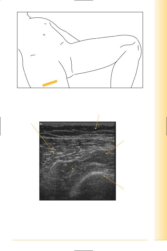
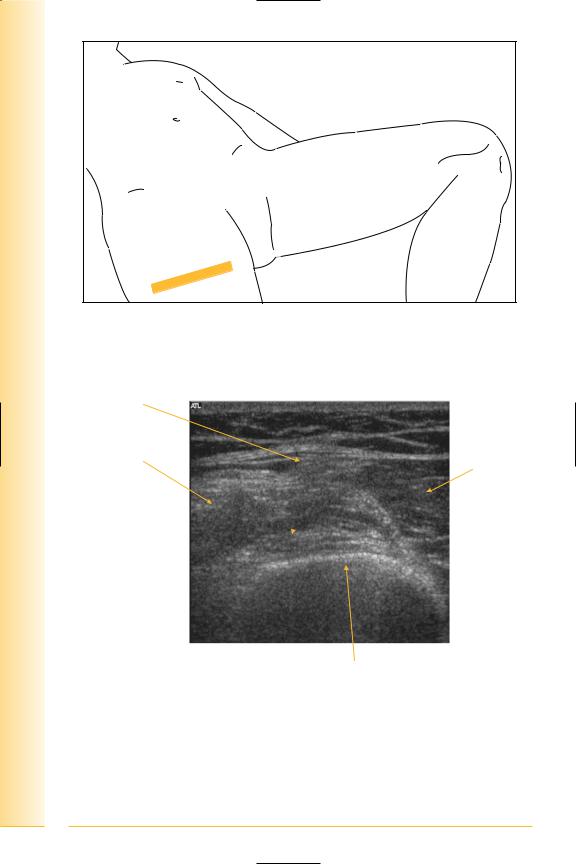
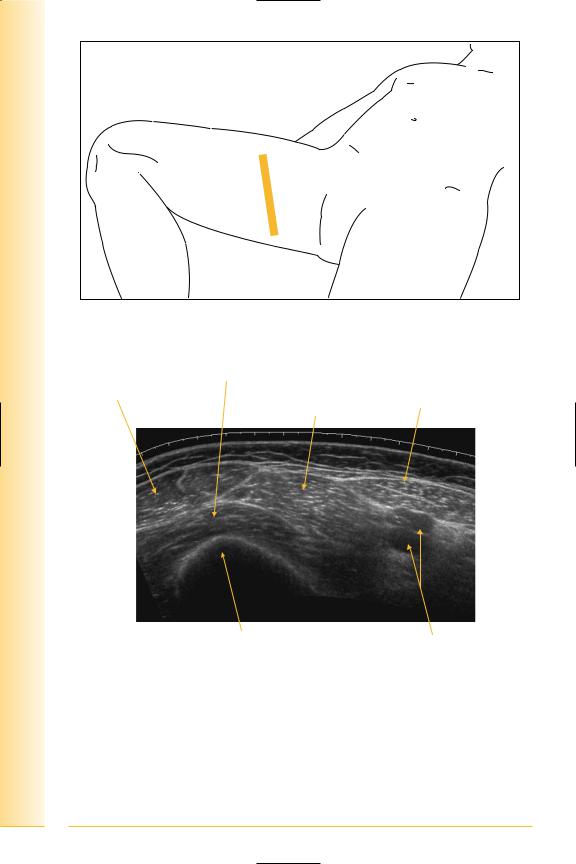
Thigh
Anterior
(Figures 210–217)
•Sartorius
Origin: anterior superior iliac spine insertion, antero-medial tibia.
•Quadriceps
Rectus femoris: origin – anterior inferior iliac spine and ilium.
Vastus lateralis: origin – greater trochanter and linea aspera.
Vastus medialis: origin – linea aspera and lesser trochanter.
Vastus intermedius: origin – anterior and lateral surface of femur.
The quadriceps muscles insert onto the upper pole of the patella.
Notes
limb Lower
Thigh
FIG. 210 TS, supine, probe over anterior thigh
Fat
Vastus lateralis
Vastus medialis
Lateral |
Medial |
|
 Vastus intermedius
Femur
FIG. 211 TS, antero-lateral mid-thigh
173
of Atlas
ultrasound musculoskeletal anatomy
FIG. 212 TS anterior thigh
Rectus femoris tendon
Vastus lateralis
Vastus intermedius 
Lateral
Femur
FIG. 213 TS, anterior thigh
174
Vastus medialis
Medial

limb Lower
Thigh
FIG. 214 TS panorama, anterior thigh
Vastus lateralis |
Fat |
Recus femoris |
Sartorius |
Lateral |
Medial |
Vastus medialis
Vastus intermedius |
Femur |
FIG. 215 TS, mid-thigh
175
of Atlas
ultrasound musculoskeletal anatomy
FIG. 216 TS panorama, antero-medial thigh
Vastus intermedius
Rectus femoris
Sartorius
Vastus medialis
Medial
Lateral
Femur |
Femoral vessels |
|
FIG. 217 TS panorama, antero-medial thigh
Ilio-tibial tract
(Figures 218 and 219)
Broad thickening of the fascia lata arising from the outer lip of iliac crest and inserting on the antero-lateral aspect of tibia. Gluteus maximus and tensor
176 fasciae latae are attached to it.

limb Lower
Thigh
FIG. 218 LS, knee flexed, lateral aspect
Ilio-tibial band
Proximal |
Femur |
FIG. 219 LS, Ilio-tibial band, distal
Gerdy’s tubercle
Tibia Distal
177
of Atlas
ultrasound musculoskeletal anatomy
178
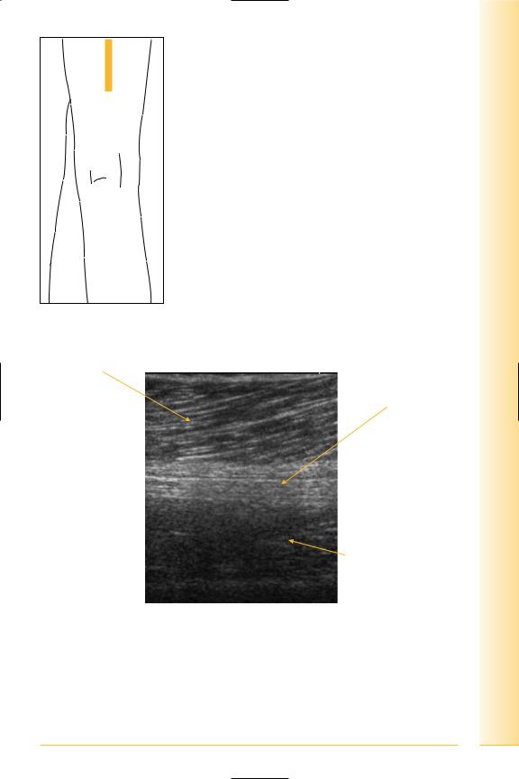
Posterior thigh
(Figures 220 and 221)
Hamstrings
•Semimembranosus
insertion: postero-medial tibial condyle.
•Semitendinosus
insertion: medial tibia (pes anserinus).
•Biceps femoris
insertion: fibular apex.
Notes

limb Lower
Thigh
FIG. 220 TS, prone, posterior thigh
Biceps femoris
Lateral intermuscular |
Semitendinosus |
septum |
Vastus lateralis 
Adductor magnus
Lateral
Medial
Sciatic nerve
FIG. 221 TS, mid-posterior thigh
179

of Atlas
ultrasound musculoskeletal anatomy
180
Sciatic nerve
(Figures 222 and 223)
This nerve is covered by gluteus maximus and hamstring muscles and lies on ischium, obturator internus, quadratus femoris, and adductor magnus.
Notes
limb Lower
Thigh
FIG. 222 LS, prone, posterior thigh
Biceps femoris
Sciatic nerve
Proximal |
Distal |
Adductor magnus
FIG. 223 LS, mid-posterior thigh
181

of Atlas
ultrasound musculoskeletal anatomy
182
Adductor canal
(Figures 224 and 225)
Femoral vessels pass through opening in adductor magnus just above adductor tubercle to the posterior knee.
Notes

limb Lower
Thigh
FIG. 224 TS, supine, probe antero-medial
Vastus medialis
Sartorius
|
Adductor magnus |
Anterior |
Posterior |
Femur |
|
Femoral artery |
Femoral vein |
FIG. 225 Distal anterior medial thigh, adductor canal
183